한국인에서의 후골간신경의 분지 위치 및 순서
가톨릭대학교 의과대학 재활의학교실, 제주대학교 의과대학 재활의학교실�
고영진∙이종인∙박주현∙최은석∙신지남�∙홍현택∙강현규∙원선재
– Abstract –
The Anatomy of the Posterior Interosseous Nerve in Korean:
A Cadaver Dissection Study
Young Jin Ko, M.D., Jong In Lee, M.D., Joo Hyun Park, M.D., Eun Seok Choi, M.D., Ji Nam Shin, M.D.*, Hyun Taek Hong, M.D., Hyun Kyu Kang, M.D., and Sun Jae Won, M.D.
Department of Rehabilitation Medicine, College of Medicine, The Catholic University of Korea and Department of Rehabilitation Medicine, College of Medicine, Cheju National University*
Objectives: The aim of this study was to describe the most common order and location of branching patterns of the posterior interosseous nerve (PIN) in Koreans.
Methods: Eight limbs from four male cadavers were dissected. The order and location of the branching from PIN were identified in relation to the bony landmarks. Bony landmarks used were lateral epicondyle of humerus and Lister’ s tubercle of radius. The length of the forearm was defined as the distance from lateral epicondyle to Lister’ s tubercle. The locations of the branching points were measured from lateral epicondyle to the branching point, and were expressed as percentage in comparison to the total length of the forearm.
Results: Though it was variable between individual specimens, the most typical branching order of PIN from proximal to distal was ECRB, supinator, EDC, ECU, EDQ, APL, EPB, EPL and EIP. ECRB was innervated by radial nerve in two specimens (25%). Branching to the muscle was completed within 80%
of forearm length. PIN was detected to run beneath the supinator muscle at a location of between 9.8%
and 22.7% of the length of forearm.
Conclusion: The identification of the branching order and location of PIN in relation to the bony land- mark would be useful in diagnosing and evaluating the PIN injury.
Key Words: Posterior interosseous nerve, Radial nerve, Posterior interosseous syndrome
Address reprint requests to Sun Jae Won, M.D.
442-723 Department of Rehabilitation Medicine, Holy Family Hospital, College of Medicine, The Catholic University, Sosabon 2-dong, Sosa-gu, Bucheon-si, Gyeonggi-do, Korea TEL : 82-32-340-2170, FAX : 82-32-340-2173, E-mail : [email protected]
서 론
후골간신경(Posterior interosseous nerve)은 요골 신경(Radial nerve)의 신경분지로, 주관절 외상과의 상부에서 천요골신경(Superficial radial nerve)을 분 지한 이후를 말하는데, 이후 주관절의 외측을 지난 뒤 외회근 하부를 거쳐 대부분의 상완의 신전근에 분지를 내고 손목관절까지 주행한다. 장요측수근신근(Exten-
sor carpi radialis longus)을 제외한 전완부의 모든 신전근을 지배하며, 지배근육으로는 단요측수근신근 (Extensor carpi radialis brevis), 외회근(Supina- tor), 지신근(Extensor digitorum), 척측수근신근 (Extensor carpi ulnaris), 소지신근(Extensor digi- ti quinti), 장무지외전근(Abductor pollicis longus), 단무지신근(Extensor pollicis brevis), 장 무지신근(Extensor pollicis longus), 그리고 시지신
근(Extensor indicis proprius)이 있다. 후골간신경증 후군이나, 상완의 골절, 종양 등에 의해 후골간신경의 손상이 일어날 수 있는데, 신경손상의 진단과 치료에 있어 후골간신경의 분지 순서 및 위치를 정확히 아는 것이 중요하다. 후골간신경에 대한 해부학적 연구는 여 러 차례 보고되었는데,1-5 연구에 따라 신경의 분지 순서 및 위치는 차이가 있다. 인종에 따른 차이는 보고된 바 없으며, 아직 한국인에서의 연구도 보고된 바가 없다.
이에 저자들은 한국인에서의 후골간신경의 분지 위치 및 그 순서를 해부학적 구조물에서 알아보고자 본 연구 를 시행하였다.
연구대상 및 방법
상지의 외상 및 수술한 흔적이 없는 총 4구의 성인 사체의 8개의 상지를 대상으로 시행하였으며, 모두 남 자이었다. 모든 사체를 앙와위로 눕히고, 먼저 견관절, 주관절, 완관절을 해부학적 자세를 취한 뒤 상완을 내 회전시켜 손바닥이 바닥에 모두 닿도록 하였다. 상완근 (Brachialis)과 완요골근(Brachioradialis)사이를 박 리하여 요골신경을 확인한 후 주행을 따라 내려가면서 신경분지를 박리하여3 신경분지의 분지순서와 위치를 구 하였다. 신경분지의 위치는 주관절외상과로부터 신경분 지까지의 거리를 상완의 길이에 대한 백분율로 표기하 였고, 상완의 길이는 주관절외상과와 요골의 Lister’s
Fig. 1. The length of the forearm was defined as the distance from lateral epicondyle to Lister’s tubercle; ECRB, extensor carpi radi- alis brevis; EDC, extensor digitorum communis; ECU, extensor carpi ulnaris.
Fig. 2. Example of branching of the postrior interosseous nerve (PIN). Black arrow shows the branching point to extensor digitorum communis (EDC), extensor carpi ulnaris (ECU) and extensor digiti quinti (EDQ). The dotted line is the distance of the branching point; APL, abductor pollicis longus; AS, aponeurosis of supinator muscle; LE, Lateral epicondyle of humerus;
EIP, extensor indicis proprius; EPB, extensor pollicis brevis; EPL, extensor pollicis longus.
tubercle 까지로 하였으며(Fig. 1)(Fig. 2), 후골간신 경이 외회근 하부로 들어가는 지점과 나오는 지점을 같 은 방법으로 표기하였다. 운동신경분지의 위치는 신경 줄기의 신경주막(perineurium)에서 신경이 갈라져서 나가는 점으로 정의하였다.
결 과
1. 후골간신경의 분지순서
전형적인 신경분지 순서는 단요측수근신근, 외회근,
지신근, 척측수근신근, 소지신근, 장무지외전근, 단무 지신근, 장무지신근, 시지신근의 순이었다. 총 8지 중 2지에서 단요측수근신근이 요골신경에서 분지되었고, 또한 3지에서 장무지신근이 단무지신근보다 먼저 분지 되었다(Table 1).
2. 후골간신경의 분지위치
상완의 길이는 평균 20.0±0.76 cm이었다. 천요골신 경과 단요측수근신근으로 가는 분지는 모두 주관절외상 과의 근위부에서 분지하였고, 지신근이하의 분지는 외 회근보다 원위부에서 분지하였다. 모든 신경이 상완 길
Fig. 3. The aponeurosis of supinator was dissected and the posterior interosseous nerve (PIN) was exposed. PIN was detected to run beneath the supinator muscle at a location between 9.8% and 22.7% of the length of forearm. Black arrowhead; upper border of supinator. White arrowhead; lower border of supinator.
Table 1. Proximal-to-distal innervation order of PIN in eight specimens
Specimen No. Innervation Order
1 ECRB, SRN, SUP, EDC, EDQ, ECU, APL, EPB, EPL, EIP
2 SRN, ECRB, SUP, EDC, ECU, EDQ, APL, EPB, EPL, EIP
3 ECRB, SRN, SUP, EDC, ECU, EDQ, APL, EPB, EPL, EIP
4 SRN, ECRB, SUP, ECU, EDC, EDQ, APL, EPB, EPL, EIP
5 SRN, ECRB, SUP, EDC, ECU, EDQ, APL, EPL, EPB, EIP
6 SRN, ECRB, SUP, EDC, ECU, EDQ, APL, EPB, EPL, EIP
7 SRN, ECRB, SUP, EDC, EDQ, ECU, APL, EPL, EPB, EIP
8 SRN, ECRB, SUP, EDC, ECU, EDQ, APL, EPL, EPB, EIP
SRN, superficial radial nerve; ECRB, extensor carpi radialis brevis; SUP, supinator; EDC, extensor digitorum communis; ECU, extensor carpi ulnaris; EDQ, extensor digiti quinti; APL, abductor pollicis longus; EPB, extensor pollicis brevis; EPL, extensor polli- cis longus; EIP, extensor indicis proprius
이의 80% 이내에서 분지되었다(Table 2).
3. 후골간신경과 외회근의 관계
후골간신경은 주관절외상과로부터 상완의 길이의 9.8
±4.3% 가 되는 지점에서 외회근 하부로 들어갔으며 22.7±3.4% 지점에서 외회근을 벗어났다(Fig. 3).
고 찰
전형적인 분지순서는 단요측수근신근, 외회근, 지신 근, 척측수근신근, 소지신근, 장무지외전근, 단무지신 근, 장무지신근, 시지신근의 순이었으며, 이는 다른 여 러 연구와 비슷한 결과이다.4-8 후골간신경의 정확한 분 지순서는 복합 외상이나 척수손상등으로 인한 상지 마 비에서 정확한 진단과 손상부위의 규명에 중요하다. 단 요측수근신근의 경우 후골간신경에서 분지하는 것으로 알려져 있으나,6 Liu 등4은 10%에서 요골신경에서 분 지하는 것으로 보고하였고, Thomas 등1은 45%는 요 골신경, 29%는 천요골신경에서 분지하고, 후골간신경 에서는 26%만이 분지하는 것으로 보고하였다. 저자들 의 연구에서는 25%에서 요골신경에서 분지하였고, 천 요골신경에서의 분지는 관찰되지 않았다.
후골간신경의 분지에 대한 기존의 연구는 대부분이 그 길이를 직접 재어 보고하였으나,2,4,6 개개인에 따라
상완의 길이가 다르고, 그에 따라 신경의 길이도 차이 가 나기 때문에 임상적으로 이용하기에는 어려움이 있 다. 따라서 저자들은 주관절의 외상과와 Lister’s tubercle 까지의 길이를 기준으로 신경분지 위치를 표 기하였다.
외회근 이하 부위의 신경분지는 그 분지순서가 더욱 다양한데, 지신근, 척측수근신근, 소지신근 으로 가는 분지를 1군, 장무지외전근, 단무지신근, 장무지신근으 로 가는 분지를 2군, 시지신근으로 가는 분지를 3군으 로 하였을 때, 후골간신경이 외회근을 벗어난 이후 1군 이 분지되고, 이어서 2군, 3군의 순서로 분지된다. 구 체적인 부위를 보면 지신근, 척측수근신근, 소지신근은 25~27% 부위에서 분지되었고, 장무지외전근, 단무지 신근, 장무지신근은 37~52% 부위에서, 시지신근은 65~80% 부위에서 분지되었다. 각 군내에서는 그 분지 순서가 다양하게 바뀌었지만 2군에서는 장무지외전근이 항상 가장 먼저 분지되었다. 이러한 결과는 기존의 보 고에서도 확인할 수 있었다.3,4,6 전체적으로 볼 때 외회 근, 장무지외전근, 시지신근의 순서는 바뀌지 않았다.
Suematsu 등9은 후골간신경증후군을 3 가지 형태로 제안하였는데, 1 형은 무지를 포함한 모든 수지의 마 비, 2형은 무지의 마비, 3형은 무지를 제외한 수지의 마비로 구분하여, 후골간신경이 외회근의 근위부에서 포착될 경우 1형이 많고, 외회근의 원위부에서 포착될 경우 2형과 3형이 많다고하여, 후골간신경손상과 외회 근의 연관성을 보고하였다. 저자들의 연구에서 후골간 신경은 평균적으로 외상과로부터 상완길이의 9,8% 지 점에서 외회근내로 들어가 22.7% 지점에서 외회근을 벗어났다. 이러한 결과는 후골간신경손상의 진단과 손 상부위에 규명에 도움이 될 것이다.
후골간신경은 시지신근으로 가는 마지막 분지를 낸후 계속 주행하여 완관절낭의 배측부에 대한 감각을 담당 한다.10 시지신근으로 분지를 낸 후의 후골간신경은 감 각신경만으로 이루어져 주로 신경이식시에 공여부로 이 용되기 때문에 몇몇 연구에서 그 주행에 관해 보고하였 는데, 평균적으로 6.2 cm 길이의 신경을 얻을 수 있다 고 하였다.2,11 본 연구에서도 시지신근으로 가는 분지이 후에는 근육을 지배하는 분지는 관찰할 수 없었으며, 모두 주관절 외상과로부터 상완길이의 80% 이내의 거 리에서 분지되어, 시지신근으로의 분지 이후 완관절낭 의 배측부로 들어가기까지 약 4.0 cm 길이의 신경은 신경이식시 공여부로 이용할 수 있을 것으로 생각된다.
강직 조절을 위한 운동점 차단술의 경우 각 근육의 운동점의 위치와 갯수가 중요한데, 본 연구에서는 이에 대한 연구는 이뤄지지 못했다. 또한 후골간신경은 그 분지순서가 매우 다양한데, 표본의 수가 8개밖에 되지 않은 점도 본 연구의 부족한 점이다. 향후 좀더 많은 표본에서의 연구가 필요할 것으로 생각된다.
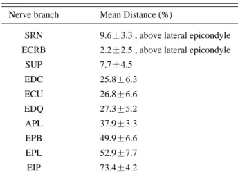
Table 2. Mean pertinent distances (%) in nerve branches of PIN
Nerve branch Mean Distance (%)
SRN 9.6±3.3 , above lateral epicondyle ECRB 2.2±2.5 , above lateral epicondyle
SUP 7.7±4.5 EDC 25.8±6.3 ECU 26.8±6.6 EDQ 27.3±5.2 APL 37.9±3.3 EPB 49.9±6.6 EPL 52.9±7.7 EIP 73.4±4.2
SRN, superficial radial nerve; ECRB, extensor carpi radialis brevis; SUP, supinator; EDC, extensor digitorum communis;
ECU, extensor carpi ulnaris; EDQ, extensor digiti quinti; APL, abductor pollicis longus; EPB, extensor pollicis brevis; EPL, extensor pollicis longus; EIP, extensor indicis proprius Values are mean±SD
결 론
저자들은 상지의 외상 및 수술한 흔적이 없는 총 4구 의 한국인 성인 사체의 8개의 상지를 대상으로 해부학 적 연구을 통하여 다음과 같은 결과를 얻었다.
1. 전형적인 분지순서는 단요측수근신근, 외회근, 지 신근, 척측수근신근, 소지신근, 장무지외전근, 단무지 신근, 장무지신근, 시지신근의 순이었다.
2. 단요측수근신근으로 가는 분지는 25%가 요골신경 에서 분지되었으며, 37.5%에서 장무지신근이 단무지신 근보다 먼저 분지되었다.
3. 분지순서는 개인차가 있으나, 외회근, 장무지외전 근, 시지신근의 분지순서는 항상 일정하였다.
본 연구에서 확인한 후골간신경의 분지순서 및 위치 는 한국인에서 후골간신경의 손상을 진단 및 평가하는 데 도움이 될 것으로 생각된다.
참고문헌
01. Thomas SJ, Yakin DE, Parry BR, Lubahn JD: The anatomical relationship between the posterior interosseous nerve and the supinator muscle. J Hand Surg 2000: 25:
936-941.
02. Elgafy H, Ebraheim NA, Yeasting RA: The anatomy of the posterior interosseous nerve as a graft. J Hand Surg 2000: 25: 930-935.
03. Missankov AA, Sehgal AK, Mennen U: Variations of the posterior interosseous nerve. J Hand Surg 2000: 25: 281- 282.
04. Liu J, Pho RW, Pereira BP, Lau HK, Kumar VP: Distribu- tion of primary motor nerve branches and terminal nerve entry points to the forearm muscles. Anat Rec 1997: 248:
456-463.
05. Seradge H, Tian W, Baer C, Seradge A: Anatomical varia- tion of the posterior interosseous nerve: a cadaver dissec- tion study. Orthopedics 2000: 23: 1195-1196.
06. Abrams RA, Ziets RJ, Lieber RL, Botte MJ: Anatomy of the radial nerve. Motor branches in the forearm. J Hand Surg 1997: 22: 232-237.
07. Branovacki G, Hanson M, Cash R, Gonzalez M: The innervation pattern of the radial nerve at the elbow and in the forearm. J Hand Surg 1998: 23: 167-169.
08. Sunderland S: Nerve grafting. Nerves and nerve injuries.
2nd ed. Baltimore: Williams & Wilkins, 1978, pp603-636.
09. Suematsu N, Hirayama T: Posterior interosseous nerve palsy. J Hand Surg 1998: 23: 104-106.
10. Dellon AL: Partial dorsal wrist denervation: resection of the distal posterior interosseous nerve. J Hand Surg 1985:
10: 527-533.
11. Waters PM, Schwartz JT: Posterior interosseous nerve: an anatomic study of potential nerve grafts. J Hand Surg 1993: 18: 743-745.