ORIGINAL ARTICLE
담도암 및 담낭암 환자에서 18 F-Fluorodeoxyglucose
양전자방출단층촬영으로 측정한 원발 종양 최대 표준화섭취계수의 생존에 대한 예후적 가치
이지용, 김홍주, 임서형, 신동석, 유정희, 주덕윤, 박정호, 박동일, 조용균, 손정일, 전우규, 김병익
성균관대학교 의과대학 강북삼성병원 내과학교실
Primary Tumor Maximum Standardized Uptake Value Measured on
18F-Fluorodeoxyglucose Positron Emission Tomography-Computed Tomography Is a Prognostic Value for Survival in Bile Duct and Gallbladder Cancer
Ji Yong Lee, Hong Joo Kim, Seo Hyung Yim, Dong Suk Shin, Jung Hee Yu, Deok Yun Ju, Jung Ho Park, Dong Il Park, Yong Kyun Cho, Chong Il Sohn, Woo Kyu Jeon and Byung Ik Kim
Department of Internal Medicine, Kangbuk Samsung Medical Center, Sungkyunkwan University School of Medicine, Seoul, Korea
Background/Aims: Few studies have assessed the prognostic value of the primary tumor maximum standardized uptake value (SUVmax) measured by 2-[18F]-fluoro-2-deoxy-D-glucose PET-CT for patients with bile duct and gallbladder cancer.
Methods: A retrospective analysis of 61 patients with confirmed bile duct and gallbladder cancer who underwent FDG PET-CT in Kangbuk Samsung Medical Center (Seoul, Korea) from April 2008 to April 2011. Prognostic significance of SUVmax and other clinicopathological variables was assessed.
Results: Twenty-three patients were diagnosed as common bile duct cancer, 17 as hilar bile duct cancer, 12 as intrahepatic bile duct cancer, and nine as gallbladder cancer. In univariate analysis, diagnosis of intrahepatic cholangiocarcinoma and gallbladder cancer, mass forming type, poorly differentiated cell type, nonsurgical treatment, advanced American Joint Committee on Cancer (AJCC) staging and primary tumor SUVmax were significant predictors of poor overall survival. In multivariate analysis adjusted for age and sex, primary tumor SUVmax (hazard ratio [HR], 4.526; 95% CI, 1.813-11.299), advanced AJCC staging (HR, 4.843; 95% CI, 1.760-13.328), and nonsurgical treatment (HR, 6.029; 95% CI, 1.989-18.271) were independently associated with poor overall survival.
Conclusions: Primary tumor SUVmax measured by FDG PET-CT is an independent and significant prognostic factor for overall survival in bile duct and gallbladder cancer. (Korean J Gastroenterol 2013;62:227-233)
Key Words: Prognosis; Fluorodeoxyglucose F18; Positron-emission tomography and computed tomography; Bile duct neoplasms;
Gallbladder neoplasms
Received June 22, 2013. Revised August 8, 2013. Accepted August 9, 2013.
CC This is an open access article distributed under the terms of the Creative Commons Attribution Non-Commercial License (http://creativecommons.org/licenses/
by-nc/3.0) which permits unrestricted non-commercial use, distribution, and reproduction in any medium, provided the original work is properly cited.
교신저자: 김홍주, 110-746, 서울시 종로구 새문안로 29, 성균관대학교 의과대학 강북삼성병원 내과학교실
Correspondence to: Hong Joo Kim, Department of Internal Medicine, Kangbuk Samsung Medical Center, Sungkyunkwan University School of Medicine, 29 Saemunan-ro, Jongno-gu, Seoul 110-746, Korea. Tel: +82-2-2001-2060, Fax: +82-2-2001-2049, E-mail: [email protected]
Financial support: None. Conflict of interest: None.
INTRODUCTION
The diagnosis of bile duct and gallbladder cancer is diffi- cult and the prognosis of both types of cancer is poor.1 The
overall 5-year survival rate of gallbladder cancer is <5%, and the natural history of unresectable cholangiocarcinoma re- sults in no survival at year 5.2-4 Currently, only surgical re- section with appropriate lymph node dissection remains as
the curative approach in some patients.5-7 An extensive work-up that includes diagnostic imaging modalities, such as ultrasonography, CT, MRI, and endoscopic measures, is es- sential for accurate tumor staging and treatment decisions.8 However, differential diagnosis based solely on structural imaging information remains challenging because of its vari- able reliability and lack of information on its effect on predict- ing and improving prognosis.
2-[18F]-Fluoro-2-deoxy-D-glucose (FDG) PET-CT that pro- vides metabolic and anatomic information of malignant cells simultaneously is a relatively new and accurate diagnostic approach in various malignancies. Several studies have de- scribed the usefulness of PET-CT in the detecting and staging of bile duct and gallbladder cancer.8-10 In these studies, the FDG PET-CT measured the maximum standardized uptake value (SUVmax), a semiquantitative simplified measurement of tissue deoxyglucose metabolic rate. A few retrospective studies have explored the survival prognostic significance of the primary tumor SUVmax in various malignancies.11-13 In bile duct and gallbladder cancer, fewer studies have been con- ducted to evaluate the prognostic value of primary tumor SUVmax and most of these reports included only a small num- ber of patients.14-16
So, we performed a retrospective analysis from pro- spective data to determine the prognostic value of primary tu- mor SUVmax for patients with bile duct and gallbladder cancer.
MATERIALS AND METHODS
1. Patients
Sixty-one patients who were diagnosed with bile duct and gallbladder cancer and underwent FDG PET-CT in Kangbuk Samsung Medical Center (Seoul, Korea) from April 2008 to April 2011 were included in this study. The prospectively col- lected data and medical records of each patient were re- viewed retrospectively. The diagnosis of bile duct and gall- bladder cancer was confirmed by pathological analysis of surgical specimens in resectable patients (n=16) and patho- logical specimens that obtained from percutaneous biopsy or endoscopic biopsy (n=29) or clinical follow-up (n=16) in unresectable patients. Bile duct cancer was classified as in- trahepatic, hilar or extrahepatic lesions according to the site of lesion shown on conventional imaging including multi- dector row CT (MDCT), MRCP, ERCP or EUS. All cancers were
classified according to their morphologic features revealed by imaging as mass-forming, periductal infiltrating, or intra- ductal-growing type.17 Histologic typing was classified as well, moderately, or poorly differentiated according to the World Health Organization classification.18 Staging criteria were based on the clinical staging system of the American Joint Committee on Cancer (AJCC) TNM classification.19 We also classified AJCC staging as early (I, II) and advanced (III, IV) stage. Treatment method was classified as surgical treat- ment in cases of curative surgery and non-surgical treatment in cases of exploratory laparotomy and palliative treatment.
We considered the patient with fever (body temperature
>38oC) or laboratory evidence of inflammatory response (white blood cell count >10 or <4×10⁹/L, or CRP ≥1 mg/dL), accompanied by jaundice (total bilirubin ≥2 mg/dL ) or ab- normal liver function tests (when serum AST, ALT, ALP and GGT levels are more than 1.5 times of upper normal limits) 24 hours before and after performing PET-CT, as cholangitis patient.20 The institutional review board approved this retro- spective study and waived patient consent for this specific study (KBC13065).
2. FDG PET-CT
All patients fasted for at least 6 hours before 18F-FDG PET-CT. The blood glucose level was checked before 18F-FDG administration and the patient was rescheduled if the blood glucose level exceeded 130 mg/dL. A range of 370-555 MBq for 18F-FDG was injected intravenously. Scanning began 60 minutes later after voiding. No intravenous contrast agent was used for the CT scans. Imaging and data acquisition were performed using a Discovery STE combined PET-CT system (General Electric Medical Systems, Milwaukee, WI, USA). A PET scanner composed of 13,440 bismuth germinate crys- tals arranged in 24 rings, integrated with a 16 multislice heli- cal CT scanner, acquired the co-registration of the PET and CT images in one session. A total of 6-8 bed positions were avail- able, and the acquisition time per bed position was 2 minutes. All patients were examined in the supine position with their arms raised. The PET-CT images were recon- structed using iterative reconstruction with two iterations and 14 subsets, resulting in 47 two-dimensional sections spaced 3.27 mm apart, over each axial field-of-view incre- ment of 157 mm. The attenuation-corrected PET images, CT images, and co-registered PET-CT images were analyzed si-
Table 1. Baseline Clinicopathologic Characteristics of the Patients
Characteristic Data
Total number 61 (100.0)
Age (yr) 68.5±8.9
Gender (male) 32 (52.5)
Final diagnosis CBDC Hilar IBDC GBC
23 (37.7) 17 (27.9) 12 (19.7) 9 (14.8) Morphologic type
Mass forming Periductal infiltrating Intraductal growing
31 (50.8) 30 (49.2)
0 (0) Tumor differentiation
Well differentiated Moderately differentiated Poorly differentiated
16 (45.7) 13 (37.1) 6 (17.1) AJCC stage
Early (I/II) Advanced (III/IV)
49 (80.3) 12 (19.7)
Primary tumor size (cm) 4.3±3.1
Diagnostic sensitivity of FDG PET-CT 51 (83.6)
SUVmax of primary tumor 6.4±4.6
Cholangitis 7 (11.5)
Values are presented as mean±SD or n (%).
CBDC, common bile duct cancer; IBDC, intrahepatic bile duct cancer;
GBC, gallbladder cancer; AJCC, American Joint Committee on Cancer;
FDG PET-CT, 2-[18F]-fluoro-2-deoxy- D-glucose PET-CT; SUVmax, maximum standardized uptake value.
multaneously by a board-certified nuclear medicine physi- cian on an eNTEGRA workstation with viewing-dedicated software (ELGEMS, Haifa, Israel). Image interpretation was based on identifying regions with increased FDG uptake on PET images and the anatomic delineation of all FDG-avid le- sions on the coregistered PET-CT images. Tumors were de- fined as positive FDG uptake if the radioactivity of the tumor was higher than that of the surrounding liver tissue in the vis- ual analysis. Images of each biliary tumor were assessed semiquantitatively by measuring and calculating the SUVmax
normalized to lean body mass. The SUVmax was calculated for the quantitative analysis of tumor 18F-FDG uptake, as follows:
SUVmax=C (kBq/mL)/ID (kBq)/body weight (kg) where C is the tissue activity concentration measured by PET and ID is the injected dose. All CT images were viewed separately to identi- fy additional lesions without FDG uptake using soft tissue, lung, and bone window leveling.
3. Statistical analyses
Patients were stratified and analyzed using with regard to sex, age, diagnosis, morphological feature, histologic typing, treatment method, AJCC stage, primary tumor size and pri- mary tumor SUVmax. To increased statistical power, we sub- grouped AJCC stage as early (I, II) and advanced (III, IV), and treatment as surgical (curative surgery) and non-surgical (exploratory laparotomy and palliative treatment). The You- den index was used to determine the cutoff value of SUVmax
that yielded the optimal sensitivity and specificity. Survival time was defined as the time interval from the date of patho- logic diagnosis or clinically confirmed diagnosis until death or the last follow-up date. Overall cumulative survival was an- alyzed by the Kaplan-Meier method, and univariate analysis was performed with a log rank comparison. A value of p<0.05 was considered to be statistically significant. Variables with p<0.05 in univariate analysis of factors affecting survival were included in a subsequent multivariate analysis, using a Cox proportional hazard regression analysis. The relation- ship between primary tumor SUVmax and cholangitis was ana- lyzed by Fisher’s Exact test. All statistical analyses were per- formed using IBM SPSS Statistics ver. 18.0 (IBM, Armonk, NY, USA).
RESULTS
1. Characteristics of all patients
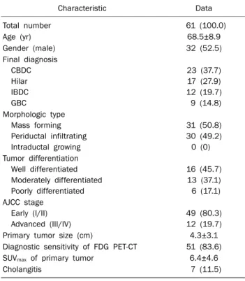
The patient characteristics are summarized in Table 1.
Twenty-nine (47.5%) patients were female. Final diagnosis was confirmed by curative resection, percutaneous and/or endoscopic biopsy, and follow-up in 16 (26.2%), 29 (47.5%) and 16 (26.2%) patients, respectively. Fourteen (23.0%) pa- tients received with curative surgery, three (4.9%) patients received exploratory laparotomy and 44 (72.1%) patients were palliatively treated. No patient had cancer with intra- ductal growing morphology. Seven (11.5%) patients who had cholangitis at 24 hours before and after undergoing PET-CT.
The Youden index showed that the optimal cutoff value of pri- mary tumor SUVmax for predicting survival was 5.5. Using pri- mary tumor SUVmax of 5.5 yielded a sensitivity 69.2% and a specificity 77.3%. There was no significant association be- tween primary tumor SUVmax and cholangitis (p=0.699).
Fig. 1. Univariate analysis of the patient population, tumor characteristics, and overall survival. Kaplan-Meier survival curves with log rank comparisons between each group of final diagnosis (A), morphological type (B), histologic grade (C), treatment method (D), American Joint Committee on Cancer stage (E) and primary tumor standardized uptake value (SUVmax) (F). Log rank p-values were <0.05 for each comparison, respectively.
Fig. 2. Axial FDG PET-CT images of two patients with common bile duct cancer who received nonsurgical treatment. Primary tumor maximum standardized uptake value is 3.1 (arrow) in 69-year-old man who was alive 26 months after diagnosis (A), and 9.9 (arrow) in 76-year-old man who died 10 months after diagnosis (B), respectively.
2. Prognostic factors of bile duct and gallbladder cancer In all 61 patients, the overall median survival was 10.0 months (95% CI, 7.39 -12.60). In univariate analysis with log
rank comparisons, diagnoses of intrahepatic bile duct can- cer (IBDC) (p<0.001) and gallbladder cancer (p=0.004), mass forming type (p=0.009), poorly differentiated cell type (p<0.001), nonsurgical treatment (p=0.004), advanced
Table 2. Cox Proportional Hazard Regression Analysis of Factors Associated with Overall Survival of Bile Duct and Gallbladder Cancer
Factor Patient (n) Unadjusted Adjusteda
HR 95% CI p-value HR 95% CI p-value
Primary tumor SUVmax
<5.5 32 1 1
≥5.5 29 3.927 1.968-7.836 <0.001 4.526 1.813-11.299 0.001
AJCC stage
Early (I/II) 49 1 1
Advanced (III/IV) 12 10.646 4.428-25.596 <0.001 4.843 1.760-13.328 0.002
Treatment
Surgical 14 1 1
Nonsurgical 47 4.053 1.559-10.537 0.004 6.029 1.989-18.271 0.001
Morphology
Mass forming 31 1 1
Periductal infiltrating 30 0.418 0.217-0.803 0.009 0.828 0.335-2.050 0.684
Age (yr)
≥65 41 1 1
<65 20 0.544 0.262-1.128 0.102 1.017 0.976-1.061 0.417
Sex
Male 32 1 1
Female 29 1.424 0.744-2.726 0.286 1.19 0.566-2.504 0.646
Diagnosis
CBDC 23 1
Hilar 17 2.159 0.889-5.242 0.089
IBDC 12 6.354 2.520-16.019 <0.001
GBC 9 4.153 1.585-10.883 0.004
Histology
Well 16 1
Moderate 13 2.651 0.979-7.182 0.055
Poor 6 27.532 6.494-116.730 <0.001
HR, hazard ratio; SUVmax, maximum standardized uptake value; AJCC, American Joint Committee on Cancer; CBDC, common bile duct cancer; IBDC, intrahepatic bile duct cancer; GBC, gallbladder cancer.
aMultivariate analysis.
stage (p<0.001), and higher primary tumor SUVmax than 5.5 (p<0.001) were significant associated factors of poor overall survival (Figs. 1, 2). However, primary tumor size (p=0.104), age (p=0.087) and sex (p=0.268) were not significantly asso- ciated with overall survival.
3. Primary tumor SUVmax is an independent prognostic factor of bile duct and gallbladder cancer
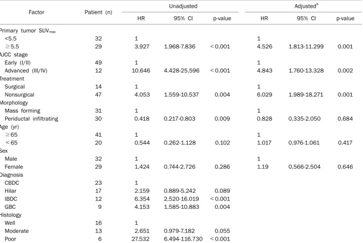
In Cox proportional hazard regression analysis, primary tu- mor SUVmax (hazard ratio [HR], 4.526; 95% CI, 1.813- 11.299; p=0.001) was an independent and significant pre- dictive factor associated with poor overall survival, regard- less of age, sex, morphological feature, AJCC stage or treat- ment. Advanced stage (HR, 4.843; 95% CI, 1.760-13.328;
p=0.002) and nonsurgical treatment (HR, 6.029; 95% CI, 1.989-18.271; p=0.001) were also independent prognostic factors of overall survival, too (Table 2). Whereas mass-form-
ing morphological feature was not an independent sig- nificant variable for predicting poor prognosis.
DISCUSSION
The usefulness of FDG PET-CT is not confined to diagnos- ing and staging cancers, but extended to the evaluation of the proliferative activity and/or malignant potential of tumors that influence prognosis. Several studies involving detecting, staging, and differentiating tumors have been conducted for bile duct and gallbladder cancer,8-10 but these did not fully as- sess the prognostic significance of this technique.
The results of our study show that a high FDG uptake in the primary tumor, measured by FDG PET-CT, was independently correlated with poor overall survival in patients with all types of bile duct and gallbladder cancer. The advanced clinical stage and nonsurgical treatment were also independent and
significant predictors of survival on multivariate analysis.
There is a possibility of inflammatory effect because the coexisting active inflammatory condition can result in a false-positive FDG uptake.9 In the present study, we found no significant correlation between primary tumor SUVmax and presence of cholangitis at the time of performing PET-CT.
Kitamura et al.15 reported there are no marked inflammatory effect on FDG uptake in patients with extrahepatic bile duct cancer.
Various studies have assessed whether the primary tumor SUVmax can be used to predict the survival of patients with oth- er malignancies. In lung cancer, breast cancer, malignant lymphoma, endometrial cancer and early cervical cancer, studies showed that primary tumor SUVmax on FDG-PET was an independent and significant prognostic value on multi- variate analysis.11-13,21-23 A similar study evaluated the corre- lation between FDG uptake and prognosis for patients with bile duct and gallbladder cancer.14 The results showed that high SUVmax was associated with poor overall survival on uni- variate analysis but was not independently associated with prognosis on multivariate analysis. Kitamura et al.15 reported that SUVmax on FDG PET is an independent predictor of surviv- al in patients with extrahepatic bile duct cancer on multi- variate analysis. Seo et al.16 described that SUVmax is a sig- nificant predictor of disease free survival, not overall survival, for patients with mass-forming IDBC. In all of these studies, SUVmax was evaluated solely by FDG PET. Our study is the first to measure SUVmax by FDG PET-CT; the results reveal the in- dependent and significant correlation between primary tu- mor SUVmax and overall survival in all types of bile duct and gallbladder cancer.
The association of high FDG uptake with poor prognosis may occur because FDG accumulates more in metabolically active cells with aggressive malignant potential. In bile duct cancer, FDG accumulation appears primarily to result from the increased expression of glucose transporter, especially glucose transporter-1 (Glut-1).24 The overexpression of Glut-1 is associated with depth of invasion and increased in- cidence of lymph node and hepatic metastasis in colorectal cancer,25 increased proliferation of glioma26 and worsened prognosis in non-small cell lung cancer.27 However, no report has assessed the association of up-regulation of Glut-1 with the prognosis in bile duct and gallbladder cancer. Further study is needed to clarify the relationship between the over-
expression of Glut-1 and cell proliferation, malignant poten- tial, and prognosis of bile duct and gallbladder cancer.
Increased FDG uptake may be associated with advanced stage. Some authors reported that a high SUVmax of primary tumors is significantly related to higher stage in various cancers.12,22,23,25 In bile duct cancer, one study reported a significant association between high SUVmax and advanced pathologic stage.14 In this study, the pathologic stage was al- so independently associated poor overall survival in multi- variate analysis. However, in our study, high SUVmax was asso- ciated with poor overall survival regardless of advanced stage in multivariate analysis.
Some evidence indicates that high FDG uptake is asso- ciated with recurrence in lung cancer and IBDC.16,22 In mass-forming IBDC, the disease-free survival of patients with a high primary tumor SUV was significantly worse than those with a low SUV.16 Although we have no more detailed in- formation for postoperative recurrence in our study, re- currence might be a contributory factor of poor survival.
Recurrent malignant cancer cells tend to be more aggressive and resistant to palliative treatment.
There are several limitations in this study. Although we used prospective database by using a single PET-CT instru- ment, this study is a retrospective study. We could not eval- uate the prognostic value of primary tumor SUVmax for each types of final diagnosis because of the small number of in- cluded patients. Additional larger and prospective studies are needed to elucidate the clinical significance of FDG PET-CT and tumor SUV for bile duct and gallbladder cancer, and for each type of them.
In conclusion, primary tumor SUVmax measured using FDG PET-CT is an independent and significant prognostic factor for overall survival in patients with bile duct and gallbladder cancer. Prospective analyses with larger numbers of cases are warranted for confirmation.
REFERENCES
1. Okuda K, Nakanuma Y, Miyazaki M. Cholangiocarcinoma: re- cent progress. Part 1: epidemiology and etiology. J Gastroenterol Hepatol 2002;17:1049-1055.
2. Farley DR, Weaver AL, Nagorney DM. “Natural history” of un- resected cholangiocarcinoma: patient outcome after non- curative intervention. Mayo Clinic Proc 1995;70:425-429.
3. Park J, Kim MH, Kim KP, et al. Natural history and prognostic fac-
tors of advanced cholangiocarcinoma without surgery, chemo- therapy, or radiotherapy: a large-scale observational study. Gut Liver 2009;3:298-305.
4. Miyazaki M, Ito H, Nakagawa K, et al. Does aggressive surgical resection improve the outcome in advanced gallbladder carci- noma? Hepatogastroenterology 1999;46:2128-2132.
5. Jarnagin WR, Fong Y, DeMatteo RP, et al. Staging, resectability, and outcome in 225 patients with hilar cholangiocarcinoma.
Ann Surg 2001;234:507-517; discussion 517-519.
6. Endo I, Gonen M, Yopp AC, et al. Intrahepatic cholangiocar- cinoma: rising frequency, improved survival, and determinants of outcome after resection. Ann Surg 2008;248:84-96.
7. Ercolani G, Vetrone G, Grazi GL, et al. Intrahepatic cholangio- carcinoma: primary liver resection and aggressive multimodal treatment of recurrence significantly prolong survival. Ann Surg 2010;252:107-114.
8. Petrowsky H, Wildbrett P, Husarik DB, et al. Impact of integrated positron emission tomography and computed tomography on staging and management of gallbladder cancer and cholangio- carcinoma. J Hepatol 2006;45:43-50.
9. Kim JY, Kim MH, Lee TY, et al. Clinical role of 18F-FDG PET-CT in suspected and potentially operable cholangiocarcinoma: a pro- spective study compared with conventional imaging. Am J Gastroenterol 2008;103:1145-1151.
10. Lee SW, Kim HJ, Park JH, et al. Clinical usefulness of 18F-FDG PET-CT for patients with gallbladder cancer and cholangiocar- cinoma. J Gastroenterol 2010;45:560-566.
11. Lee YY, Choi CH, Kim CJ, et al. The prognostic significance of the SUVmax (maximum standardized uptake value for F-18 fluo- rodeoxyglucose) of the cervical tumor in PET imaging for early cervical cancer: preliminary results. Gynecol Oncol 2009;115:
65-68.
12. Kitajima K, Kita M, Suzuki K, Senda M, Nakamoto Y, Sugimura K. Prognostic significance of SUVmax (maximum standardized uptake value) measured by [18F]FDG PET/CT in endometrial cancer. Eur J Nucl Med Mol Imaging 2012;39:840-845.
13. Ahmadzadehfar H, Rodrigues M, Zakavi R, Knoll P, Mirzaei S.
Prognostic significance of the standardized uptake value of pre-therapeutic (18)F-FDG PET in patients with malignant lymphoma. Med Oncol 2011;28:1570-1576.
14. Furukawa H, Ikuma H, Asakura K, Uesaka K. Prognostic im- portance of standardized uptake value on F-18 fluorodeoxy- glucose-positron emission tomography in biliary tract carci- noma. J Surg Oncol 2009;100:494-499.
15. Kitamura K, Hatano E, Higashi T, et al. Prognostic value of (18)F-fluorodeoxyglucose positron emission tomography in pa- tients with extrahepatic bile duct cancer. J Hepatobiliary Pancreat
Sci 2011;18:39-46.
16. Seo S, Hatano E, Higashi T, et al. Fluorine-18 fluorodeoxyglucose positron emission tomography predicts lymph node metastasis, P-glycoprotein expression, and recurrence after resection in mass-forming intrahepatic cholangiocarcinoma. Surgery 2008;
143:769-777.
17. Lim JH. Cholangiocarcinoma: morphologic classification accord- ing to growth pattern and imaging findings. AJR Am J Roentgenol 2003;181:819-827.
18. Bosman FT, Carneiro F, Hruban RH, et al. WHO classification of tumours of the digestive system. 4th ed. Geneva: World Health Organization, 2010.
19. Edge SB, Byrd DR, Compton CC, Fritz AG, Greene FL, Trotti A. AJCC cancer staging manual. 7th ed. New York: Springer, 2010.
20. Kiriyama S, Takada T, Strasberg SM, et al; Tokyo Guidelines Revision Committee. TG13 guidelines for diagnosis and severity grading of acute cholangitis (with videos). J Hepatobiliary Pancreat Sci 2013;20:24-34.
21. Vansteenkiste JF, Stroobants SG, Dupont PJ, et al. Prognostic im- portance of the standardized uptake value on (18)F-fluoro-2-de- oxy-glucose-positron emission tomography scan in non-small- cell lung cancer: An analysis of 125 cases. Leuven Lung Cancer Group. J Clin Oncol 1999;17:3201-3206.
22. Sasaki R, Komaki R, Macapinlac H, et al. [18F]fluorodeox- yglucose uptake by positron emission tomography predicts out- come of non-small-cell lung cancer. J Clin Oncol 2005;23:
1136-1143.
23. Oshida M, Uno K, Suzuki M, et al. Predicting the prognoses of breast carcinoma patients with positron emission tomography using 2-deoxy-2-fluoro[18F]-D-glucose. Cancer 1998;82:2227- 2234.
24. Lee JD, Yang WI, Park YN, et al. Different glucose uptake and gly- colytic mechanisms between hepatocellular carcinoma and in- trahepatic mass-forming cholangiocarcinoma with increased (18)F-FDG uptake. J Nucl Med 2005;46:1753-1759.
25. Furudoi A, Tanaka S, Haruma K, et al. Clinical significance of hu- man erythrocyte glucose transporter 1 expression at the deep- est invasive site of advanced colorectal carcinoma. Oncology 2001;60:162-169.
26. Chung JK, Lee YJ, Kim SK, Jeong JM, Lee DS, Lee MC. Compar- ison of [18F]fluorodeoxyglucose uptake with glucose trans- porter-1 expression and proliferation rate in human glioma and non-small-cell lung cancer. Nucl Med Commun 2004;25:11-17.
27. Younes M, Brown RW, Stephenson M, Gondo M, Cagle PT.
Overexpression of Glut1 and Glut3 in stage I nonsmall cell lung carcinoma is associated with poor survival. Cancer 1997;80:
1046-1051.