ARTICLE
Int J Thyroidol 2020 May 13(1): 37-42 https://doi.org/10.11106/ijt.2020.13.1.37Received April 16, 2020 / Revised May 15, 2020 / Accepted May 18, 2020
Correspondence: Dong Kun Lee, MD, PhD, Department of Otolaryngology-Head and Neck Surgery, Dong-A University College of Medicine, 26 Daesingongwon-ro, Seo-gu, Busan 49201, Korea
Tel: 82-51-240-5423, Fax: 82-51-253-0712, E-mail: [email protected]
Copyright ⓒ the Korean Thyroid Association. All rights reserved.
This is an open-access article distributed under the terms of the Creative Commons Attribution Non-Commercial License (http://creative- commons.org/licenses/by-nc/4.0/), which permits unrestricted non-commercial use, distribution, and reproduction in any medium, provided the original work is properly cited.
갑상선유두암에서 18F-FDG PET/CT의 진단적 유용성
동아대학교 의과대학 이비인후-두경부외과학교실1, 핵의학교실2
김현기
1, 이동근
1, 박헌수
1, 정영진
2Diagnostic Value of SUV in 18F-FDG PET/ CT for Papillary Thyroid Cancer
Hyungi Gim1, Dong Kun Lee1, Heon Soo Park1 and Yeong Jin Jeong2
Departments of Otolaryngology-Head and Neck Surgery1, Nuclear Medicine2, Dong-A University College of Medicine, Busan, Korea
Background and Objectives: PET/CT is widely used to determine whether metastasis or recurrence will occur following initial treatment of thyroid cancer. However, there are not much research on diagnostic usefulness of preoperative PET/CT imaging for papillary thyroid cancer (PTC). We analyzed the correlation between the preoperative maximum standard uptake value (SUVmax) of PET/CT and prognostic factors of PTC to evaluate the diagnostic usefulness of PET/CT. Materials and Methods: Of the 133 patients who underwent surgery for PTC, 88 patients who had an increased uptake in thyroid mass in preoperative PET/CT were enrolled. They were divided into two groups according to B-RAF gene mutation, extrathyroidal extension (ETE), lymph node metastasis, and recurrence. The average of the SUVmax for each group was analyzed through multiple regression analysis. Correlation analysis were performed on changes in SUVmax according to the size of the thyroid tumor. Statistical analysis was performed to determine whether there were stage differences between the 47 patients who had no increased uptake in PET/CT and the others who had. Results: There were no statistical correlation between recurrence, the presence of central/lateral lymph node metastasis, ETE, stage and the SUVmax in PET/CT (p=0.513, p=0.8, p=0.73, p=0.01, p=0.9). There were statistical correlations between the size of tumor (p=0.001), the presence of the B-RAF mutation (p=0.024) and SUVmax. Statistical correlations between the presence of the hypermetabolic uptake and high stage were found by the chi-square test (p=0.012). Conclusion: The size of tumor, B-RAF are major factors in determining prognosis of PTC. There is a possibility that there is a correlation between preoperative SUVmax and prognosis of PTC.
Key Words: Papillary thyroid carcinoma, 18F-FDG PET/CT, Standardized uptake value
서 론
18F-불화디옥시포도당 양전자컴퓨터단층촬영(18F- Fluoro-2-Deoxy-D-glucose-PET/CT, 18F-FDG PET/CT)
은 갑상선유두암종 환자들의 국소 또는 원격 전이 및 재 발 여부를 알기 위해 사용되고 있다. 특히 갑상선 수술 이후 혈장 내 갑상선글로불린(thyroglobulin)이 상승되어 재발이 의심되지만 전신 스캔(whole body radioactive iodine scans)에서 음성 반응이 나와 재발 유무를 판별하
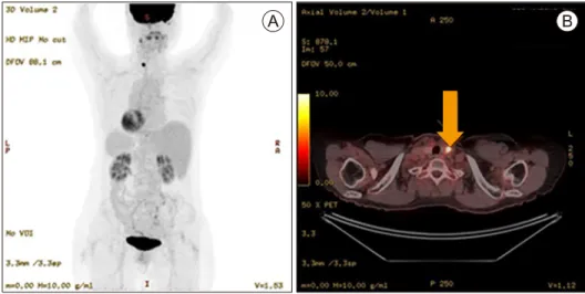
Fig. 1. Positron emission tomography/computed tomography (PET/CT) images of a patient with papillary thyroid cancer.
(A) A typical Torso 18F-fluorodeoxy-glucose (FDG)-PET image of a patient with papillary thyroid cancer. (B) A 62-year-old woman with Left papillary thyroid cancer. 18F-FDG-PET/CT image clearly shows an increased focal 18F-FDG accumulation (SUVmax=16.86) in the tumor (arrow).
기에 모호할 때 감별검사로 유용하다.1) 또한 갑상선 암 이외에, 이차성으로 발생하는 다른 암이나 원격전이의 여부를 판단하는데 도움이 된다. 갑상선유두암종이 의 심되는 갑상선 결절 환자에서, 최대 표준섭취화계수 (maximum standardized uptake value, SUVmax) 값이 양 성과 악성 결절 간에 유의한 차이가 난다는 보고가 있으 며, 최대 표준섭취화계수를 이용한 국소적 전이 여부 판 단에 대한 국외 보고는 다수 있다.2-4) 하지만 수술 전 최 대 표준섭취화계수와 갑상선암의 재발, 국소적 전이 같 은 예후인자와 최대 표준섭취화계수와의 관계에 대한 국내 연구는 없었다. 이에 저자들은 갑상선유두암으로 진단받고 18F-FDG PET/CT를 촬영한 환자들에서 최대 표준섭취화계수 값에 따른, 재발, 전이 여부 등을 분석하 였고, 수술 후 병리 결과와 비교하여, 18F-FDG PET/CT 의 유용성에 대해 알아보고자 하였다.
대상 및 방법
2010년 7월에서 2015년 12월 사이에, OO대학교병원 에서 갑상선유두암으로 갑상선절제술을 받고 수술 후 병리검사 상 갑상선유두암종으로 확진된 환자에서, 의 무 기록을 후향적으로 검토하였다. 수술 전에 18F-FDG PET/CT (Discovery 710 SE1300, GE Healthcare, Milwaukee, WI, USA)를 촬영한 환자들을 대상으로 하였다. 수술의 범위는 미국 갑상선 학회 권고기준에 따라 갑상선 절제 술만을 시행한 경우와 임파선 곽청술(neck dissection)을 병행한 경우도 포함되었다.
갑상선 종물에서 18F-FDG의 대사 항진 여부를 판별 하여 대사 항진이 관찰된 환자들 중에서 종물의 피막 침 범 여부, 임파선 전이 여부, B-type RAF kinase (B-RAF) 유전자 변이 여부, 종물의 병기를 분석하여 최대 표준섭 취화계수 값의 상승이 예후에 미치는 영향을 분석하였 다(Fig. 1). PET/CT에서 대사 항진이 관찰되지 않은 환 자에 대해서는 최대 표준섭취화계수를 측정할 수 없기 때문에 대사항진이 있는 환자들과 병기에 따른 집단별 차이가 있는지 분석을 추가로 시행하였다.
대상은 총 88명(여자 68명, 남자 20명)으로 갑상선 절 제술 및 필요시 경부임파선 곽청술을 추가로 시행하였 으며, 모든 수술은 두 명의 이비인후과 전문의에 의해 행해졌다(Table 1). 대상 중 (1) 갑상선 이외의 다른 암이 중복된 경우, (2) 이전에 갑상선유두암 수술을 받은 후 재수술하는 경우는 배제하였다.
최대 표준섭취화계수(maximum standardized uptake value, SUVmax)
표준섭취화계수는 질병 부위의 당대사율이 얼마인지 를 나타내는 반정량적 표준지표다. 반정량적인 방법으 로 정상조직과 이상 조직 간의 주입한 방사성핵종이 균 등하게 펴져 있다는 가정하에 병소 내 얼마나 평균보다 높게 섭취하는지를 아래와 같은 공식으로 표준섭취계수 를 측정하였다. SUVmax는 질병부위의 측정된 표준섭취 화계수 값 중 최대 표준섭취화계수 값이다.
SUV=r/(a’/w)
r: radioactivity activity concentration [kBq/mL],
Table 1. Characteristics of patients
Characteristics Number
Gender
Male 20
Female 68
Lobectomy 7
CCND (+) 2
CCND (−) 5
Total thyroidectomy 81
CCND (+) 65
CCND (-) 16
MRND
Done 20
Not done 68
CCND: central compartment neck dissection, MRND:
modified radical neck dissection
Fig. 2. SUVmax according to tumor size of papillary thyroid cancer.
a’: decay-corrected amount of injected radiolabeled FDG [kBq]
W: weight of the patient [g]
통계 분석
결과는 SPSS (version 22. statistical package for the social sciences by IBM)를 사용하여, B-RAF 유전자 변이 여부, 종물의 피막 침범 여부, 임파선 전이 여부, 재발 유무에 따라 두 그룹을 설정하고, 두 그룹의 최대 표준섭 취화계수의 평균을 이용하여 다중회귀분석을 통해 검토 하였다. 그리고 갑상선 종물에서 18F-FDG의 대사 항진 이 관찰된 환자들과 관찰되지 않은 환자들은 카이-제곱 검정을 통해 병기 차이가 있는지 분석하였다. 모든 자료 는 p-value가 0.05 미만일 때 유의하다고 판정하였다.
결 과
갑상선의 국소적 18F-FDG 섭취 증가를 보인 88명 중 여자는 68명, 남자는 20명이었으며, 평균 연령은 53.3세 (13-81세)였다. 모두 갑상선 종물 내에서 대사항진이 관 찰되었고, 이 중 3명은 경부임파선에서도 18F-FDG의 섭취증가가 관찰되었다. 성별과 최대 표준섭취화계수 측정값 간의 통계적 유의미한 차이는 없었다(p=0.36).
종물 병변의 평균 크기는 11.90 mm (범위, 2-60 mm)이 며, 환자들의 최대 표준섭취화계수의 평균값은 6.49 (1.18-33.69)로 나왔다. 갑상선 암의 크기에 따른 최대 표 준섭취화계수 값의 변화는 통계적으로 유의미한 상관 관계가 있었다(p=0.001) (Fig. 2). 최대 표준섭취화계수 값과 갑상선 암의 크기와의 관련성에 대해 상관분석을
실시하였으며, 상관계수는 0.512 (p<0.001)로 높은 관련 성이 있다. 수술 후 평균 경과관찰 기간은 52개월이고 (17-74개월), 경과 관찰 기간 중 갑상선유두암으로 인해 사망한 환자는 없었다.
전체 검사군 88명 중 74명은 중심 경부 곽청술 또는 측면 경부 곽청술을 시행 받았다(93.1%). 중심 경부임파 선 곽청술(central compartment neck dissection)을 시행한 환자 74명에서 36명이 수술 후 병리검사상 임파선 전이 가 확인되었으며(48.64%), 측면 경부임파선 곽청술을 시 행한 환자 21명 중 18명이 수술 후 병리검사상 임파선 전이가 확인되었다(85.7%). 중심 경부임파선 곽청술을 시행한 환자 내에서, 중심 경부에서 임파선의 전이의 유 무와 갑상선 종양의 최대 표준섭취화계수 값 간에는 통 계적 유의성이 없었다(p=0.8). 측경부 임파선의 전이 환 자도 임파선 전이 여부와 갑상선 종양의 최대 표준섭취 화계수 간의 통계적 유의성이 없었다(p=0.73). 수술을 시행한 88명의 환자 중 5명은 갑상선암 재발이 확인되었 다. 5명 모두 원격전이는 없었으며, 경부임파선 전이가 확인되었다. 갑상선암이 재발한 환자의 평균 최대 표준 섭취화계수는 7.30이고, 재발 안 한 경우의 평균 최대 표 준섭취화계수는 6.44이었다. 갑상선암 재발 유무와 최대 표준섭취화계수 간의 통계적 상관성은 없었다(p=0.513).
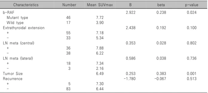
18F-FDG의 대사 항진이 관찰된 환자 88명 중 63명은 수술 후 병리검사에서 B-RAF 검사를 시행하였고, 그중 46명이(73%) 양성(돌연변이형, mutant type)으로 결과가 나왔다. B-RAF 검사가 양성인 환자의 평균 최대 표준섭 취화계수 값은 7.70으로, B-RAF 음성(wild type)의 환자 평균인 3.90보다 높았으며 두 군 간의 통계적으로 유의 미한 상관관계가 있었다(p=0.024) (Table 2).
18F-FDG의 대사 항진이 관찰된 환자 88명 중 55명이
Table 4. 18F-FDG uptake according to PTC stage (AJCC 8th)
Stage 18F-FDG uptake
Chi-square p-value
+ -
6.32 0.012
I 46 35
II 42 12
Stage and 18F-FDG uptake are statistically significantly related to the existence of uptake by stage 1 and stage2.
Chi-square test was performed.
*p<0.05.
Table 2. SUVmax according to characteristics and factors for PTC
Characteristics Number Mean SUVmax B beta p-value
b-RAF 2.922 0.238 0.024
Mutant type 46 7.72
Wild type 17 3.90
Extrathyroidal extension 2.438 0.192 0.100
+ 55 7.18
- 33 5.34
LN meta (central) 0.353 0.028 0.802
+ 36 7.88
- 38 6.22
LN meta (lateral) 0.586 0.038 0.736
+ 18 7.34
- 3 2.16
Tumor Size 6.49 0.253 0.383 0.001
Recurrence -1.780 -0.067 0.513
+ 5 7.30
- 83 6.44
F=6.201, p=0.000, R2=0.21.
Two groups were set according to the presence or absence of each item. We reviewed through multiple regression analysis using the average of the maximum standard ingestion coefficients of the two groups.
B-RAF: B-type RAF kinase, ETE: extrathyroidal extension, LN: lymph node, PTC: papillary thyroid cancer, SUVmax: the maximum standardized uptake value
Table 3. SUVmax according to PTC stage (AJCC 8th)
Stage Number Mean
SUVmax F p-value
0.122 0.903
I 43 6.32
II 42 5.89
III 1 10.62
IV 2 9.58
As a result of comparing the average SUV value for each stage, the SUV value for each stage is not statistically different, so the SUV value for each stage is not relevant.
ANOVA test was performed.
수술 후 병리검사 상 갑상선 외 피막 침범(extra-thyroidal extension)이 있는 것으로 결과가 나왔다. 갑상선 외 피막 침범이 있는 환자의 최대 표준섭취화계수의 평균은 7.18 로 피막 침범이 없는 환자의 평균 최대 표준섭취화계수 값인 5.34보다 높았으나, 통계적 유의성은 없었다 (p=0.100). 대상군 88명 중 43명은 AJCC 8판 기준으로 갑상선유두암종 병기 1기에 해당하였고, 42명이 2기, 1 명이 3기, 2명이 4기에 해당하였다. 병기와 최대 표준섭 취화계수 크기의 통계적 상관성은 없었다(p=0.903) (Table 3).
본 연구에서는 18F-FDG의 대사 항진이 관찰 안 된 환자 47명과 대사 항진이 관찰된 환자 88명의 병기 또한 비교 분석하였다. 대사 항진이 없는 군 47명 중 43명이
1기, 42명이 2기에 해당하였다. 카이-제곱 검정을 통해 대사 항진과 높은 병기 간의 통계적 상관성을 구했다 (p=0.012) (Table 4).
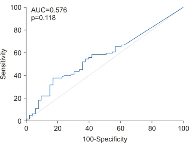
갑상선 외 피막 침범이 있는 환자의 최대 표준섭취화 계수의 ROC curve를 분석하였으며, AUC=0.576이며 통 계적으로 유의하지 않았지만, cut-off value는 SUV가 4.52 이상일 때 갑상선 외 피막 침범을 예측할 수 있었다 (Fig. 3).
고 찰
갑상선암에서 18F-FDG PET/CT는 종전의 수술 이후 발생하는 재발 및 전이에 대한 검사 이외, 수술 전 암의
Fig. 3. ROC curves of ETE and SUVmax. The ROC curves of ETE and SUVmax were analyzed, AUC=0.576, and were not statistically significant (p=0.118). The cut-off value was 4.52, and it can be predicted that there is ETE when it is 4.52 or more. ETE: Extrathyroidal extension, SUVmax:
the maximum standardized uptake value
예후를 예측하는 용도로는 그 유용성에 대해 아직 연구 가 많지 않다.2,5-11) 로빈슨의 연구에서, 갑상선 분화암의 2년 생존율과 18F-FDG PET/CT에서 대사항진 양성 여 부는 통계학적으로 상관관계가 있다는 보고가 있었다 (p<0.001).5,12) 그러나 갑상선유두암 환자의 사망률은 매 우 낮아(1% 미만), 적은 수의 표본에서는 사망률을 평가 하기 어렵다.1,2) 본 논문에서도 사망자가 없어 사망률과 최대 표준섭취화계수 값 간의 상관관계는 검토할 수 없 었다.
갑상선유두암의 병기 설정은 갑상선 내 결절의 크기, 종물의 피막 침범 여부, 임파선 전이 여부, 갑상선 주위 조직으로의 침범 여부를 주로 고려한다.13)
본 연구에서, 최대 표준섭취화계수 값에 따른 종물의 피막 침범 여부, 임파선 전이 여부에는 통계적 연관성이 없었다. 갑상선암 환자에서 갑상선암의 크기와 최대 표 준섭취화계수 간의 유의미한 통계적 연관성이 있다는 보고가 있지만,2) 양성 결절의 크기에는 최대 표준섭취화 계수 간에는 연관성이 없다고 보고되었다. 본 연구에서 도 갑상선유두암의 경우, 종물의 크기와 연관성이 있다 는 동일한 결과를 알 수 있었다.
암 환자에 있어 B-RAF나 neuroblastoma RAS (NRAS) 같은 유전변이 지표가 생존율을 포함한, 암의 예후에 영 향을 미친다는 보고가 있다.14) 유방암 및 기타 고형암에 서 안 좋은 예후를 시사하는 Triple-negative breast cancer 및 hormone receptor negativity와 18-불화디옥시포도당의 대사항진 여부 간의 연관성이 있다고 보고되었다.15,16) 본 논문에서, B-RAF의 돌연변이형 여부는 통계학적으로 최
대 표준섭취화계수 값과 유의미한 연관성을 보였다.
B-RAF 돌연변이 여부와 갑상선유두암의 예후 간의 연 관성에 대해서는 아직 논란이 많다. 그러나 최근 6087명 을 대상으로 시행한 메타분석에서, B-RAF의 돌연변이는 갑상선유두암 환자의 임파선 전이, 병기 및 생존율에 영 향을 미치는 것으로 보고되었다.17)
갑상선암의 분화도가 좋지 않을수록 최대 표준섭취화 계수는 증가한다고 보고되었다.18) 저자들은 갑상선유두 암 만을 대상으로 선정하였기에 분화도에 따른 최대 표 준섭취화계수의 차이는 논의할 수 없었다.
본 논문의 결과에서, PET/CT에서 갑상선 종물에 대 사 항진이 있는 경우에는 갑상선유두암의 예후를 직접 적으로 예측할 수 있는 재발률과 최대 표준섭취화계수 값 간의 통계적인 유의성은 없었다. 하지만 갑상선유두 암이라도 18F-FDG의 대사 항진이 없는 환자들보다 대 사 항진이 있는 환자들의 병기가 통계적으로 유의하게 높아 병기가 높을수록 PET/CT에서 대사 항진이 더 잘 보이는 패턴을 알 수 있었다. 재발한 경우는 5명으로 그 수가 적어 통계 결과의 의의가 적었다. 그러나 최대 표준 섭취화계수 값과 결절의 크기, B-RAF 돌연변이형의 유 무 간에 높은 상관관계를 보였다. 이에 수술 전 최대 표 준섭취화계수 값과 갑상선유두암의 예후 간의 연관성이 있을 가능성이 높다. 본 연구를 통해, 수술 전 18F-FDG PET/CT 검사와 갑상선유두암의 예후에 영향을 주는 요 인들과의 연관성에 대해 알 수 있었고, 갑상선유두암에 서 18F-FDG PET/CT의 진단적 유용성에 대한 확정적 인 결론은 내릴 수 없으나, 진단적으로 유용할 가능성이 높아, 추후 장기간의 경과 관찰 및 많은 수의 연구가 필 요하다는 결론을 내릴 수 있었다.
결 론
본 논문은 갑상선유두암종이 확진된 환자군 내에서, 환자의 특징, 종양의 크기, 종물의 피막 침범 여부, 임파 선 전이 여부, 갑상선유두종의 재발에 따른 최대 표준섭 취화계수 값의 상관관계를 분석한 연구이다. 이전의 연 구들은 최대 표준섭취화계수와 갑상선 결절의 악성 여 부 간의 관계를 알기 위한 연구였지만, 본 연구는 예후에 영향을 주는 병리학적 결과들과 수술 전 최대 표준섭취 화계수값과의 관계를 비교하였다. 예후에 영향을 미치 는 요인인 임파선 전이 및 갑상선외 피막 침범은 수술 전 최대 표준섭취화계수 값과 유의미한 관련성이 없었 다. 하지만, 18F-FDG의 대사 항진 유무와 갑상선유두암 의 병기간 통계학적 연관성이 있었고, 최대 표준섭취화
계수값과 종물의 크기 및 B-RAF type 사이에는 통계학 적 연관성이 있었다. 갑상선유두암종에서 종물의 크기 와 B-RAF 돌연변이 여부는 이미 알려진 대로 예후에 영 향을 미치는 주요 인자로서 임상적인 의미가 있다. 수술 전 최대 표준섭취화계수 값과 갑상선유두암의 예후 간 의 연관성이 있을 가능성이 높다. 연관성을 명확히 파악 하기 위해서, 향후 더 많은 수의 환자를 대상으로 18F-FDG PET/CT를 이용한 연구가 필요할 것으로 생 각된다.
중심 단어: 갑상선 유두암, 18F-FDG PET/CT, 표준섭취계수.
Acknowledgments
This study was supported by research funds from Dong-A University.
Conflicts of Interest
No potential conflict of interest relevant to this article was reported.
Orcid
Hyungi Gim: https://orcid.org/0000-0003-3443-6200 Dong Kun Lee: https://orcid.org/0000-0002-7296-1420 Heon Soo Park: https://orcid.org/0000-0002-4567-7272 Yeong Jin Jeong: https://orcid.org/0000-0001-7611-8185
References
1) Kim TH, Ji YB, Song CM, Kim JY, Choi YY, Park JS, et al. SUVmax of 18F-FDG PET/CT in the differential diagnosis of benign and malignant thyroid nodules according to tumor volume. World J Surg Oncol 2015;13:217.
2) Stangierski A, Wolinski K, Czepczynski R, Czarnywojtek A, Lodyga M, Wyszomirska A, et al. The usefulness of standardized uptake value in differentiation between benign and malignant thyroid lesions detected incidentally in 18F-FDG PET/CT examination. PLoS One 2014;9(10):e109612.
3) Kim BS, Kang WJ, Oh SW, Lee DS, Chung JK, Lee MC.
Usefulness of SUV ratio for differentiating benign from malignant focal thyroid lesions incidentally detected by F-18 FDG PET/ CT: comparison with SUV analysis. J Korean Thyroid Assoc 2009;2(2):120-7.
4) Kim BS, Ryu HS, Kang KH. The value of preoperative PET-CT in papillary thyroid cancer. J Int Med Res 2013;
41(2):445-56.
5) Marcus C, Whitworth PW, Surasi DS, Pai SI, Subramaniam RM. PET/CT in the management of thyroid cancers. AJR Am J Roentgenol 2014;202(6):1316-29.
6) Dong MJ, Liu ZF, Zhao K, Ruan LX, Wang GL, Yang SY, et al. Value of 18F-FDG-PET/PET-CT in differentiated thyroid carcinoma with radioiodine-negative whole-body scan: a meta-analysis. Nucl Med Commun 2009;30(8):639-50.
7) Weber T, Ohlhauser D, Hillenbrand A, Henne-Bruns D, Reske SN, Luster M. Impact of FDG-PET computed tomography for surgery of recurrent or persistent differentiated thyroid carcinoma. Horm Metab Res 2012;44(12):904-8.
8) Kim TY, Kim WB, Ryu JS, Gong G, Hong SJ, Shong YK.
18F-fluorodeoxyglucose uptake in thyroid from positron emission tomogram (PET) for evaluation in cancer patients: high prevalence of malignancy in thyroid PET incidentaloma. Laryngoscope 2005;115(6):1074-8.
9) de Geus-Oei LF, Pieters GF, Bonenkamp JJ, Mudde AH, Bleeker-Rovers CP, Corstens FH, et al. 18F-FDG PET reduces unnecessary hemithyroidectomies for thyroid nodules with inconclusive cytologic results. J Nucl Med 2006;47(5):770-5.
10) Are C, Hsu JF, Schoder H, Shah JP, Larson SM, Shaha AR.
FDG-PET detected thyroid incidentalomas: need for further investigation? Ann Surg Oncol 2007;14(1):239-47.
11) Eloy JA, Brett EM, Fatterpekar GM, Kostakoglu L, Som PM, Desai SC, et al. The significance and management of incidental [18F]fluorodeoxyglucose-positron-emission tomography uptake in the thyroid gland in patients with cancer. AJNR Am J Neuroradiol 2009;30(7):1431-4.
12) Robbins RJ, Wan Q, Grewal RK, Reibke R, Gonen M, Strauss HW, et al. Real-time prognosis for metastatic thyroid carcinoma based on 2-[18F]fluoro-2-deoxy-D-glucose-positron emission tomo- graphy scanning. J Clin Endocrinol Metab 2006;91(2):498-505.
13) Yeh MW, Bauer AJ, Bernet VA, Ferris RL, Loevner LA, Mandel SJ, et al. American Thyroid Association statement on preoperative imaging for thyroid cancer surgery. Thyroid 2015;25(1):3-14.
14) Heppt MV, Siepmann T, Engel J, Schubert-Fritschle G, Eckel R, Mirlach L, et al. Prognostic significance of BRAF and NRAS mutations in melanoma: a German study from routine care.
BMC Cancer 2017;17(1):536.
15) Soussan M, Orlhac F, Boubaya M, Zelek L, Ziol M, Eder V, et al. Relationship between tumor heterogeneity measured on FDG-PET/CT and pathological prognostic factors in invasive breast cancer. PLoS One 2014;9(4):e94017.
16) Takagi H, Sakamoto J, Osaka Y, Shibata T, Fujita S, Sasagawa T. Usefulness of the maximum standardized uptake value for the diagnosis and staging of patients with cervical cancer undergoing positron emission tomography/computed tomography. Medicine (Baltimore) 2018;97(7):e9856.
17) Wang Z, Chen JQ, Liu JL, Qin XG. Clinical impact of BRAF mutation on the diagnosis and prognosis of papillary thyroid carcinoma: a systematic review and meta-analysis. Eur J Clin Invest 2016;46(2):146-57.
18) Treglia G, Annunziata S, Muoio B, Salvatori M, Ceriani L, Giovanella L. The role of fluorine-18-fluorodeoxyglucose positron emission tomography in aggressive histological subtypes of thyroid cancer: an overview. Int J Endocrinol 2013;2013:856189.