R E S E A R C H
Open Access
SUVmax of
18
F-FDG PET/CT in the
differential diagnosis of benign and malignant
thyroid nodules according to tumor volume
Tae Heon Kim
1, Yong Bae Ji
1, Chang Myeon Song
1, Ji Young Kim
2, Yun Young Choi
2, Jeong Seon Park
3and Kyung Tae
1*Abstract
Background: The aim of this study was to investigate whether the maximum standardized uptake value (SUVmax) measured on fluorine-18 fluorodeoxyglucose positron emission tomography/computed tomography (18F-FDG PET/CT) could be used as the primary means of differential diagnosis of thyroid nodules when tumor volume is assessed. Methods: We studied 192 patients who underwent preoperative18F-FDG PET/CT followed by thyroidectomy. We evaluated the correlation between the volume of thyroid nodules,18F-FDG uptake on visual analysis, and the mean
SUVmax measured on18F-FDG PET/CT.
Results: When stratified by tumor volume, the mean SUVmax was higher in malignant than in benign nodules in nodules≥1 cm3(p < 0.001). However, it did not differ between benign and malignant nodules smaller than 1 cm3. At a cut-off value of SUVmax of 6, the respective sensitivities of18F-FDG PET/CT, ultrasonography, and fine needle aspiration cytology were 60.8, 96.4, and 99.1 %, and the respective specificities were 95.9, 98.2, and 96.8 %.
Conclusions:18F-FDG PET/CT is limited as a primary modality in the differential diagnosis of benign and malignant thyroid nodules because of its low sensitivity.
Keywords:18F-FDG PET/CT, Thyroid nodule, Thyroid cancer, Differential diagnosis, SUVmax Background
Unlike normal tissue, malignant tumors are character-ized by increased glycolysis, which leads to elevated glu-cose uptake. Fluorine-18 fluorodeoxygluglu-cose positron emission tomography/computed tomography (18F-FDG PET/CT) makes use of this to aid the diagnosis and sta-ging of various human malignancies.
In recent years,18F-FDG PET/CT has become widely used to diagnose thyroid cancer; it is especially useful in identifying sites of recurrence in patients with elevated serum thyroglobulin and negative whole body radio-active iodine scans after thyroidectomy for differentiated thyroid carcinoma [1–3]. 18F-FDG PET/CT also has po-tential advantages in the detection of distant metastasis
or synchronous secondary tumor although its cost-effectiveness is not determined. However, the role of18 F-FDG PET/CT in the differential diagnosis of benign and malignant thyroid nodules is not yet well-established in clinical practice because some benign thyroid nodules show high18F-FDG uptake, while some malignant nodules are not visualized at all. Consequently, there is a paucity of data regarding the role of18F-FDG PET/CT in the dif-ferential diagnosis of thyroid nodules; the few studies that exist were conducted in small series of patients [4–6] or in patients whose nodules were identified incidentally dur-ing routine health checks [7–9]. In addition, while these studies estimated the mean maximum standardized up-take value (SUVmax) of 18F-FDG, they failed to consider the effect of nodule size. Other studies have enrolled pa-tients with metastatic thyroid cancer or those with diffuse thyroid uptake of18F-FDG.
Our aim in the present study was to establish whether the SUVmax measured on 18F-FDG PET/CT could be a * Correspondence:[email protected]
1Department of Otolaryngology–Head and Neck Surgery, College of
Medicine, Hanyang University, 222 Wangsimni-ro, Seongdong-gu, Seoul 133-792, Korea
Full list of author information is available at the end of the article
© 2015 Kim et al. This is an Open Access article distributed under the terms of the Creative Commons Attribution License (http://creativecommons.org/licenses/by/4.0), which permits unrestricted use, distribution, and reproduction in any medium, provided the original work is properly credited. The Creative Commons Public Domain Dedication waiver (http:// creativecommons.org/publicdomain/zero/1.0/) applies to the data made available in this article, unless otherwise stated.
primary modality in the differential diagnosis of benign and malignant thyroid nodules according to tumor volume.
Methods
Patients
We retrospectively studied 192 patients who underwent thyroidectomy for thyroid nodules and received a pre-operative 18F-FDG PET/CT scan out of about 3500 pa-tients who were diagnosed with thyroid nodule in our institute between January 2007 and December 2009. Of the tumors, 152 were malignant and 40 benign. Among the malignant cases, there were 146 papillary carcinomas, 3 follicular carcinomas, and 3 anaplastic carcinomas. The benign cases comprised 38 nodular hyperplasias and 2 fol-licular adenomas. Of the patients, 23 (15 %) were men and 129 (85 %) women. The mean age was 56 years, and all patients had normal thyroid function. The study proto-col was approved by the Institutional Review Board of Hanyang University Hospital.
In this study, 141 patients with malignant nodule under-went 18F-FDG PET/CT scan to evaluate distant or re-gional metastasis after diagnosis of thyroid cancer by fine needle aspiration cytology (FNAC) according to the pa-tient’s request or the recommendation of the doctors. Thirty-two patients with incidentally found benign and malignant nodule underwent 18F-FDG PET/CT scan for the purpose of routine health examination or evaluation of metastasis from other malignancy, and 19 patients of study group underwent 18F-FDG PET/CT for research purposes after submitting their written informed consent, especially in patients with follicular neoplasm or indeter-minate cases on FNAC.
Exclusion criteria were recurrent thyroid cancer, dif-fuse 18F-FDG uptake by the thyroid that could not be differentiated from thyroid nodule on18F-FDG PET/CT, and a history of thyroid surgery, or radiotherapy in the head and neck region for other diseases.
Patients with benign nodules underwent thyroidec-tomy: (1) for cosmetic purposes, (2) at their request, (3) if malignancy was suspected on ultrasonography (US), or (4) if their FNAC results were classified as inadequate or indeterminate.
Thyroid US and ultrasound-guided FNAC were per-formed by an experienced board-certified specialist in all cases. The FNAC results were categorized as (1) inad-equate, (2) benign, (3) indeterminate (e.g., follicular neo-plasm or atypia), (4) suspected malignancy, or (5) malignancy. In cases of microcalcification, irregular margins, marked hupoechogenity, or a taller-than-wide shape on US, the nodules were interpreted as malignant. Nodule diameter on US was used to calculate nodule volume. If there were multiple nodules on US, we mea-sured a single nodule that was also examined by FNAC.
18
F-FDG PET/CT
Whole body PET/CT images were acquired using a ded-icated PET/CT system (Biograph 6, Siemens Medical Systems, Knoxville, TN, USA). Hard exercise was pro-hibited for several days, and the patients fasted for at least 6 h prior to the scan. Blood glucose levels were tested to confirm they were below 200 mg/dL. After an intraven-ous injection of18F-FDG (0.21 mCi/kg), the patients were asked to wait for 1 h in a dimly lit room and encouraged to drink 1 L of water. A CT scan (80 mA and 140 kVp) was performed to correct attenuation prior to the PET scan. CT scanning was performed in 5-mm sections from the base of the skull to the proximal thigh; the images were reconstructed using a 512 × 512 matrix and a 50-cm field of view. PET scans were obtained from the proximal thigh to the base of the skull (six to seven bed positions, 2 min 30 s per each position), and the images were recon-structed with a 168 × 168 matrix, using the ordered subset expectation maximum iterative reconstruction algorithm, a 5-mm Gaussian filter, and a 50-cm field of view.
18
F-FDG uptake by thyroid nodules was evaluated by vis-ual analysis using the naked eye and quantitative measure-ment using SUVmax. If there is any focal increased uptake lesion detected in anterior neck on maximum intensity projection images by visual analysis, the uptake of thyroid nodule was verified by thorough examination of the axial PET/CT images by two nuclear medicine physicians. Then, SUVmax was measured for each thyroid nodule using volume of interest (VOI). SUV was calculated as follows: SUV = [FDG activity concentration (Bq/mL)] × [total lean body mass (kg)]/[injected FDG activity (Bq)].
Statistical analysis
Statistical analyses were carried out with SPSS 17.0 (SPSS Inc., Chicago, IL, USA). We compared the uptake of 18F-FDG analyzed visually and SUVmax determined on18F-FDG PET/CT between malignant and benign thy-roid nodules. The same comparisons were made when the nodules were stratified by volume using a cut-off volume of 1 cm3. An independent Studentt-test was performed to identify differences in SUVmax between the malignant and benign thyroid nodules. Pearson’s correlation coefficients were calculated to determine the correlation between nod-ule volume and the SUVmax.
Results
Differences in volume and SUVmax between malignant and benign nodules
The mean volumes of the malignant and benign nodules were not significantly different (11.63 ± 7.27 mL vs. 10.98 ± 4.56 mL;p = 0.968). On the other hand, the mean SUVmax was significantly higher in malignant than in be-nign nodules (5.11 ± 0.49 vs. 2.32 ± 0.25;p < 0.001). Among the malignant cases, the mean SUVmax was 4.80 ± 0.46 for
papillary carcinoma, 17.66 ± 7.65 for follicular carcinoma, and 7.72 ± 3.44 for anaplastic carcinoma. In the benign cases, it was 2.45 ± 0.27 for nodular hyperplasia and 1.05 ± 1.05 for follicular adenoma.
Differences in18F-FDG uptake on visual analysis and SUVmax between malignant and benign nodules stratified by nodule volume
The differences between malignant and benign nodules in terms of visual18F-FDG uptake and SUVmax, stratified by nodule volume, are shown in Tables 1 and 2. In nodules smaller than 1 cm3(98 cases),18F-FDG uptake on visual analysis was not different between the malignant and be-nign cases (p = 0.572). However, in nodules ≥1 cm3
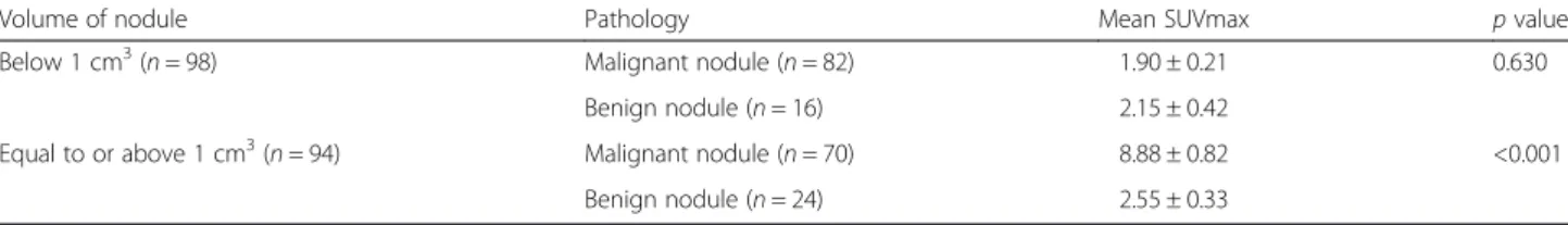
(94 cases), it was significantly higher in malignant than in be-nign nodules (all patients with malignant nodules vs. 79.2 % of patients with benign nodules; p < 0.001) (Table 1). In nodules <1 cm3, mean SUVmax was 1.90 ± 0.21 in the malignant cases and 2.15 ± 0.42 in the benign cases (p = 0.630). However, it was significantly higher in malignant nodules with volumes ≥1 cm3 (8.88 ± 0.82 for malignant vs. 2.55 ± 0.33 for benign nodules, p < 0.001) (Table 2).
Correlation between nodule volume and SUVmax
There was no significant correlation between the volumes of all thyroid nodules and mean SUVmax (Pearson’s cor-relation coefficient,r = 0.149). Likewise, there was no sig-nificant correlation between the volume of malignant nodules only and mean SUVmax (r = 0.151). When strati-fied by volume, a weak correlation (r = 0.452) with
SUVmax was observed in the case of small (<1 cm3) ma-lignant nodules, but not in the case of larger (≥1 cm3
) ma-lignant nodules (r = 0.076).
Diagnostic accuracy of18F-FDG PET/CT, US, and FNAC
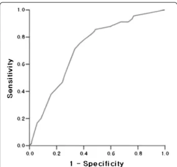
In the case of volumes ≥1 cm3, a receiver operating characteristic (ROC) curve analysis determined the opti-mal value of SUVmax for differential diagnosis between benign and malignant thyroid nodules (Fig. 1). The diag-nostic power of 18F-FDG PET/CT was the highest at SUVmax = 6: at that value, it had 60.8 % sensitivity, 96.8 % specificity, a positive predictive value of 97.7 %, a negative predictive value of 48.4 %, and 70.2 % diagnos-tic accuracy.
We compared the results of thyroid US, FNAC, and 18
F-FDG PET/CT at that cut-off value. Their respective sensitivities were 96.4, 99.1, and 60.8 %, and respective specificities were 95.9, 98.2, and 96.8 %. Similarly, their respective positive predictive values were 98.9, 99.4, and 97.7 %, and respective negative predictive values were 86.8, 95.2, and 48.4 %. Diagnostic accuracy was 97.3 % for thyroid US, 98.8 % for FNAC, and 70.2 % for 18 F-FDG PET/CT.
Diagnostic accuracy of18F-FDG PET/CT in cases undiagnosed by FNAC
FNAC diagnosed 151 cases of malignancy or suspected malignancy, 35 benign cases, 4 cases classified as inde-terminate, and 2 as inadequate. Of the 151 supposed malignancies, 1 was found to be benign after thyroidec-tomy. Of the four indeterminate cases, two were
Table 118F-FDG uptake by visual analysis of18F-FDG PET/CT images in malignant and benign thyroid nodules according to nodule volume
Volume of nodule Pathology Visual analysis of18F-FDG uptake Number (%)
p value
Below 1 cm3(n = 98) Malignant nodule (n = 82) Uptake 55 (67.1 %) 0.572
No uptake 27 (32.9 %)
Benign nodule (n = 16) Uptake 11 (68.8 %)
No uptake 5 (31.2 %)
Equal to or above 1 cm3(n = 94) Malignant nodule (n = 70) Uptake 70 (100 %) <0.001
No uptake 0 (0 %)
Benign nodule (n = 24) Uptake 19 (79.2 %)
No uptake 5 (20.8 %)
Table 2 Mean SUVmax of malignant and benign nodules according to nodule volume
Volume of nodule Pathology Mean SUVmax p value
Below 1 cm3(n = 98) Malignant nodule (n = 82) 1.90 ± 0.21 0.630
Benign nodule (n = 16) 2.15 ± 0.42
Equal to or above 1 cm3(n = 94) Malignant nodule (n = 70) 8.88 ± 0.82 <0.001
Benign nodule (n = 24) 2.55 ± 0.33
confirmed as follicular carcinomas and two as follicular adenomas (Figs. 2 and 3). The cases classified as inad-equate were found to be nodular hyperplasia (Table 3).
When the six patients with indeterminate or inad-equate findings on FNAC were examined by 18F-FDG PET/CT (with a cut-off SUVmax = 6), the method had 100 % sensitivity and specificity, 0 % false positives and false negatives, positive and negative predictive values of 100 %, and 100 % diagnostic accuracy.
Discussion
In our study, the mean SUVmax was on the whole sig-nificantly higher in malignant than in benign thyroid nodules (p < 0.001). However, when the nodules were stratified by volume, we observed no significant differ-ences in18F-FDG uptake on visual analysis and in mean SUVmax between the malignant and benign nodules when the nodules were below 1 cm3. This might be due to the partial-volume effect, i.e., underestimation of small volumes. The resolution of18F-FDG PET/CT is es-timated at approximately 6 mm3 [10]. When 18F-FDG PET/CT is performed on smaller lesions, the adjacent normal tissue that does not show increased uptake is also present in the visual field, and this may lead to in-accurate assessment of FDG uptake. Hence, in this study, we evaluated the diagnostic accuracy of18F-FDG PET/CT only in nodules with a volume ≥1 cm3except for those nodules <1 cm3in which neither visual analysis of uptake nor mean SUVmax had any power to differen-tiate between malignant and benign nodules.
As a tool in the differential diagnosis of benign and malignant thyroid nodules, thyroid US has been shown to have sensitivity of 30–75 %, specificity of 80–95 %, positive predictive value of 15–94 %, negative predictive value of 70–80 %, and diagnostic accuracy of approxi-mately 75 % [11, 12]. By comparison, the sensitivity and specificity of FNAC have been estimated at approxi-mately 85–90 % and 85–97 %, respectively, although these values may vary depending on the examiner’s tech-nical skills [13, 14]. In our study, both thyroid US and FNAC had a higher diagnostic accuracy than suggested in the previous reports (the corresponding values for US
Fig. 1 Receiver operating characteristic (ROC) curve of SUVmax. The diagnostic power of18F-FDG PET/CT is the highest at SUVmax = 6
Fig. 2 Images of follicular adenoma. a On ultrasonography, there is an 8-cm, markedly hypoechoic nodule. b Mildly increased18F-FDG uptake in
in our study were 96.4, 95.9, 98.9, 86.8, and 97.3 %; and for FNAC: 99.1, 98.2, 99.4, 95.2, and 98.8 %). One of the contributing factors could be our study criteria, which excluded patients with negative findings on FNAC des-pite having intermediate findings or a suspected malig-nancy on US.
In the current study, the diagnostic ability of18F-FDG PET/CT to differentiate benign and malignant thyroid nodules was lower than that of US or FNAC, even in lar-ger (≥1 cm3
) nodules. This implies that 18F-FDG PET/ CT is less likely to be a primary modality in the differen-tial diagnosis of all thyroid nodules. Furthermore, the role of18F-FDG PET/CT as the primary modality in the differential diagnosis of thyroid carcinoma may be lim-ited at the present time because the small thyroid cancer with a volume of <1 cm3is common, and the diagnostic accuracy of 18F-FDG PET/CT is especially low in those cases. In addition, considering cost-effectiveness, 18 F-FDG PET/CT is not recommended routinely.
However, the SUVmax of 18F-FDG PET/CT might be useful in the differential diagnosis of thyroid nodule de-tected incidentally by 18F-FDG PET/CT scan which was performed for the purpose of routine health examination or evaluation of metastasis from other malignancy. Our findings suggest that malignancy can be suspected when nodules ≥1 cm3show FDG uptake on visual analysis of PET/CT images, because all the malignant thyroid nod-ules with a volume of≥1 cm3showed the uptake on vis-ual analysis. In particular, there is a very high probability that thyroid nodules with a volume of≥1 cm3and SUV-max of >6 on 18F-FDG PET/CT are thyroid carcinoma because of a higher specificity of 96.8 % and a positive predictive value of 97.7 %.
According to reports, differential diagnosis by FNAC can be difficult in approximately 20 % of thyroid nod-ules, particularly in the case of non-diagnostic or unsat-isfactory samples, follicular lesions of undetermined significance, or follicular neoplasms [15]. The diagnostic power of18F-FDG PET/CT in such cases is still contro-versial [16–19]. Here, we evaluated the role of18
F-FDG PET/CT in six patients whose preoperative ultrasound-guided FNAC findings were not conclusively malignant or benign. In these patients, the sensitivity and specificity of 18
F-FDG PET/CT were 100 %, indicating that it might be a useful modality in cases undiagnosed by FNAC. However, before these findings can be considered conclusive, they should be verified in a larger sample of patients.
Inevitably, this study has some limitations. The pa-tients were not randomized, and there was selection bias. We included only those who underwent thyroidec-tomy and 18F-FDG PET/CT for benign or malignant
Fig. 3 Images of follicular carcinoma. a On ultrasonography, there is a 1.7-cm, hypoechoic nodule. b Focal, intense18F-FDG uptake in the left isthmic nodule on18F-FDG PET/CT (SUVmax = 11.97)
Table 3 Pathology results and SUVmax on18F-FDG PET/CT in cases that were inconclusive on fine needle aspiration cytology
Results of FNAC Group SUVmax Pathology
Indeterminate (n = 4) Malignant (n = 2) 11.97 Follicular carcinoma 8.2 Follicular carcinoma Benign (n = 2) 2.10 Follicular adenoma
0 Follicular adenoma Inadequate (n = 2) Benign (n = 2) 2.6 Nodular hyperplasia
0 Nodular hyperplasia
FNAC fine needle aspiration cytology, SUVmax maximum standardized uptake value of18F-FDG PET/CT
nodules. We excluded any benign or indeterminate cases where thyroidectomy was not indicated and those cases in which18F-FDG PET/CT was not performed.
Conclusions 18
F-FDG PET/CT is limited as a primary modality in the differential diagnosis of benign and malignant thyroid nodules because of its low sensitivity. However, malig-nancy can be suspected when a visual analysis of 18 F-FDG PET/CT images shows increased F-FDG uptake in nodules with volumes≥1 cm3. In addition, if SUVmax is greater than 6 in nodules≥1 cm3, the probability of ma-lignancy is very high.
Abbreviations
18F-FDG PET/CT:fluorine-18 fluorodeoxyglucose positron emission
tomography/computed tomography; FNAC: fine needle aspiration cytology; SUVmax: maximum standardized uptake value; US: ultrasonography. Competing interests
The authors declare that they have no competing interests. Authors’ contributions
KT and THK designed the study. THK, YBJ, CMS, JYK, YYC, and JSP participated in acquisition of data and interpretation of data. YBJ and CMS performed statistical analysis. THK wrote the draft. KT, YYC, and JSP revised the manuscript. All authors read and approved the final manuscript. Acknowledgements
No acknowledgements to make. Author details
1Department of Otolaryngology–Head and Neck Surgery, College of
Medicine, Hanyang University, 222 Wangsimni-ro, Seongdong-gu, Seoul 133-792, Korea.2Department of Nuclear Medicine, College of Medicine,
Hanyang University, Seoul, Korea.3Department of Radiology, College of
Medicine, Hanyang University, Seoul, Korea.
Received: 9 April 2015 Accepted: 25 June 2015 References
1. Feine U, Lietzenmayer R, Hanke JP, Held J, Wohrle H, Muller-Schauenburg W. Fluorine-18-FDG and iodine-131-iodide uptake in thyroid cancer. J Nucl Med. 1996;37:1468–72.
2. Wang W, Macapinlac H, Larson SM, Yeh SD, Akhurst T, Finn RD, et al. [18F] 2-Fluoro-2-deoxy-D glucose positron emission tomography localizes residual thyroid cancer in patients with negative diagnostic (131)I whole body scans and elevated serum thyroglobulin levels. J Clin Endocrinol Metab. 1999;84:2291–302.
3. Dietlein M, Scheidhauer K, Voth E, Theissen P, Schicha H. Fluorine-18 fluorodeoxyglucose positron emission tomography and iodine-131 whole-body scintigraphy in the follow up of differentiated thyroid cancer. Eur J Nucl Med. 1997;24:1342–8.
4. Kresnik E, Gallowitsch HJ, Mikosch P, Stettner H, Igerc I, Gomez I, et al. Fluorine-18 fluorodeoxyglucose positron emission tomography in the preoperative assessment of thyroid nodules in an endemic goiter area. Surgery. 2003;133:294–9.
5. Bloom AD, Adler LP, Shuck JM. Determination of malignancy of thyroid nodules with positron emission tomography. Surgery. 1993;114:728–34. 6. Mitchell JC, Grant F, Evenson AR, Parker JA, Hasselgren PO, Parangi S.
Preoperative evaluation of thyroid nodules with18FDG-PET/CT. Surgery.
2005;138:1166–74.
7. Cohen MS, Arslan N, Dehdashti F, Doherty GM, Lairmore TC, Brunt LM, et al. Risk of malignancy in thyroid incidentalomas identified by fluorodeoxyglucose-positron emission tomography. Surgery. 2001;130:941–6.
8. Kang KW, Kim SK, Kang HS, Lee ES, Sim JS, Lee IG, et al. Prevalence and risk of cancer of focal thyroid incidentaloma identified by 18-F-fluorodeoxyglucose positron emission tomography for metastasis evaluation and cancer screening in healthy subjects. J Clin Endocrinol Metab. 2003;88:4100–4.
9. Choi JY, Lee KS, Kim HJ, Shim YM, Kwon OJ, Park K, et al. Focal thyroid lesions incidentally identified by integrated18F-FDG PET/CT: clinical significance and improved characterization. J Nucl Med. 2006;47:609–15. 10. Hoffman EJ, Huang SC, Phelps ME. Quantitation in positron emission
computed tomography: 1. Effect of object size. J Comput Assist Tomogr. 1979;3:299–308.
11. Frates MC, Benson CB, Charboneau JW, Cibas ES, Clark OH, Coleman BG, et al. Management of thyroid nodules detected at US: society of radiologists in ultrasound consensus conference statement. Radiology. 2005;237:794–800. 12. Kim EK, Park CS, Chung WY, Oh KK, Kim DI, Lee JT, et al. New sonographic criteria for recommending fine-needle aspiration biopsy of nonpalpable solid nodules of the thyroid. AJR. 2002;178:687–91.
13. Regina CM, Gharib H. Continuing controversies in the management of thyroid nodules. Ann Intern Med. 2005;142:926–31.
14. Gardner HAR, Ducatman BS, Wang HH. Predictive value of fine-needle aspiration of the thyroid in the classification of follicular lesions. Cancer. 1993;71:2598–603. 15. Cibas ES, Ali SZNCI, Thyroid FNA. State of the Science Conference 2009: the
Bethesda system for reporting thyroid cytopathology. Am J Clin Pathol. 2009;132:658–65.
16. Kim JM, Ryu JS, Kim TY, Kim WB, Kwon GY, Gong G, et al.18 F-fluorodeoxyglucose positron emission tomography does not predict malignancy in thyroid nodules cytologically diagnosed as follicular neoplasm. J Clin Endocrinol Metab. 2007;92:1630–4.
17. Sebastianes FM, Cerci JJ, Zanoni PH, Soares Jr J, Chibana LK, Tomimori EK, et al. Role of18F-fluorodeoxyglucose positron emission tomography in
preoperative assessment of cytologically indeterminate thyroid nodules. J Clin Endocrinol Metab. 2007;92:4485–8.
18. Traugott AL, Dehdashti F, Trinkaus K, Cohen M, Fialkowski E, Quayle F, et al. Exclusion of malignancy in thyroid nodules with indeterminate fine-needle aspiration cytology after negative18F-fluorodeoxyglucose positron emission tomography: interim analysis. World J Surg. 2010;34:1247–53.
19. Deandreis D, Al Ghuzlan A, Auperin A, Vielh P, Caillou B, Chami L, et al. Is
18F-fluorodeoxyglucose-PET/CT useful for the presurgical characterization of
thyroid nodules with indeterminate fine needle aspiration cytology? Thyroid. 2012;22:165–72.
Submit your next manuscript to BioMed Central and take full advantage of:
• Convenient online submission
• Thorough peer review
• No space constraints or color figure charges
• Immediate publication on acceptance
• Inclusion in PubMed, CAS, Scopus and Google Scholar
• Research which is freely available for redistribution
Submit your manuscript at www.biomedcentral.com/submit