서론
18F-fluorodeoxyglucose (이하 FDG)를 이용한 양전자방출 단층촬영술(positron emission tomography; 이하 PET)은 당 대사를 반영하는 분자영상 기법으로 종양의 대사 활성도를 정 량화 할 수 있는 것이 장점이다. 대부분의 악성 종양은 탄수화 물 대사가 높은데 여기에는 조직학적 유형이나 분화도 같은 종 양의 자체 요인, 당대사나 저산소증 같은 종양의 생화학적 특 성, 동반된 염증반응 같은 종양 외적인 요인 등 여러 가지 요소
가 관여한다(1). 일반적으로 PET/CT 양성도에 영향을 미치는 인자들은 유방암의 조직학적 유형, 종양의 등급, 크기 등이며 이에 비하여 호르몬 수용체 여부, 액와 임파선 전이 여부, 폐경 여부 등은 영향이 없는 것으로 알려져 있다(2-7). 그러므로 PET/CT는 분화도가 낮거나 병변의 크기가 큰 유방암의 발견 에 유용하지만, 등급이 낮거나, 1 cm 이하의 작은 유방암에서 는 민감도가 낮다고 알려져 있다(3-5).
저자들은 이전 연구에서 유방암의 조직학적 유형에 상관없 이 병변이 클수록 peak standardized uptake value (pSUV)가
J Korean Soc Radiol 2012;66(2):169-175
Received September 15, 2011;
Accepted November 19, 2011 Corresponding author: Eun Hye Lee, MD
Department of Radiology, Soonchunhyang University Bucheon Hospital, Soonchunhyang University College of Medicine, 170 Jomaru-ro, Wonmi-gu,
Bucheon 420-767, Korea.
Tel. 82-32-621-5851 Fax. 82-32-621-5874 E-mail: [email protected]
Copyrights © 2012 The Korean Society of Radiology
Purpose: To evaluate the factors associated with variations in
18F-fluorodeoxyglu- cose positron emission tomography/CT (18F-FDG PET/CT) uptake in ductal carcino- mas of the breast.Materials and Methods: We enrolled 216 ductal carcinoma cases that underwent
18F-FDG PET/CT. We evaluated the positivity and measured peak standardized uptake value (pSUV) of lesions that underwent 18F-FDG PET/CT. We analyzed the correlation between pSUV and invasiveness, lesion size, and the histologic factors of invasive ductal carcinoma (IDC).
Results: In the
18F-FDG PET/CT of ductal carcinomas, sensitivity was 90.2%, positive and negative predictive values were 99.5% and 25.0%, respectively. In ductal carci- noma in situ (DCIS) and IDC, the sensitivities were 68.8% and 92.0%, respectively.The mean pSUV of true positive (TP) DCIS and IDC were 2.6 and 5.1 (p < 0.05), re- spectively, whereas the false negative (FN) were 1.3 and 1.2 (p > 0.05), respectively, and that of false positive (FP) and true negative (TN) lesions were 2.2 and 0.9, re- spectively. The mean size of TP DCIS and IDC were 4.5 cm and 2.7 cm (p < 0.05), re- spectively, whereas the mean size of FN DCIS and IDC were 1.5 cm and 1.4 cm (p >
0.05), respectively, and that of FP and TN lesions were 1.8 cm and 1.2 cm respec- tively. Among the histological factors affecting IDC, mitosis showed the best corre- lation with pSUV (rho = 0.5).
Conclusion: For
18F-FDG PET/CT of ductal carcinomas, the positive predictive value was 99.5% and the FN rate was 9.7%. False negative factors included DCIS and an IDC < 1.5 cm, whereas mitosis was the TP factor.Index terms
Carcinoma, Ductal, Breast Positron-Emission Tomography Fluorodeoxyglucose F18
18
F-Fluorodeoxyglucose Positron Emission Tomography/CT Scan Findings for Ductal Carcinomas of Breast: Association of
Standardized Uptake Value and Histological Findings
1유방암의
18F-Fluorodeoxyglucose Positron Emission Tomography/CT 소견: Standardized Uptake Value와 조직학적 소견의 연관 관계
1So Young Bae, MD
1, Eun Hye Lee, MD
1, Jung Mi Park, MD
2, Jeong Ja Kwak, MD
3Departments of 1Radiology, 2Nuclear Medicine, 3Pathology, Soonchunhyang University Bucheon Hospital, Soonchunhyang University College of Medicine, Bucheon, Korea
유방영상검사의 소견이나 결과를 모르는 상태에서 한 명의 핵의학과 전문의가 18F-FDG PET/CT를 판독하였다. 주변 유 방조직보다 섭취가 높은 경우를 양성으로 판정했고, 이 부위에 서 pSUV를 측정한 후 해당 영상을 저장하였다. 주변 조직과 비교하여 섭취도가 비슷한 경우는 음성으로 판독하였으며 영 상의학과 의사와 함께 영상 소견을 감안하여 18F-FDG PET/
CT의 해당 부위에서 pSUV를 측정하였다. 이상의 결과를 토대 로 유방의 관상피암에서 PET/CT의 민감도, 양성 및 음성예측 도(positive/negative predictive value), 위음성률(false nega- tive rate)을 구하였다.
관상피암의 크기 분석은 병리 결과지를 기준으로 작성하였 다. 관내암과 침윤성관암을 18F-FDG PET/CT 진양성군과 위음성군으로 각각 나누었으며, 병변의 크기와 pSUV의 관계 를 분석하였다. 또한 침윤성관암의 등급을 분석하였으며, 관 형성(tubular formation), 핵 다양성(nuclear pleomorphism), 유사분열(mitosis), 그리고 광범위한 관내상피성분(extensive intraductal component) 여부를 파악하고 이들 조직학적 인자 들과 pSUV의 관계를 분석하였다.
침윤성관암에서 병변의 크기, 관 형성, 핵 다양성, 유사분열, 그리고 광범위한 관내상피성분 등과 pSUV의 관계를 보기 위 하여 Spearman rank 상관분석(correlation analysis)을 시행하 였으며 Statistical Package for Social Sciences (SPSS), ver- sion 14.0(SPSS Inc., Chicago, IL, USA)을 이용하였다. 침윤 성관암과 관내암에서 pSUV의 평균값에 차이가 있는지, 또한 각각에서 크기에 따른 pSUV의 평균값에 차이가 있는지 알아 보기 위하여 Mann-Whitney test를 이용하였다. p value 0.05 미만인 경우를 통계학적으로 유의성이 있는 것으로 하였다.
결과
216개의 유방 관상피암 중 195예(90.2%)가 18F-FDG PET/
CT에서 양성으로 판정되었고 그 중 관내암은 11개(31.2%), 침 윤성관암은 184개(92.0%)였다. 나머지 21예(9.8%)는 음성으 로 판정되었는데 그 중 관내암이 5개(68.8%), 침윤성관암이 16개(8.0%)였다(Table 1). 전체 유방 관상피암의 평균 pSUV 는 4.6(범위, 0.6~32.0)이었다. 관내암의 평균 pSUV는 높음을 보고하였으나, 고등급의 침윤성관암은 크기와 pSUV
간에 상관관계가 없었다(8). 그렇다면 저등급이지만 종괴가 크 거나, 고등급이지만 크기가 작은 유방암의 pSUV에 대한 의문 이 생겼다. 또한, 저자들이 알기로는 조직학적 유형이나 등급 외에 침윤성관암의 등급을 결정하는 세부 조직학적 인자들 - 관형성(tubular formation), 핵 다양성(nuclear pleomor- phism), 유사분열(mitosis) - 과 pSUV의 연관성에 대한 연구 는 없었다.
따라서 본 연구에서는 유방의 관상피암(ductal carcinomas of breast)에서 18F-FDG PET/CT의 섭취도에 영향을 미치는 요인들을 알아보고자 하였으며, 18F-FDG PET/CT에서 위음 성 결과를 초래하는 인자들을 알아보았다.
대상과 방법
2006년 6월부터 2010년 2월까지 본원에서 유방암으로 진 단되어 PET/CT를 시행한 환자는 259명이었다. 그 중 관상피 암(ductal carcinomas of breast)이 아닌 환자 30명, PET/CT 시행 전에 수술 혹은 항암치료를 받은 환자 13명, 다른 병원에 서 수술 받은 환자 7명은 대상에서 제외하였다. 최종적으로 209명의 유방암 환자들을 대상으로 하였고, 그 중 다발성이 6 명, 양측성이 1명이었다. 216개 관상피암(ductal carcinomas of breast) 중 관내암은 16개, 침윤성관암은 200개였다. 대상 군은 모두 여자였으며 평균 나이는 49세(범위 25~83세)였다.
PET/CT는 이중 검출기(dual head detector)가 장착된 장비 (Biograph2, Siemens, IL, USA)를 이용하였다. 환자들은 촬 영 전 최소 6시간 금식하였으며, 18F-FDG를 7.9 MB/kg 용 량으로 정맥주사하고 약 1시간 뒤에 촬영하였다. 검사 소요시 간은 약 30분이었으며 조영증강은 시행하지 않았다. 촬영범위 는 두개골 기저에서 대퇴부 또는 하지까지 포함하였다. 한 bed 당 2.5분씩 방출영상(emission tomography)을 얻고, CT (2 slice, 5 mm slice thickness, 130 Kvp, 60~70 mAs)를 이용 하여 감쇠보정(attenuation equalization)을 하였다. 일차적으로 PET-corrected 영상을 얻은 후, 필요에 따라 재구성(recon- struction)하여 축상(axial) 및 관상(coronal)의 융합 영상(fu- sion image)을 얻었다.
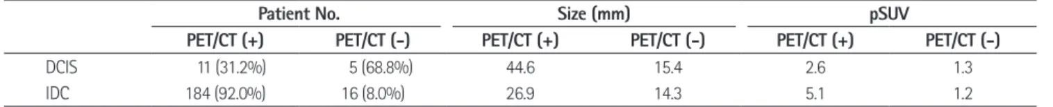
Table 1. Comparison of Histology, Size, and pSUV in Ductal Carcinomas of Breast
Patient No. Size (mm) pSUV
PET/CT (+) PET/CT (-) PET/CT (+) PET/CT (-) PET/CT (+) PET/CT (-)
DCIS 11 (31.2%) 5 (68.8%) 44.6 15.4 2.6 1.3
IDC 184 (92.0%) 16 (8.0%) 26.9 14.3 5.1 1.2
Note.-DCIS = ductal carcinoma in situ, IDC = invasive ductal carcinoma, PET = positron emission tomography, pSUV = peak standardized uptake value
진양성인 경우에 침윤성관암의 pSUV가 관내암보다 높았으나
18F-FDG PET/CT에서 위음성인 경우는 침윤성관암과 관내 암 간에 차이가 없었다(Table 1). 이상의 결과에서 전체 유방 관상피암(ductal carcinomas of breast)에 대한 18F-FDG PET/CT의 민감도는 90.2%(195/216), 양성예측도는 99.5%(195/196), 위음성률은 9.7%(21/216)였다. 관내암과 침윤성관암에 대한 18F-FDG PET/CT의 민감도는 각각 68.8%(11/16), 92.0%(184/200)였다.
유방 관상피암의 평균 크기는 2.7 cm(범위, 0.3~13.0 cm) 2.2(범위, 0.9~4.8), 침윤성관암은 4.8(범위, 0.6~32.0)이
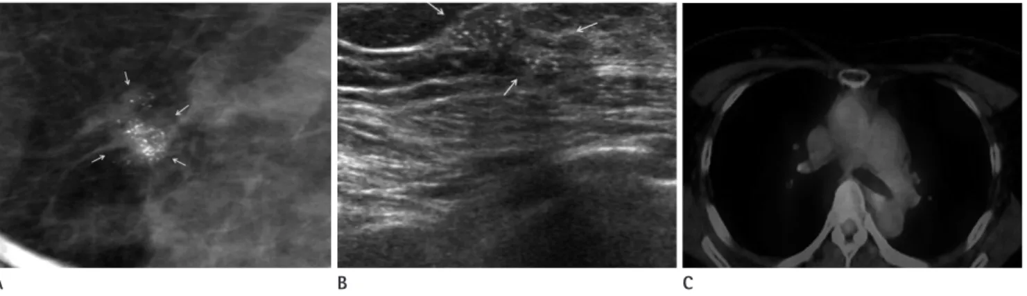
었으며 유의한 차이를 보였다(p < 0.01)(Table 2). 18F-FDG PET/CT에서 진양성인 195예의 평균 pSUV는 5.0(범위, 0.8~32.0)이었고, 관내암의 평균 pSUV는 2.6(범위, 1.6~4.8)(Fig. 1), 침윤성관암은 5.1(범위, 0.8~32.0)이었다 (p < 0.05)(Fig. 2). 18F-FDG PET/CT에서 위음성인 관상 피암 21예의 평균 pSUV는 1.2(범위, 0.6~2.0)였고, 그 중 관 내암은 1.3(범위, 0.9~2.0)(Fig. 3), 침윤성관암은 1.2(범위 0.6~1.7)였다(p > 0.05)(Fig. 4). 18F-FDG PET/CT에서 Fig. 1. A 56-year-old woman having 45 mm DCIS with PET/CT positive.
A. In the mammography, asymmetry and about 7 cm area of linear and linear-branching microcalcifications are noted in the left upper outer breast (arrows).
B. On USG, there is an irregular indistinct mass with microcalcifications (arrows).
C. On PET/CT, the mass shows pSUV of 4.8 (arrow).
Note.-DCIS = ductal carcinoma in situ, PET = positron emission tomography, pSUV = peak standardized uptake value, USG = ultrasonography
A B C
Table 2. Correlation of Size and pSUV in All Cases of Ductal Carcinomas of Breast
pSUV Size (mm) Rho p value
DCIS 2.2 35.5 0.73 < 0.01
IDC 4.8 25.9 0.56 < 0.01
Note.-DCIS = ductal carcinoma in situ, IDC = invasive ductal carcinoma, pSUV = peak standardized uptake value, Rho = Spearman’s correlation coeffi- cient
Fig. 2. A 49-year-old woman having 23 mm IDC with PET/CT positive.
A. In the mammography, asymmetry is noted in the right upper central breast (arrow).
B. On USG, there is an oval partially indistinct mass (arrows).
C. On PET/CT, pSUV of the mass was 11.4 (arrow).
Note.-IDC = invasive ductal carcinoma, PET = positron emission tomography, pSUV = peak standardized uptake value, USG = ultrasonography
A B C
각각 등급 1에서 32, 27, 20이었고, 등급 2은 134, 97, 56이었 으며, 등급 3에서는 87, 80, 77이었다. 조직학적 등급이 높아 질수록 관 형성, 핵 다양성, 유사분열의 점수가 높았다(Table 3). 18F-FDG PET/CT에서 위음성 침윤성관암에서도 조직학 적 등급이 높아질수록 관 형성, 핵 다양성, 유사분열의 점수가 높아졌으나 등급 3 병변 중에는 위음성 증례가 없었다(Table 4). 침윤성관암의 조직학적 인자들 중 관 형성(rho = 0.33), 핵 다양성(rho = 0.34), 유사분열(rho = 0.51)의 점수가 높을 수록 pSUV가 유의하게 높았으며(p < 0.01) 그 중 유사분열이 가장 높은 상관관계를 보였다. 광범위한 관내상피성분 음성인 병변은 침윤성관암 중 저등급(등급 1, 2)에서 61.7%(111/180), 고등급에서 38.3%(69/180)로 저등급에서 광범위한 관내상 피성분 음성이 많았다. 광범위한 관내상피성분 양성인 병변은 저등급 중 90%(18/20), 고등급에서 10%(2/20)였다. 광범위 한 관내상피성분과 pSUV는 음의 상관관계를 보였으나 상관 였고 그 중 관내암은 3.6 cm(범위, 0.5~12.0 cm), 침윤성관
암은 2.6 cm(범위, 0.3~13.0 cm)였다. 관내암이 침윤성관암 보다 약간 컸으나 유의한 차이는 없었다(p = 0.68). 18F-FDG PET/CT에서 진양성인 관상피암의 평균 크기는 3.0 cm(범 위, 0.3~13.0 cm)였고, 그 중 관내암은 4.5 cm, 침윤성관암 은 2.7 cm였다(p < 0.05). 18F-FDG PET/CT에서 위음성 관내암의 평균 크기는 1.5 cm(범위, 0.4~4.5 cm)였으며, 관 내암과 침윤성관암의 평균 크기는 각각 1.5 cm, 1.4 cm였다 (p > 0.05)(Table 1).
침윤성관암의 모든 예에서 병리학적 등급과 광범위한 관내 상피성분 유무를 알 수 있었으나 병리결과지에 관 형성, 핵 다 양성, 유사분열, 광범위한 관내상피성분이 기록된 병변은 200 개 중 102개였다. 그 중 18F-FDG PET/CT 진양성이 92개, 위 음성이 10개였다. 18F-FDG PET/CT에서 진양성인 침윤성관 암에서 조직학적 등급별 관 형성, 핵 다양성, 유사분열 점수는 Fig. 3. A 56-year-old woman having 13 mm DCIS with PET/CT negative.
A, B. In the mammography, compact cluster of linear and granular microcalcifications with irregular indistinct isodense mass are noted in the left upper central breast (A) and USG (B) (arrows).
C. On PET/CT, there is no evidence of hypermetabolism in the left breast. But considering radiologic findings, pSUV of estimated area was 0.9.
Note.-DCIS = ductal carcinoma in situ, PET = positron emission tomography, pSUV = peak standardized uptake value, USG = ultrasonography
Fig. 4. A 49-year-old woman having 7 mm IDC with PET/CT negative.
A. In the mammography, there is no abnormal finding.
B. But on USG, there is an oval indistinct mass in the left mid outer breast (arrow).
C. On PET/CT, there is no evidence of hypermetabolism in the left breast. But considering radiologic finding, pSUV of estimated area was 1.1.
Note.-IDC = invasive ductal carcinoma, PET = positron emission tomography, pSUV = peak standardized uptake value, USG = ultrasonography A
A
B
B
C
C
더 나쁜 예후를 보이므로 FDG-PET를 예후지표로 사용할 수 있다고 하였다.
지금까지 알려진 바에 따르면 FDG-PET는 유방암에서 48~89% 정도의 다양한 민감도와 80~100%의 특이도를 보 였다(7, 11, 12). 본 연구에서는 유방암에서 18F-FDG PET/
CT의 민감도는 90.2%, 양성예측도와 위음성률은 각각 99.5%, 9.7%였다. 이는 기존의 보고들과 일치하는 결과였다.
Kumar 등(5)은 종양의 크기와 등급이 FDG-PET의 결과를 예측하는 독립적인 인자라고 하였고, 암의 등급과 SUV값에 비 례적인 상관관계가 있다고 하였다. 또한 종양의 크기가 10 mm 이하이거나 저등급 종양은 FDG-PET의 위음성률을 높이 는 주요 인자라고 하였다. 본 연구에서도 침윤성관암이 관내암 보다 높은 pSUV를 보였고(4.78 vs. 2.16, p < 0.01), 침윤성 관암에서는 등급이 높을수록 pSUV가 증가하였는데(1등급 2.91, 2등급 3.90, 3등급 6.88; rho = 0.23, p < 0.001) 이 와 같은 결과는 이제까지 알려진 내용과 일치하였다(2, 7, 12).
Jeong 등(8)은 유방암의 조직학적 유형에 관계없이 병변의 크기가 클수록 pSUV가 높아지는 양상을 보였고(p < 0.001), 정도는 높지 않았다(rho = -0.22, p = 0.03)(Table 5).
고찰
FDG는 포도당과 함께 세포 내로 이동한 후 헥소키나아제 (hexokinase)에 의해 인산화과정(phosphorylation)을 거치지 만 더 이상 대사에 이용되지 않고 세포 내에 머물러 있게 되어 대사적으로 활성화된 조직에서는 포도당 섭취와 함께 FDG의 섭취가 증가하게 된다(9). 또한 종양이 성장함에 따라 저산소 증에 적응하기 위하여 당의 이동이 촉진되고 헥소키나아제의 활성도가 증가하므로 당대사가 증가한다(1). FDG-PET는 악 성 병변에서 대사 활성도가 증가하는 점을 이용한 기능적 검사 로, 유방촬영이나 초음파검사와 달리 해부학적 변화가 있기 전 에 진단이 가능한 것으로 알려져 있다. 최근에 개발된 PET/CT 는 CT와 PET 영상을 혼합하여 해부학적 영상과 기능적인 영 상을 동시에 제공함으로써 고전적인 PET보다 병변의 정확한 위치를 쉽게 파악할 수 있게 되었다(9). Oshida 등(10)에 의하 면 FDG 섭취가 높은 유방암은 FDG 섭취가 낮은 종양에 비해
Table 3. Comparison of Histologic Details in PET/CT (+) Invasive Ductal Carcinomas
Grade Tubular Formation Nuclear Pleomorphism Mitosis
1 2 3 1 2 3 1 2 3 pSUV
Low grade (n = 63) 5 (7.9%) 13 (20.6%) 45 (71.4%) 9 (14.3%) 47 (74.6%) 7 (11.1%) 52 (82.5%) 9 (14.3%) 2 (3.2%) 3.74 High grade (n = 29) 0 (0%) 0 (0%) 29 (100%) 0 (0%) 7 (24.1%) 22 (75.9%) 1 (3.4%) 8 (27.6%) 20 (69.0%) 6.32 Note.-PET = positron emission tomography, pSUV = peak standardized uptake value
Table 4. Comparison of Histologic Details in PET/CT (-) Invasive Ductal Carcinomas
Grade Tubular Formation Nuclear Pleomorphism Mitosis
1 2 3 1 2 3 1 2 3 pSUV
Low grade (n = 10) 2 (20%) 5 (50%) 3 (30%) 2 (20%) 8 (80%) 0 (0%) 10 (100%) 0 (0%) 0 (0%) 1.15
High grade (n = 0) 0 (0%) 0 (0%) 0 (0%) 0 (0%) 0 (0%) 0 (0%) 0 (0%) 0 (0%) 0 (0%) 0
Note.-PET = positron emission tomography, pSUV = peak standardized uptake value
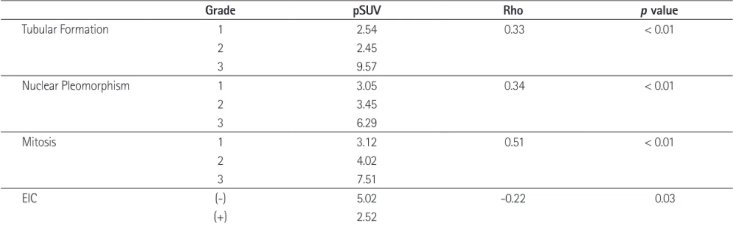
Table 5. Correlation of Histopathologic Factors and pSUV in Invasive Ductal Carcinomas
Grade pSUV Rho p value
Tubular Formation 1 2.54 0.33 < 0.01
2 2.45
3 9.57
Nuclear Pleomorphism 1 3.05 0.34 < 0.01
2 3.45
3 6.29
Mitosis 1 3.12 0.51 < 0.01
2 4.02
3 7.51
EIC (-) 5.02 -0.22 0.03
(+) 2.52
Note.-EIC = extensive intraductal component, pSUV = peak standardized uptake value, Rho = Spearman’s correlation coefficient
2. Avril N, Menzel M, Dose J, Schelling M, Weber W, Jänicke F, et al. Glucose metabolism of breast cancer assessed by 18F-FDG PET: histologic and immunohistochemical tissue analysis. J Nucl Med 2001;42:9-16
3. Buck AK, Schirrmeister H, Mattfeldt T, Reske SN. Biological characterisation of breast cancer by means of PET. Eur J Nucl Med Mol Imaging 2004;31 Suppl 1:S80-S87
4. Avril N, Rosé CA, Schelling M, Dose J, Kuhn W, Bense S, et al. Breast imaging with positron emission tomography and fluorine-18 fluorodeoxyglucose: use and limitations. J Clin Oncol 2000;18:3495-3502
5. Kumar R, Chauhan A, Zhuang H, Chandra P, Schnall M, Alavi A. Clinicopathologic factors associated with false negative FDG-PET in primary breast cancer. Breast Cancer Res Treat 2006;98:267-274
6. Crowe JP Jr, Adler LP, Shenk RR, Sunshine J. Positron emis- sion tomography and breast masses: comparison with clinical, mammographic, and pathological findings. Ann Surg Oncol 1994;1:132-140
7. Crippa F, Seregni E, Agresti R, Chiesa C, Pascali C, Bogni A, et al. Association between [18F]fluorodeoxyglucose up- take and postoperative histopathology, hormone receptor status, thymidine labelling index and p53 in primary breast cancer: a preliminary observation. Eur J Nucl Med 1998;25:1429-1434
8. Jeong SH, Lee EH, Park JM, Lee HK, Yi BH, Choi N. Factors affecting 18F-fluorodeoxyglucose (FDG) uptake in breast cancer. J Korean Soc Radiol 2010;63:287-292
9. Rosen EL, Eubank WB, Mankoff DA. FDG PET, PET/CT, and breast cancer imaging. Radiographics 2007;27 Suppl 1:
S215-S229
10. Oshida M, Uno K, Suzuki M, Nagashima T, Hashimoto H, Ya- gata H, et al. Predicting the prognoses of breast carcinoma patients with positron emission tomography using 2-deoxy- 2-fluoro[18F]-D-glucose. Cancer 1998;82:2227-2234 11. Dehdashti F, Mortimer JE, Siegel BA, Griffeth LK, Bonasera
TJ, Fusselman MJ, et al. Positron tomographic assessment of estrogen receptors in breast cancer: comparison with FDG-PET and in vitro receptor assays. J Nucl Med 1995;36:
1766-1774
12. Samson DJ, Flamm CR, Pisano ED, Aronson N. Should FDG PET be used to decide whether a patient with an abnor- 또한 저등급 침윤성관암은 크기에 따라 pSUV가 비례하여 증
가하는 반면에 고등급에서는 종양의 크기와 pSUV에 상관관 계가 없다고 하였다. 본 연구는 유방암 중 관상피암에 국한하 였는데 역시 관상피암의 크기가 클수록 pSUV가 높아지는 양 상을 보였다. 이번 연구에서 새롭게 발견한 사실은 관내암과 저등급 침윤성관암에서 종양의 크기와 pSUV의 상관관계가 더 높았으며, 특히 관내암이 크기와 pSUV 간의 상관관계가 가 장 높았다(rho = 0.56; p < 0.01)는 점이다.
본 연구에서는 침윤성관암의 분화도에 영향을 주는 병리학 적 인자를 세분화하여 pSUV와의 상관관계를 알아보았는데, 관 형성, 핵 다양성, 유사분열은 높은 값을 가질수록 pSUV가 더 높게 나타났다(rho = 0.33, 0.34, 0.51; p < 0.01). 그 중 유사 분열과 pSUV의 상관관계가 가장 높았는데 이는 S-phase가 FDG 축적과 연관된다는 기존의 연구와 일치하는 내용으로 생 각된다(12). 광범위한 관내상피성분은 음의 상관관계를 보였 으나 상관의 정도는 미약하였다. 이는 광범위한 관내상피성분 이 고등급 침윤성암보다 저등급 침윤성암에 더 자주 동반되기 때문으로 생각된다.
본 연구의 제한점은 첫째, 18F-FDG PET/CT를 시행하지 않은 대부분의 관내암 환자들이 포함되지 않았기 때문에 18F- FDG PET/CT의 민감도가 다소 높게 나타났다는 점이다. 그러 나, 관내암은 정의상 원격전이를 하지 않기 때문에 수술 전에 PET/CT를 시행하는 경우가 많지 않으므로 피할 수 없는 문제 라고 생각된다. 둘째, 침윤성관암의 크기가 작으면서 광범위한 관내상피성분을 동반하고 있는 경우는 그 병변의 pSUV가 광 범위한 관내상피성분에 의한 것인지, 아니면 작은 침윤성관암 에 의한 것인지 판단하기 어렵다는 점이다. 그러나 현재의 병리 기준에 의하면 광범위한 관내상피성분이 있거나 혹은 관상피 암이 넓게 동반되더라도 침윤 부위가 확실히 있다면 그 크기에 상관없이 침윤성관암으로 진단되기 때문에 현재의 병리학적 진단 기준하에서는 어쩔 수 없는 문제라고 생각된다.
결론적으로, 유방의 관상피암에서 18F-FDG PET/CT의 양 성예측도는 99.5%, 위음성률은 9.7%였다. 유방 관상피암의
18F-FDG PET/CT에서 위음성 관련인자는 관내암과 크기가 1.5 cm 이하인 침윤성관암이었으며 반면에 진양성 관련인자는 높은 유사분열 점수였다.
참고문헌
1. Jadvar H, Alavi A, Gambhir SS. 18F-FDG uptake in lung, breast, and colon cancers: molecular biology correlates and disease characterization. J Nucl Med 2009;50:1820-1827
tion should undergo biopsy? Acad Radiol 2002;9:773-783 mal mammogram or breast finding at physical examina-
유방암의
18F-Fluorodeoxyglucose Positron Emission Tomography/CT 소견: Standardized Uptake Value와
조직학적 소견의 연관 관계
1배소영
1· 이은혜
1· 박정미
2· 곽정자
3목적: 유방의 관상피암(ductal carcinomas of breast)의 18F-fluorodeoxyglucose positron emission tomography/CT (이 하 18F-FDG PET/CT)에서 섭취도에 영향을 미치는 관련 인자들을 알아보고자 하였다.
대상과 방법: 본원에서 최근 4년간 18F-FDG PET/CT를 시행하고 수술 받은 유방의 관상피암 216예를 대상으로 하였 다. PET/CT 양성 여부를 판정한 후 병변의 pSUV를 측정하였고 침윤성 여부, 병변 크기, 침윤성 암의 조직학적 인자 등 과 peak standardized uptake value (이하 pSUV)의 연관성을 분석하였다.
결과: 전체 관상피암에서 18F-FDG PET/CT의 민감도는 90.2%, 양성 및 음성예측도는 99.5%, 25.0%였다. 관내암과 침윤성관암의 민감도는 68.8%, 92.0%였다. 진양성 관내암과 침윤성관암의 평균 pSUV는 2.6, 5.1이었고(p < 0.05), 위음성 관내암과 침윤성관암의 평균 pSUV는 1.3, 1.2였다(p > 0.05). 위양성 및 진음성 병변의 평균 pSUV는 2.2, 0.9 였다. 진양성 관내암과 침윤성관암의 평균 크기는 4.5 cm, 2.7 cm였고(p < 0.05), 위음성 관내암과 침윤성관암의 평 균 크기는 1.5 cm, 1.4 cm였다(p > 0.05). 위양성 및 진음성 병변의 평균 크기는 1.8 cm, 1.2 cm였다. 침윤성관암의 조직학적 인자 중 pSUV와 상관도가 가장 높은 것은 유사분열이었다(rho = 0.5).
결론: 유방의 관상피암에서 18F-FDG PET/CT의 양성예측도는 99.5%, 위음성률은 9.7%였다. 유방암의 18F-FDG PET/CT에서 위음성 관련인자는 관내암과 크기가 1.5 cm 이하인 침윤성관암이었다. 반면에 진양성 관련인자는 높은 유 사분열 점수였다.
순천향대학교 의과대학 부천병원 1영상의학과학교실, 2핵의과학교실, 3진단병리과학교실