CASE REPORT
Copyright © 2011, the Korean Surgical Society
Journal of the Korean Surgical Society
JKSS
pISSN 2233-7903ㆍeISSN 2093-0488 J Korean Surg Soc 2011;81:225-228
http://dx.doi.org/10.4174/jkss.2011.81.3.225
Received October 28, 2010, Revised December 6, 2010 Accepted December 29, 2010 Correspondence to: Woo-Hyung Kwun
Department of Surgery, Yeungnam University College of Medicine, 317-1 Daemyeong-dong, Nam-gu, Daegu 705-717, Korea Tel: +82-53-620-3591, Fax: +82-53-624-1213, E-mail: [email protected]
cc Journal of the Korean Surgical Society is an Open Access Journal. All articles are distributed under the terms of the Creative Commons Attribution Non-Commercial License (http://creativecommons.org/licenses/by-nc/3.0/) which permits unrestricted non-commercial use, distribution, and reproduction in any medium, provided the original work is properly cited.
Tacrolimus related neurologic complication after pediatric kidney transplantation
Woo-Hyung Kwun
Department of Surgery, Yeungnam University College of Medicine, Daegu, Korea
Recently significant neurotoxicity has been reported with the use of carcineurin inhibitors. An 11-year-old-girl had under- gone a transplantation of kidney from her mother. On post-operative day 12, hypertension, headache, and left motor weak- ness (grade I) suddenly occurred. The brain-magnetic resonance imaging and magnetic resonance angiography showed acute cerebral infarction at subcortical white matter of the right hemisphere and multiple stenoses of both anterior cerebral artery and middle cerebral artery. While stopping tacrolimus treatment, we experienced clinical and radiological improvement. So, the neurological complications of this patient seem to have been caused by the use of tacrolimus.
Key Words: Neurotoxicity syndromes, Tacrolimus
INTRODUCTION
The increasing use of immunosuppressive agents after transplantations has made more complications. Among these, neurological side effects of carcineurin inhibitor are becoming controversial recently. The neurological change related to tacrolimus was first presented as posterior re- versible leukoencephalopathy in 1996 [1]. Until now, there have been many clinical symptoms reported and they are referred to various names including posterior reversible encephalopathy syndrome. We report an 11-year-old girlʹs reversible cerebral infarction and cerebral vascular steno- sis after using tacrolimus for her kidney transplantation.
CASE REPORT
An 11-year-old girl with end-stage renal disease had un- derwent a transplantation of kidney from her mother. Pre‐
transplant donor-specific T- and B-cell cross-matches were negative. Initial immunosuppressive treatment consisted of tacrolimus (0.2 ng/kg/day orally, target level 15 to 20 ng/mL), mycophenolate mofetil (1,000 mg twice a day or- ally), and methylprednisolone (25 mg twice a day orally).
Tacrolimus levels remained within the target range during the first ten days. Renal function was quite good after kid- ney transplantation. On post-operative day 12, hyper- tension (160/100), headache, and left motor weakness (grade I) suddenly occurred. The brain-magnetic reso- nance imaging (MRI) and magnetic resonance angiog- raphy (MRA) findings showed acute cerebral infarction of
Woo-Hyung Kwun
226 thesurgery.or.kr
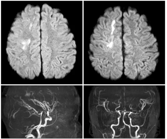
Fig. 1. Brain-magnetic resonance imaging and magnetic resonance angiography examination on post- operative day 12 showed acute cerebral infarction at subcortical white matter of right hemisphere and multiple stenoses of both anterior cerebral artery and middle cerebral artery.
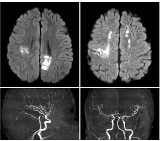
subcortical white matter of the right hemisphere and mul- tiple stenosis of both anterior cerebral artery (ACA) and middle cerebral artery (MCA) (Fig. 1). Serum tacrolimus level at symptom onset was 19.7 ng/mL. Conservative treatment was done for several days and tacrolimus con- tinued with dosage adjusted to maintain a serum level of 5 to 10 ng/mL. The repeated brain-MRI and MRA scaning was performed at post-operative day 19. The findings showed newly developed acute cerebral infarction on the subcortical white matter of the left hemisphere, cortex of the left parietal lobe and mildly improving status of steno- sis in both ACA and MCA (Fig. 2). This neurological change seems to be acute cerebral infarction due to cere- bral vascular stenosis caused by either vasculitis or vas- cular spasm, which is highly suspected to be the con- sequence of tacrolimus. We decided to change from tacro- limus to cyclosporine. Two days after the change, the cy- closporine level was 232.64 ng/mL. Afterwards, neuro- logic symptoms improved and follow-up (post-operative day # 29) brain MRI and MRA findings showed an improv- ing status of multifocal acute infarction and no stenosis of
either ACA or MCA (Fig. 3). At the 40th postoperative day, left motor weakness improved to grade IV, and rehabi- litation treatment was ongoing.
DISCUSSION
In 1996, Hinchey et al. [1] described a syndrome of acute but reversible clinical findings including headache, men- tal status alteration, seizures, hypertension, and acute vis- ual changes associated with abnormalities seen on MRI of symmetric white matter lesions, usually in bilateral parie- tal and occipital lobes. They termed this as reversible pos- terior leukoencephalopathy syndrome. Although it was initially thought to affect the subcortical white matter on- ly, additional studies supported by improved radiologic imaging modalities have revealed that the cortical gray matter may also be involved. The term “posterior rever- sible encephalopathy syndrome (PRES)” proposed by Casey et al. [2] is widely used recently because it expresses its clinical manifestation and radiologic findings appro priately.
Neurologic complication, tacrolimus
thesurgery.or.kr 227
Fig. 2. Brain-magnetic resonance imaging and magnetic resonance angiography examination on post- operative day 19 showed newly developed acute cerebral infarction on subcortical white matter of left hemisphere, cortex of left parietal lobe and mildly improving status of stenosis in both anterior cerebral artery and middle cerebral artery.
Fig. 3. Brain-magnetic resonance imaging and magnetic resonance angiography examination on post- operative day 29 showed improved status of multifocal acute infarction, no stenosis of both anterior cerebral artery and middle cerebral artery.
Woo-Hyung Kwun
228 thesurgery.or.kr
There have been many reports of this syndrome in the lit- erature since its initial definition.
As PRES has become better recognized, many con- tributing factors have been identified. For example, re- ports have linked PRES to hypertension, immunosup- pressive/chemotherapeutic agents, eclampsia, porphyria, and renaldysfunction [3,4]. The incidence of neurotoxicity, which is one of the major adverse events of calcineurin in- hibitors, was higher in patients receiving tacrolimus rath- er than cyclosporine. As both sensory and motor functions may be adversely affected, patients thus present with a wide range of neurological and psychiatric disorders.
Mild symptoms include tremor, neuralgia, and peripheral neuropathy. Severe symptoms could manifest as psy- choses, hallucinations, cortical blindness, seizures, cer- ebellar ataxia, motor weakness, or PRES. MRI is the stand- ard modality in diagnosing this syndrome. Fluid-attenu- ated inversion recovery is the most sensitive sequence for recognition of cortical and subcortical edema in PRES where hyperintense signal alterations are more prevalent than in conventional sequences [5,6]. This girl showed slightly different radiologic findings from those of typical PRES what causes vasogenic edema with vasculopathy, such as vasoconstriction and vasodilation. This girl's case was unique since there was cytotoxic edema with vaso- spasm. However, we could experience clinical and radio- logical improvement when we stopped tacrolimus treat- ment. Thus, the neurological complication of this patient seems to have been caused by the use of tacrolimus. PRES induced by tacrolimus can usually be diagnosed on the ba- sis of a characteristic clinical and radiographic pattern and is usually reversible by reducing the dosage or with hold- ing the drug for a few days [7]. Failure to recognize its her- alding symptoms may potentially increase morbidity in
organ transplantation. Therefore, careful examination of patients receiving immunosuppressive agents was need- ed to discover this uncommon but good prognostic com- plication.
CONFLICTS OF INTEREST
No potential conflict of interest relevant to this article was reported.
REFERENCES
1. Hinchey J, Chaves C, Appignani B, Breen J, Pao L, Wang A, et al. A reversible posterior leukoencephalopathy synd- rome. N Engl J Med 1996;334:494-500.
2. Casey SO, Sampaio RC, Michel E, Truwit CL. Posterior re- versible encephalopathy syndrome: utility of fluid-attenu- ated inversion recovery MR imaging in the detection of cortical and subcortical lesions. AJNR Am J Neuroradiol 2000;21:1199-206.
3. Singh N, Bonham A, Fukui M. Immunosuppressive-asso- ciated leukoencephalopathy in organ transplant reci- pients. Transplantation 2000;69:467-72.
4. Shin RK, Stern JW, Janss AJ, Hunter JV, Liu GT. Reversible posterior leukoencephalopathy during the treatment of acute lymphoblastic leukemia. Neurology 2001;56:388-91.
5. Bartynski WS, Boardman JF. Catheter angiography, MR an- giography, and MR perfusion in posterior reversible ence- phalopathy syndrome. AJNR Am J Neuroradiol 2008;29:
447-55.
6. McKinney AM, Short J, Truwit CL, McKinney ZJ, Kozak OS, SantaCruz KS, et al. Posterior reversible encephalop- athy syndrome: incidence of atypical regions of involve- ment and imaging findings. AJR Am J Roentgenol 2007;
189:904-12.
7. Lee VH, Wijdicks EF, Manno EM, Rabinstein AA. Clinical spectrum of reversible posterior leukoencephalopathy syndrome. Arch Neurol 2008;65:205-10.