Vol. 23, No. 3, 2011 405
Received March 9, 2010, Revised August 6, 2010, Accepted for publication September 14, 2010
*This paper was supported by research funds of Chonbuk National University in 2009.
Corresponding author: Seok-Kweon Yun, M.D., Department of Derma- tology, Chonbuk National University Medical School, 634-18 Geom- am-dong, Deokjin-gu, Jeonju 561-712, Korea. Tel: 82-63-250-1894, Fax: 82-63-250-1970, E-mail: [email protected].
This is an Open Access article distributed under the terms of the Creative Commons Attribution Non-Commercial License (http://
creativecommons.org/licenses/by-nc/3.0) which permits unrestricted non-commercial use, distribution, and reproduction in any medium, provided the original work is properly cited.
Ann Dermatol Vol. 23, No. 3, 2011 DOI: 10.5021/ad.2011.23.3.405
CASE REPORT
Basal Cell Carcinoma on the Pubic Area: Report of a Case and Review of 19 Korean Cases of BCC from Non-sun-exposed Areas
Jin Park, M.D., Yong-Sun Cho, M.D., Ki-Hun Song, M.D., Jong-Sun Lee, M.D., Seok-Kweon Yun, M.D., Han-Uk Kim, M.D.
Department of Dermatology, Chonbuk National University Medical School, Jeonju, Korea
Basal cell carcinoma (BCC) is one of the most commonly diagnosed malignant skin tumors and develops characteri- stically on sun-exposed areas, such as the head and neck.
Ultraviolet light exposure is an important etiologic factor in BCCs, and BCCs arising from non-sun- exposed areas are, therefore, very rare. In particular, the axilla, nipple, the genital and perianal areas are not likely to be exposed to ultraviolet light; thus, if BCC develops in these areas, other predisposing factors should be considered. Herein, we report a case of BCC arising on the pubic area in a 70-year-old man. We also performed a survey of the literature and discussed the 19 cases of BCC from non-sun-exposed areas reported to date in Korea. (Ann Dermatol 23(3) 405∼408, 2011)
-Keyword-
Basal cell carcinoma
INTRODUCTION
Basal cell carcinoma (BCC) is the most common malignant
neoplasm of the skin. BCCs develop characteristically on sun-exposed areas, with about 85% of reported cases occurring either on the head or neck1. Nevertheless, this carcinoma can arise, albeit rarely, in non-sun-exposed areas, such as the axilla, nipple, and the genital and perianal areas2,3. BCCs of the pubic area are exceedingly rare, i.e. about 0.05% of all BCCs2.
In this report, we describe a 70-year-old man who developed a BCC on the pubic area and we review previous case reports of BCC on the non-sun- exposed areas from Korea.
CASE REPORT
A 70-year-old man was referred to our clinic from a local hospital. He presented with a painful brown-to-gray- colored nodule on his right pubic area that he had had for 4 years. The skin lesion was growing and had become prominent in the previous 4 months, causing pain and bleeding. He had a history of hypertension, diabetes mellitus, and surgical intervention for benign prostatic hyperplasia. There was no medical history of sexually transmitted diseases, radiotherapy, chemical (arsenic or tar) exposure, or trauma to the genital area. There was no remarkable family history of skin disease or skin cancer.
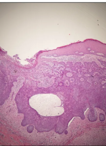
Physical examination revealed a 3.0×2.5 cm tender, brown, crusted nodule, with a gray-colored patch on the right pubic area (Fig. 1). An incisional biopsy was per- formed, as the preliminary diagnosis was skin cancer, such as squamous cell carcinoma or melanoma. Micro- scopically, retraction spaces were observed between the tumor islands and the surrounding stroma, and mucin- containing cystic spaces were present in the center of the tumor islands. The tumor was composed of basaloid cells,
J Park, et al
406 Ann Dermatol
Fig. 1. Brown crusted nodules of various sizes, with a gray patch on the right pubic area.
Fig. 2. Microscopic view of islands of cells, with peripheral palisading and haphazard arrangement of the more centrally located cells. Retraction spaces are present between the tumor islands and the surrounding stroma. Mucin-containing cystic spaces are visible at the centers of the tumor islands (H&E, ×40).
with peripheral palisading and peritumoral lacunae between the tumor mass and interstitial stroma. These histological findings were compatible with nodular BCC (Fig. 2).
Preoperative blood analysis included white cell count, platelet count, red blood cell count, and hepatic and renal biochemical profiles. They were all within normal limits.
We performed a positron emission tomography-computed tomography (PET-CT) scan to determine if the metastatic lesions were present, but no metastatic lesions were f ound.
The tumor was totally excised by Mohs micrographic sur- gery, and the skin defect was reconstructed using a local flap. After removal of the tumor, there was no evidence of either local recurrence or metastasis during the 36-month follow-up period.
DISCUSSION
Chronic exposure to ultraviolet light (UVL) is an important predisposing factor for BCC, and more than 80% of BCCs are found in sun-exposed areas of the body, such as the face. Consequently, BCCs of the non-sun-exposed areas, such as axilla, nipple, or the genital and perianal areas are extremely rare. LeSueur et al.4 investigated 10,000 BCCs and only 15 axillary BCCs (0.05%) were identified. With regard to the BCCs of the nipple, less than 30 cases were reported in the world5. Gibson and Ahmed2 reported 36 genital BCCs (0.2%) and 15 perianal BCCs (0.08%) out of a total of 18,943 investigated BCCs. Ten of the 36 genital BCCs occurred in the pubic area, representing 0.05% of the cases studied. Given that these regions are usually well- covered and not exposed to sunlight, other etiologic factors should be considered when a patient presents with a BCC of the non-sun-exposed areas. These factors include
radiation therapy, alterations in immune surveillance, exposure to coal tar or arsenics, sexually transmitted dis- eases, burns, traumatic scars, and chronic skin irritation due to chronic dermatologic conditions, such as chronic dermatitis6. Prior to this case report, only 18 cases of BCCs from non-sun-exposed area had been reported in Korea.
There were 11 cases of genital BCCs, 5 cases of axillary BCCs, 2 cases of nipple BCCs and no case of perianal BCC. Only two cases of scrotal BCC due to trauma have been reported so far (Choi et al.7 and Park et al.8);
however, no other etiologic factors have been determined in the rest of Korean cases of BCCs on the non-sun- exposed area reported thus far, as was the case in our study (Table 1)5,7-19.
According to Betti et al.20 and Bastiaens et al.21, different histologic subtypes of BCC can be found at different ana- tomical sites. Thus, nodular BCC predominantly occurs on the head and neck, while superficial BCC occurs mainly on the trunk. Meanwhile, LeSueur et al.4 reported that the most frequent histopathologic type of BCC in the axilla was the nodular type (67%), and the other reported types were superficial (20%) and micronodular (13%). Of the 5 cases of axillary BCCs reported in Korea to date, 2 were of the pigmented type, 1 was of the nodular type, 1 was of the basosquamous type, and 1 was of the adenoid type.
Of the 2 cases of nipple BCC reported in Korea, one was pigmented and the other was nodular type. Gibson and Ahmed2 reported that the most frequent histopathologic type of BCC in the perianal and genital regions was the
Basal Cell Carcinoma on the Pubic Area
Vol. 23, No. 3, 2011 407 Table 1. Reported cases of basal cell carcinama on the non-sun-exposed areas in Korean case literature and present case
Authors Age Sex Site Duration Factors* Size (cm) Histologic
type Treatment Clinical course F/U
period Meta‡ Choi et al.7 (2002) 66 M Scrotum 5 yrs Trauma 1.5×1.3 Pigmented Excision No recurrence NA† None Park et al.8 (2006) 83 M Scrotum 2 yrs Trauma 2.6×2.8,
0.7×1.0, 0.4×0.6
Nodular Excision No recurrence NA† None
Jeon et al.9 (2006) 68 M Scrotum 2 yrs None 2.5 Nodular None NA† NA† None
Kim et al.10 (1994) 67 M Scrotum 5 yrs None 2.5×2.5 Nodular Excision NA† NA† None Kim et al.10 (1994) 43 F Vulva 8 yrs None 2.0×3.0 Nodular Currettage NA† NA† None Kim et al.11 (1988) 49 F Vulva 20 yrs None 1×0.8×0.6 Nodular Excision NA† NA† None Lee et al.12 (2002) 73 F Vulva 3 yrs None 0.5×1.0 Nodular Excision No recurrence 2 yrs None Lee et al.12 (2002) 75 F Vulva 10 yrs None 1.0×2.5 Infiltrative Excision No recurrence 2 yrs None Kwon et al.13
(2003) 51 F Vulva 2 yrs None 2×2×1.5 Adenoid Excision No recurrence NA† None
Ryu et al.14 (2008) 43 F Vulva NA† None 3.5×1.0 Superficial Excision No recurrence 1 yr None Lee et al.15 (2004) 76 M Penis 4 yrs None 1.3×1.3 Nodular Excision No recurrence 4 months None Choi et al.16 (2006) 85 M Axilla NA† None 2.5×1.0 Nodular Excision No recurrence 1 yr None Choi et al.16 (2006) 62 M Axilla 1 yr None NA† Basosquamous Excision No recurrence 6 months None Choi et al.16 (2006) 35 M Axilla 5 yrs None NA† Pigmented Excision No recurrence 1 yr None Lee et al.17 (2007) 67 M Axilla 1 yr None 2.0×0.5 Pigmented Excision No recurrence 1 yr None Kim et al.18 (2008) 33 F Axilla 7 months None 1.0×0.8×1.5 Adenoid Excision No recurrence NA† None Kim et al.5 (2006) 73 M Nipple 2 yrs None Bean sized Pigmented None NA† NA† None Kim JH.19 (2007) 73 M Nipple 2 yrs None 2.0×1.0 Nodular Mastectomy
with ALND§
NA† NA† None
Present case 70 M Pubic
area 4 yrs None 3.0×2.5 Nodular Excision No recurrence 3 yrs None
*Considered etiologic factors, †NA: not available, ‡Metastasis at the time of diagnosis, §ALND: axillary lymph node disssection.
nodular type (66%), followed by the superficial (18%), infiltrative (8%), micronodular (4%), basosquamous (2%) and fibroepithelioma of Pinkus (2%). Of the 12 genital BCC cases reported in Korea (including this case report), 8 were of the nodular type, 1 was of the superficial type, 1 was of the infiltrative type, 1 was of the pigmented type, and 1 was of the adenoid type. Thus, the majority of genital BCC cases reported in Korea were histopatho- logically of the nodular type. Overall, the most common histopatholgic type of BCC on the non-sun-exposed areas was the nodular type (53%), followed by pigmented (21%), adenoid (11%), infiltrave (5%), superficial (5%), and basosquamous type (5%) (Table 1).
The risk factors for BCC recurrence include the time from the lesion’s appearance, the size larger than 2 cm, lesions with a previous history of therapeutic management, and histologically infiltrative, micronodular, or morpheaform lesions3. In the 19 cases of BCCs of non-sun-exposed area reported in the Korean literature (including this case report), the average size of the tumor was about 2.0 cm (range: 0.6∼3.5 cm), and the average time since the tumor developed was 4.5 years (range: 7 months∼20 years). However, none of the cases reported in Korea
recurred or metastasized at the time of diagnosis. Derma- tological lesions in the non-sun-exposed areas tend to be neglected by patients, because these regions are the least exposed areas of the human body. Thus, tumors on such sites may be larger and have been present for longer than tumors located in other parts of the body.
The usual treatment for BCC is wide excision; the 5-year recurrence rate after wide excision of BCCs, with clear excisional margins, is 3 to 14%.
Other treatment options for BCCs are Mohs micrographic surgery, curettage and electrodesiccation, cryosurgery, radiation, or photodynamic therapy with aminolevulinic acid22. Mohs micrographic surgery is a specialized sur- gical procedure, commonly used in patients who present with large tumors, high-risk morphea-type BCC tumors, recurrent tumors, or tumors located in cosmetically sensi- tive locations, such as the face. However, in general, surgical excision is preferred to the Mohs micrographic surgery, as the latter procedure requires specialized train- ing, a longer operating time, and increased costs22.
In conclusion, the BCCs arising from the non-sun-exposed areas may be larger and may have been present for longer than those arising from other areas of the body. Also,
J Park, et al
408 Ann Dermatol
physicians might initially mistake these cancers for either inflammatory or infectious disease2,23. It is recommended that physicians must consider the skin cancer as a one of the differential diagnosis and biopsy of all suspect lesions should be performed, even if the BCC at these regions can sometimes seem innocuous.
REFERENCES
1. Gallagher RP, Hill GB, Bajdik CD, Fincham S, Coldman AJ, McLean DI, et al. Sunlight exposure, pigmentary factors, and risk of nonmelanocytic skin cancer. I. Basal cell carcinoma.
Arch Dermatol 1995;131:157-163.
2. Gibson GE, Ahmed I. Perianal and genital basal cell carci- noma: a clinicopathologic review of 51 cases. J Am Acad Dermatol 2001;45:68-71.
3. Roewert-Huber J, Lange-Asschenfeldt B, Stockfleth E, Kerl H.
Epidemiology and aetiology of basal cell carcinoma. Br J Dermatol 2007;157(Suppl 2):47-51.
4. LeSueur BW, DiCaudo DJ, Connolly SM. Axillary basal cell carcinoma. Dermatol Surg 2003;29:1105-1108.
5. Kim WH, Park EJ, Kim CW, Kim KH, Kim KJ. A case of basal cell carcinoma of the nipple. Ann Dermaol 2006;18:100- 104.
6. Noodleman FR, Pollack SV. Trauma as a possible etiologic factor in basal cell carcinoma. J Dermatol Surg Oncol 1986;
12:841-846.
7. Choi YD, Borah L, Park SW, Kang MS, Wang HY. A case of pigmented basal cell carcinoma on the scrotum. Korean J Dermatol 2002;40:1571-1573.
8. Park SH, Lee SY, Kim SM, Choi HJ, Yun SK, Kim HU, et al. A case of basal cell carcinoma on post-traumatic scar of the scrotum. Korean J Dermatol 2006;44:1151-1153.
9. Jeon JH, Song HJ, Oh CH. Basal cell carcinoma of the scro- tum. Ann Dermatol 2006;18:97-99.
10. Kim CG, Cha HG, Han EH, Kwon KS, Chung TA. Two cases of basal cell carcinoma occurring on male and female exter- nal genitalia. Korean J Dermatol 1994;32:342-346.
11. Kim SH, Oh HR, Park JS, Park YS. A case of basal cell carcinoma of vulva. Korean J Obstet Gynecol 1988;31:580- 583.
12. Lee SW, Kim YC, Park HJ, Cinn YW. Two cases of basal cell carcinoma of the vulva. Korean J Dermatol 2002;40:1160- 1162.
13. Kwon JA, Lee SW, Kim KM, Kim SY. A case of atypical basal cell carcinoma on female external genitalia. Korean J Der- matol 2003;41:1674-1676.
14. Ryu AL, Kim JS, Kim TH, Lee HH, Lee KH, Nam KH. A case of vulvar basal cell carcinoma. J Soonchunhyang Med Coll 2008;14:469-473.
15. Lee YJ, Choi HJ, Yun SK, Kim HU, Ihm CW. A case of basal cell carcinoma of the penis. Korean J Dermatol 2004;42:
350-352.
16. Choi MH, Ko NY, Kim IH, Kye YC, Kim SN. Three cases of basal cell carcinoma of the axilla. Korean J Dermatol 2006;
44:887-889.
17. Lee SY, Park SH, Hong JS, Rhee CH, Yun SK, Kim HU, et al.
A case of axillary basal cell carcinoma. Korean J Dermatol 2007;45:758-760.
18. Kim SH, Ko WT, Suh MK, Lee JI. A case of axillary adenoid basal cell carcinoma. Ann Dermatol 2008;20:22-25.
19. Kim JH, Oh JW, Shin DH, Kim SI, Park BW. Basal cell carcinoma of the nipple-areolar complex. J Korean Surg Soc 2007;72:143-146.
20. Betti R, Inselvini E, Carducci M, Crosti C. Age and site prevalence of histologic subtypes of basal cell carcinomas.
Int J Dermatol 1995;34:174-176.
21. Bastiaens MT, Hoefnagel JJ, Bruijn JA, Westendorp RG, Vermeer BJ, Bouwes Bavinck JN. Differences in age, site distribution, and sex between nodular and superficial basal cell carcinoma indicate different types of tumors. J Invest Dermatol 1998;110:880-884.
22. Ceilley RI, Del Rosso JQ. Current modalities and new advances in the treatment of basal cell carcinoma. Int J Dermatol 2006;45:489-498.
23. Collins PS, Farber GA, Hegre AM. Basal-cell carcinoma of the vulva. J Dermatol Surg Oncol 1981;7:711-714.