Relation Between Left Atrial Enlargement and Stroke Subtypes in Acute Ischemic Stroke Patients
Hye-Young Shin1, In-Hye Jeong1, Chang-Ki Kang2, Dong-Jin Shin1, Hyeon-Mi Park1, Kee-Hyung Park1, Young-Hee Sung1, Dong-Hoon Shin1, Young Noh1, Yeong-Bae Lee1
1Department of Neurology, Gachon University Gil Medical Center, Incheon, Korea
2Neuroscience Research Institute, Gachon University of Medicine and Science, Incheon, Korea
Objective : Increased atrial size is frequently seen in ischemic stroke pa- tients in clinical practice. There is controversy about whether left atrial enlargement (LAE) should be regarded as a risk factor for cerebral infarction. We investigated the association between indexed left atrial vol- ume (LAVI) and conventional stroke risk factors as well as stroke sub- types in acute ischemic stroke patients.
Methods : One hundred eighty two acute cerebral infarction patients were included in this study. Brain magnetic resonance imaging and trans- thoracic echocardiography were done for all patients within 30 days of diagnosis of acute cerebral infarction. Echocardiographic LAE was identi- fied when LAVI was more than 27 mL/m2. Stroke subtypes were classi- fied by the Trial of Org 10171 in acute stroke treatment classification.
Results : There were significant differences between subjects with normal and increased LAVI in prevalence of stroke risk factors including atrial fi- brillation (p = 0.001), hypertension (p = 0.000), valvular heart disease (p
= 0.011) and previous stroke (p = 0.031). An increased LAVI was asso- ciated with cardioembolic subtype with an adjusted odds ratio was 6.749 (p = 0.002) compared with small vessel disease.
Conclusion : Increased LAVI was more prevalent in those who had car- diovascular risk factors, such as atrial fibrillation, hypertension, valvular heart disease and history of previous stroke. LAE influenced most patients in all subtypes of ischemic stroke but was most prevalent in the car- dioembolic stroke subtype. Increased LAVI might be a risk factor of cere- bral infarction, especially in patients with cardioembolic stroke subtype.
J Cerebrovasc Endovasc Neurosurg.
2013 September;15(3):131-136 Received : 21 March 2013 Revised : 18 June 2013 Accepted : 16 July 2013 Correspondence to Yeong-Bae Lee Department of Neurology, Gachon University Gil Medical Center, 1198, Guwol-dong, Namdong-gu, Incheon 405-760, Korea Tel : 82-32-460-3346
Fax : 82-32-460-3344 E-mail : [email protected]
Funding sources of this article are from Gachon University Gil Medical Center.
Portions of this work were presented in poster form as 31th Annual meeting of the Korean Neurological Association, Busan, Korea, October 6, 2011.
This is an Open Access article distributed under the terms of the Creative Commons Attribution Non- Commercial License (http://creativecommons.org/li- censes/by-nc/3.0) which permits unrestricted non- commercial use, distribution, and reproduction in any medium, provided the original work is properly cited.
Keywords Left atrial enlargement, Ischemic stroke, Stroke subtype, Echocardiography
INTRODUCTION
Stroke is the second leading cause of death in Korea and is associated with severe disability and mortality.17) The identification and treatment of stroke risk factors has important consequences on the prognosis of stroke.4)7) Well-known definite risk factors for ischemic
stroke include dyslipidemia, hypertension, diabetes mellitus, ischemic heart disease, atrial fibrillation, valv- ular heart disease, carotid stenosis, cigarette smoking, and obesity. We have frequently seen ischemic stroke patients with left atrial enlargement (LAE) in clinical practice. This condition indicates increased left ven- tricle (LV) filling pressure and the subsequent remod-
eling process in hypertensive heart disease.3) In the past few years, LAE has been found to be a risk factor for cardiovascular disease, atrial fibrillation, systemic thromboembolism, congestive heart failure (CHF) and stroke.5)6)8)12) The Framingham study noted that LAE may be a risk factor for ischemic stroke in men parti- ally mediated by LV mass.2) However, the role and ba- sic characteristics of LAE in acute cerebral infarction patients have not been sufficiently described in the literature. Especially, the relation between stroke sub- types and LAE is not clear in patients with acute is- chemic stroke. In this study, we investigated the rela- tion of LAE at the time of acute ischemic stroke with conventional stroke risk factors and stroke subtypes.
MATERIALS AND METHODS
Subjects and clinical data
The enrolled study population included 182 patients with acute ischemic stroke (mean age: 68.01 ± 12.53 years and 106 males), who were admitted from September 2010 to June 2011 and examined with brain magnetic resonance imaging (MRI).
Clinical data were collected through medical record review or direct interview. Routine laboratory tests in- cluded complete blood counts, glucose, serum electro- lytes, coagulation studies, liver function tests, and lip- id profile. Systemic hypertension was defined as sys- tolic blood pressure values > 140 mm Hg or diastolic blood pressure > 90 mm Hg during the interview, or the presence of a medical history or antihypertensive treatment. Diabetes mellitus was identified based on of elevated fasting blood glucose level, positive medical history, or current antidiabetic treatment. Hyperlipidemia was determined by a fasting total cholesterol level higher than 220 mg/dL, or antilipid agent treatment.
Coronary artery disease included the presence of an abnormal diagnostic test (stress test or coronary an- giography), a history of typical angina or myocardial infarction, or appropriate drug treatment. Atrial fi- brillation was identified by a current or past electro-
cardiography or Holter monitoring. CHF was defined based on symptoms, clinical signs, and/or anti-failure treatment. Valvular heart disease was evaluated by 2-dimensional transthoracic echocardiography. Previous stroke was indicated by a positive history on the medical record.
Stroke subtypes
Stroke subtypes were sorted in to 5 classes based on the National Institute of Neurological Disorders and Stroke and Trial of Org 10171 in Acute Stroke Treatment (TOAST) criteria as follows:1) 1) Large ar- tery atherosclerosis included patients with clinical and brain imaging findings of either significant (> 50%) stenosis or occlusion of a major brain artery or cort- ical artery branch, probably due to atherosclerosis. 2) Cardioembolism was defined as patients with cerebral infarction probably due to an embolus originated in the heart. Possible large artery atherosclerotic sources of thrombosis or embolism should be excluded. 3) Small artery occlusion (lacuna) included patients with classical lacunar syndromes and no evidence of car- diac embolism and significant large artery stenosis.
The patient should also have a significant posterior fossa level or hemispheric subcortical lesion with a di- ameter of less than 1.5 cm on neuroimaging. 4) Patients with rare causes of stroke such as non- atherosclerotic vasculopathies, hypercoagulable states, or hematologic disorders were classified as the cat- egory of other cause. 5) Patients whose cause of a stroke cannot be identified with any degree of evi- dence were labeled as unknown cause. Diagnostic evaluations included laboratory assessments, neuro- imaging (MRI, etc.), electrocardiogram, cardiac imag- ing (echocardiography, etc.), and duplex ultrasound imaging of extracranial arteries.
Echocardiographic data
Transthoracic echocardiography was performed for all patients within 30 days of diagnosis of cerebral infarction. Studies and measurements were taken based on the guidelines of the American Society of
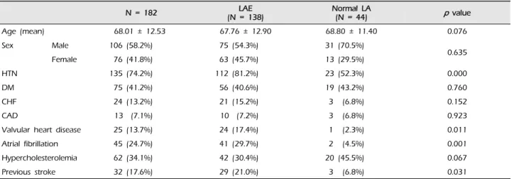
N = 182 LAE
(N = 138) Normal LA
(N = 44) p value
Age (mean) 68.01 ± 12.53 67.76 ± 12.90 68.80 ± 11.40 0.076
Sex Male 106 (58.2%) 75 (54.3%) 31 (70.5%)
0.635
Female 76 (41.8%) 63 (45.7%) 13 (29.5%)
HTN 135 (74.2%) 112 (81.2%) 23 (52.3%) 0.000
DM 75 (41.2%) 56 (40.6%) 19 (43.2%) 0.760
CHF 24 (13.2%) 21 (15.2%) 3 (6.8%) 0.152
CAD 13 (7.1%) 10 (7.2%) 3 (6.8%) 0.923
Valvular heart disease 25 (13.7%) 24 (17.4%) 1 (2.3%) 0.011
Atrial fibrillation 45 (24.7%) 41 (29.7%) 2 (4.5%) 0.001
Hypercholesterolemia 62 (34.1%) 42 (30.4%) 20 (45.5%) 0.067
Previous stroke 32 (17.6%) 29 (21.0%) 3 (6.8%) 0.031
LAE= left atrial enlargement; LA= left atrium; HTN= hypertension; DM= diabetes mellitus; CHF= congestive heart failure; CAD= coronary artery disease.
Table 1. Demographic characteristics
Echocardiography.15) Measurement of left atrial ante- roposterior diameter especially was performed ac- cording to a leading edge-to-leading edge convention at the aortic valve level. Indexed left atrial volume (LAVI) was calculated from the left atrial ante- roposterior diameter indexed to body surface area.
Echocardiographic LAE was identified when the LAVI was larger than 27 mL/m2.
Statistical analyses
The data collected included continuous variables (clinical and echocardiographic data) and categorical variables (stroke risk factors). Continuous variables are presented as the mean ± standard deviation (SD).
Differences in baseline characteristics between stroke patients with normal and increased LAVI were ana- lyzed by using an unpaired, 2-tailed Student’s t-test.
Differences of stroke risk factors between patients with and without LAE in the categorical variable val- ues were evaluated using the chi-square test. Null hy- potheses of no difference were rejected if p values were less than 0.05, or, equivalently, if the 95% con- fidence intervals (CIs) of risk point estimates excluded 1. Logistic regression analysis was employed to de- termine the correlation of stroke subtypes and in- creased LAVI. All statistical analysis has been per- formed using SPSS for Windows, version 17.0 (SPSS
Inc., Chicago, IL, USA).
RESULTS
Demographic characteristics of participants are pre- sented in Table 1. The mean LAVI of all study sub- jects was 37.62 ± 17.59 ml/m2. One hundred thirty eight (75.8 %) of patients had increased LAVI (42.58
± 17.40 ml/m2). Patients with normal LAVI were 44 (24.2 %) whose mean LAVI was 22.09 ± 3.39 ml/m2. Age and sex were not significantly different between those with increased LAVI and normal LAVI. Patients with increased LAVI tend to have more stroke risk factors than patients with normal LAVI. Patients with increased LAVI were significantly different from pa- tients with normal LAVI in the prevalence of hyper- tension (81.2% VS 52.3%; p = 0.000), history of pre- vious stroke (21% VS 6.8%; p = 0.031), atrial fi- brillation (29.7% VS 4.5%; p = 0.001), and valvular heart disease including mitral stenosis (17.4% VS 2.3%; p = 0.011) but not in the prevalence of coronary artery disease, CHF and diabetes mellitus.
Patients with increased LAVI commonly had car- dioembolic stroke subtype (41, 29.7%) compared with patients with small vessel occlusion (38, 27.5%), large vessel atherosclerosis (36, 26.0%), and unknown (20,
Stroke subtypes Mean LAVI (ml/m2) LAE n, (%) Normal LA n, (%)
All subjects (n = 182) 37.62 138 44
SVD (n = 60, 33.0%) 29.72 38 (27.5%) 22 (50%)
LAD (n = 50, 27.5%) 34.61 36 (26.0%) 14 (31.8%)
CE (n = 45, 24.7%) 53.29 41 (29.7%) 4 (9%)
Undetermined (n = 24, 13.2%) 34.47 20 (14.4%) 4 (9%)
Other cause (n = 3, 1.6%) 36.16 3 (2.1%) 0
LAE= left atrial enlargement; LA= left atrium; LAVI= indexed left atrial volume; SVD= small vessel disease; LAD= large artery disease; CE=
cardioembolism.
Table 2. Stroke subtypes and LAE
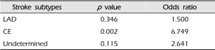
Stroke subtypes p value Odds ratio
LAD 0.346 1.500
CE 0.002 6.749
Undetermined 0.115 2.641
LAE= left atrial enlargement; LAD= large artery disease; CE=
cardioembolism.
Table 3. Statistical analyses of risk of ischemic stroke sub- types according to LAE
14.4%) subtype (Table 2). The proportion of LAE in patients with cardioembolic stroke subtype was sig- nificantly high, namely, 41 patients had increased LAVI while only 4 patients had normal LAVI.
An increased LAVI was associated with a car- dioembolic subtype compared with small vessel dis- ease (Table 3). An increased LAVI risk was present after adjustment for age and sex, and the adjusted odds ratio was 6.749 (p = 0.002) compared with small vessel disease.
DISCUSSION
The major findings of this study were as follows:
first, increased LAVI is more common in patients who had cardiovascular risk factors, such as hypertension, valvular heart disease, atrial fibrillation, and previous stroke. Second, increased LAVI influences most pa- tients in all ischemic stroke subtypes, and is especially highly prevalent in the cardioembolic stroke subtype.
Therefore, increased LAVI is more likely a risk factor of ischemic stroke, especially cardioembolic stroke, than normal LAVI.
There are suggestions that LAE is associated with the risk of stroke.5)6)9)11) A recent study in a hyper- tensive population revealed that a first-ever ischemic stroke was associated with LAE, and LAVI was the best predictor of stroke in left atrial assessment.11) They enrolled a hypertensive population with or with- out first ischemic stroke as study population and there were no differences in ischemic heart disease, parox-
ysmal atrial fibrillation and heart failure between groups. They showed, however, that paroxysmal atrial fibrillation and other stroke risk factors are associated with LAE in stroke patients with hypertension. One study reported that for patients with first-ever ische- mic stroke in an ethnically diverse population, LAE was associated with an elevated risk of cerebral in- farction after adjustment for other stroke risk factors in acute ischemic stroke patients with significantly in- creased mean left atrial anteroposterior diameter and LAVI compared with control group.5) Another study found that 75% of first ischemic stroke patients have increased LAVI.6) Ischemic stroke patients with in- creased LAVI were older and had more cardiovascular risk factors than those with normal LAVI. LAVI was also associated with mortality after adjusted for multi- ple risk factors. The current study has similar results in that patients with increased LAVI have more car- diovascular risk factors and previous stroke history.
Left atrium (LA) volume is related to severity and chronicity of diastolic dysfunction in the absence of severe valvular disease and atrial fibrillation. The poor LV compliance raises LA pressure to retain ad-
equate LV filling during ventricular diastole. Continuous exposure to abnormal LV filling pressure may result in LA remodeling and LAE. Few previous researchers have shown LAE associated with risk of CHF. The Cardiovascular Health Study observed an independent relation of LA volume in the patient group with a higher incidence and prevalence of CHF compared with control group.8) In a study of elderly subjects with preserved LV systolic function, LAVI larger than 32 mL/m2 was seen to independently predict first CHF.14) Contrary to those results, this study found no significant difference in the prevalence of CHF be- tween patients with and without LAE. This is possi- bly due to the age difference of the studied subjects.
The subject population of this study was younger than previous studies, in which subjects ≥ 65 years of age were enrolled.
The mechanisms of the relation between ischemic stroke and an LAE are not completely understood. A potential explanation is that the increased hemostasis and thrombus formation might arise as the left atrial size increases. The thrombogenicity of an enlarged LA is suggested by the results of several transesophageal echocardiographic studies.10)13) Research to determine possible intracardiac sources of cerebral emboli report that LAE is the best predictor for either clot in left at- rial appendage or spontaneous echo contrast.10) An enlarged atrial size may be the consequence of a raised intra-atrial pressure, which diminishes the flow velocity in the left atrial appendage and therefore in- creases the thrombogenicity and the embolic risk.13) LAE has been reported to be a potent risk factor for the development of atrial fibrillation. The Framingham Heart Study showed that every 5 mm increment in LA size raised the risk of atrial fibrillation by 39%.16) The Cardiovascular Health Study noted that LAE was an independent risk factor of new onset atrial fi- brillation in older adults and LA with larger than 0.5 mm diameter had a 4-fold risk of new atrial fibrillation.12) These observations may explain part of the association between stroke and LAE.
This study has a few limitations. There is potential for intrinsic selection bias in the sample because this study enrolled patients having echocardiography at the time of stroke. The study population has relatively small sample size to represent the general character- istics of ischemic stroke patients fully and there is no proper control group to strengthen the results.
Further study remains to be performed on a large population and with a normal control group.
CONCLUSION
LAE is relatively common finding in patients with ischemic stroke. However, there was a lack of inves- tigations about clinical characteristics and consid- erations as a stroke risk factor. From the results of this study, LAE was found to be a significant risk fac- tor of ischemic stroke, particularly in case of car- dioembolic stroke subtype. It may be useful in the es- timation of stroke risk and treatment of ischemic stroke in the clinical field.
REFERENCES
1. Adams HP Jr, Bendixen BH, Kappelle LJ, Biller J, Love BB, Gordon DL, et al. Classification of subtype of acute ischemic stroke. Definitions for use in a multicenter clin- ical trial. TOAST. Trial of Org 10172 in Acute Stroke Treatment. Stroke. 1993 Jan;24(1):35-41.
2. Benjamin EJ, D'Agostino RB, Belanger AJ, Wolf PA, Levy D. Left atrial size and the risk of stroke and death. The Framingham Heart Study. Circulation. 1995 Aug;92(4):835-41.
3. Bihorac A, Tezcan H, Ozener C, Oktay A, Akoglu E.
Association between salt sensitivity and target organ damage in essential hypertension. Am J Hypertens. 2000 Aug;13(8):864-72.
4. Bonita R, Beaglehole R. Does treatment of hypertension explain the decline in mortality from stroke? Br Med J (Clin Res Ed). 1986 Jan;292(6514):191-2.
5. Di Tullio MR, Sacco RL, Sciacca RR, Homma S. Left at- rial size and the risk of ischemic stroke in an ethnically mixed population. Stroke. 1999 Oct;30(10):2019-24.
6. Fatema K, Bailey KR, Petty GW, Meissner I, Osranek M, Alsaileek AA et al. Increased left atrial volume index:
Potent biomarker for first-ever ischemic stroke. Mayo Clin Proc. 2008 Oct;83(10):1107-15.
7. Gorelick PB. Stroke prevention. An opportunity for effi-
cient utilization of health care resources during the com- ing decade. Stroke. 1994 Jan;25(1):220-4.
8. Gottdiener JS, Kitzman DW, Aurigemma GP, Arnold AM, Manolio TA. Left atrial volume, geometry, and function in systolic and diastolic heart failure of persons
> or = 65 years of age (the cardiovascular health study).
Am J Cardiol. 2006 Jan;97(1):83-9.
9. Lai CL, Chien KL, Hsu HC, Su TC, Chen MF, Lee YT.
Left atrial dimension and risk of stroke in women with- out atrial fibrillation: The Chin-Shan community car- diovascular cohort study. Echocardiography. 2011 Nov;28 (10):1054-60.
10. Lee RJ, Bartzokis T, Yeoh TK, Grogin HR, Choi D, Schnittger I. Enhanced detection of intracardiac sources of cerebral emboli by transesophageal echocardiography.
Stroke. 1991 Jun;22(6):734-9.
11. Piotrowski G, Banach M, Gerdts E, Mikhailidis DP, Hannam S, Gawor R et al. Left atrial size in hyper- tension and stroke. J Hypertens. 2011 Oct;29(10):1988-93.
12. Psaty BM, Manolio TA, Kuller LH, Kronmal RA, Cushman M, Fried LP et al. Incidence of and risk fac- tors for atrial fibrillation in older adults. Circulation.
1997 Oct;96(7):2455-61.
13. Tabata T, Oki T, Fukuda N, Iuchi A, Manabe K, Kageji Y et al. Influence of left atrial pressure on left atrial ap- pendage flow velocity patterns in patients in sinus rhythm. J Am Soc Echocardiogr. 1996 Nov-Dec;9(6):857-64.
14. Takemoto Y, Barnes ME, Seward JB, Lester SJ, Appleton CA, Gersh BJ et al. Usefulness of left atrial volume in predicting first congestive heart failure in patients > or
= 65 years of age with well-preserved left ventricular systolic function. Am J Cardiol. 2005 Sep;96(6):832-6.
15. Tsang T, Barnes ME, Gersh BJ, Bailey KR, Seward JB.
Left atrial volume as a morphophysiologic expression of left ventricular diastolic dysfunction and relation to car- diovascular risk burden. Am J Cardiol. 2002 Dec;90 (12):1284-9.
16. Vaziri SM, Larson MG, Benjamin EJ, Levy D.
Echocardiographic predictors of nonrheumatic atrial fibrillation. The Framingham Heart Study. Circulation.
1994 Feb;89(2):724-30.
17. Suh OJ, Rheu SO. The Cause of death Statistics.
Department of Population Trend. Statistics Korea. 2012.