Case Report
J Gynecol Oncol Vol. 19, No. 4:261-264, December 2008 DOI:10.3802/jgo.2008.19.4.261
261
Leiomyosarcoma of the vagina:
a case report and review from the literature
Min Jung Suh, Dong Choon Park
Department of Obstetrics and Gynecology, St. Vincent's Hospital, The Catholic University of Korea, Suwon, Korea
Leiomyosarcomas comprise fewer than 2% of all malignant vaginal neoplasms. Due to their rarity, treatment for vaginal leiomyosarcomas have not been determined. We describe a 66 year old woman with vaginal leiomyosarcoma, which presented as a large palpable mass with vaginal spotting. Complete surgical excision was accomplished and after surgery, she underwent radiation therapy. Tumor recurrence was not detected for the last 5 years and now her general condition is very good. This is a rare case of leiomyosarcoma arising in vagina and we report the results of successful treatment.
Key Words: Leiomyosarcoma, Vagina
Received June 13, 2008, Revised July 24, 2008, Accepted August 20, 2008
Address reprint requests to Dong Choon Park
Department of Obstetrics and Gynecology, St. Vincent’s Hospital, The Catholic University of Korea, 93, Gi-dong, Paldal-gu, Suwon 442-723, Korea
Tel: 82-31-244-9504, Fax: 82-31-254-7481 E-mail: [email protected]
INTRODUCTION
Malignant tumors in the vagina are extremely rare, compris- ing less than 1% of cancers of the reproductive organs. The most frequently observed vaginal tumors are squamous cell carcinomas (75-90%), followed by adenocarcinoma (5-10%), melanoma, and leiomyosarcoma.1 Since only about 140 leio- myosarcomas in the vagina have been reported during the past 40 years,2 guidelines for the determination of disease stage and treatment have not yet been established. Vaginal leio- myosarcomas usually originate from the smooth muscle of the vaginal wall, but they may also develop from smooth mus- cle cells in tissues near the vagina. Malignancy of leiomyo- sarcoma is diagnosed histopathologically by assessment of cellular mitosis activity and the presence of cellular dysplasia.3
CASE REPORT
A 66-year old gravid 2-0-1-2 woman presented with vaginal spotting that had begun approximately 2 weeks earlier. Her menopause had occurred when she was at age 43 and her men- struation pattern had consisted of 30 day cycles, with normal
volume and mild dysmenorrhea. She had 2 vaginal sponta- neous deliveries and one abortion. Her family history was uneventful. She was recently diagnosed with asthma and was treated with oral agents for this disease.
On pelvic examination, a mass suspected of being a hema- tocolpos was detected, and she was transferred to the Department of Obstetrics and Gynecology of our hospital.
From the results of magnetic resonance imaging (MRI), the mass was diagnosed as a uterine smooth muscle tumor sus- pected of being malignant or a secondary degeneration, and surgery was scheduled.
1. Laboratory findings
At the time of admission, her vital sign was stable and her general condition was good. Her hemoglobin concentration was 12.4 g/dl, her red blood cell volume was 37.5%, her leu- kocyte count was 5,150 /mm3, and her platelet count was 287,000 /mm3, all within normal ranges. No glucose or pro- tein was detected in her urine. Her serum concentration of CEA, SCC, and CA125 were 2.07 ng/ml, 0.8 ng/ml, and 4.64 U/ml, respectively, all within normal ranges. Uterine cervical cytology at the time of admission was normal. Sigmoidoscopy showed that her sigmoid colon was compressed by the tumor, but it was not a rectal tumor.
2. Radiological findings
Pelvic examination showed that her uterus and both adnexae had become atrophic after menopause and were therefore small when palpated. An approximately 12.0×6.0 cm sized mass was detected in the posterior vaginal wall and it was movable without tenderness. Transvaginal ultrasound also
J Gynecol Oncol Vol. 19, No. 4:261-264, 2008 Min Jung Suh, Dong Choon Park
262
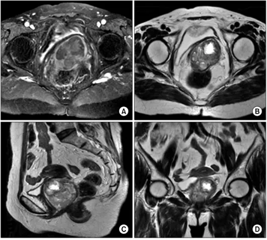
Fig. 1. Pre-operative MR images.
They show that the about 6×5.7×
5.2 cm sized heterogenous signal mass in posterolateral portion of the vagina. (A) T1-weighted axial MR image, (B) T2-weighted axial MR image, (C) T2-weighted sagi- ttal MR image, (D) T2-weighted co- ronal MR image.
Fig. 2. (A) There are marked in- creased spindle cells (H & E, ×40), (B) In focal area, we could see some foci of degeneration (H & E, ×100).
(C, D) Spindle cells revealed mode- rate nuclear atypism with frequent mitosis (H & E, ×400).
Leiomyosarcoma of the vagina: a case report and review from the literature
263 showed that her uterus and both adnexae were atrophic, and that the mass in the posterior vaginal wall was approximately 6.5×6.0 cm in size, with irregular contrast. Pelvic MRI showed a mass approximately 6×5.7×5.2 cm in volume with heterogeneous signal intensity adjacent to the rectum. The mass appeared to be leiomyoma accompanying secondary de- generation, and it could be differentiated from a smooth mus- cle tumor (Fig. 1).
3. Surgical findings
The patient was placed in the dorsal lithotomy position; spi- nal anesthesia was performed; the posterior vaginal wall tu- mor, including the capsule, was completely removed; and a drain was inserted into the area from which the tumor had been removed.
4. Pathological findings
Grossly, the tumor was black-brown in color and approx- imately 6.0×5.5×5.0 cm in size. Histologically, it was com- posed of spindle shaped cells, with partial secondary degener- ation and necrosis. When examined at high magnification (400×), the spindle cells showed evidence of dysplasia and frequent cell division (8.85/10 HPF). Based on histopatho- logical results, the tumor was diagnosed as a leiomyosarcoma (Fig. 2).
5. Surgery outcome and follow-up
After surgery, the patient did not experience any disease re- lated symptoms and was discharged. She underwent radiation therapy, consisting of 45 Gy external and 25 Gy intra-cavitary radiation. Tumor recurrence was not detected for the last 5-year follow-up and her general condition is good.
DISCUSSION
The criteria between benign leiomyoma and leiomyosarco- ma include the level of cell division and histologic evidence of cellular dysplasia. Cell division of more than 5 of 10 HPF is considered evidence of malignancy, in which the recurrence rate and metastatic potential is high.3
Due to their rarity, vaginal leiomyosarcomas are seldom de- tected prior to surgery, and typical symptoms and character- istics on physical examination have not been described. These tumors are usually diagnosed histopathologically after surgi- cal removal. Most patients with vaginal masses have no symp- toms, although some may experience vaginal or rectal bleed- ing, serous discharge, or, rarely, dypareunia. On physical ex- amination, most patients show palpable nodules surrounded by normal vaginal mucosae; in patients with advanced tu- mors, readily breakable exophytic polypoids or necrotic vagi- nal masses may be palpated. These tumors may invade the rec- tum or pelvic tissues adjacent to the vagina, requiring histo- logical diagnosis.4
Vaginal smooth muscle tumors most frequently develop in
the anterior vaginal wall, in contrast, malignant tumors most frequently develop in the posterior vaginal wall, although they can also develop elsewhere.2 Differential diagnosis of these vaginal masses include Gartner’s cyst, granuloma, epithelial inclusion cyst, other neurofibroma, rhabdomyoma, capillary hemangioma, squamous epithelial carcinoma, adenocarci- noma, rhabdomyosarcoma, melanoma, small cell carcinoma, and primary malignant tumors such as mixed müllerian sarco- ma that can develop in the vagina. In addition, leiomyosarco- ma has to be differentiated from gestational trophoblastic tu- mor, uterine endometrial cancer, and vaginal metastasis of malignant tumors developing in adjacent organs, including the uterine cervix, rectum, and bladder.2
Due to their rarity, risk for development of smooth muscle tumors have not been determined. These tumors can recur lo- cally, and have a tendency to show hematogeneous metastasis into adjacent organs. Thus, patients with this disease have a poor prognosis, and metastasis to the lungs is frequent.5 Vaginal leiomyomas can be treated by surgical resection. Due to the possibility of future transformation to sarcoma and re- currence, as well as diagnosis as malignant after surgery, these tumors have to be removed completely. The basic treat- ment for vaginal smooth muscle tumor is resection of a wide area of the primary site and of the site of possible uterine sar- coma, but the extent of resection may not be related to clinical course.4 In patients treated with postoperative chemotherapy or radiation therapy, the 5-year survival rate is only 36%.4 The differentiation grade of the tumor is the most important prog- nostic factor. In addition, younger age and lower stage at the time of diagnosis and the absence of infiltration in the re- section margin at the time of surgery were associated with good prognosis.5
Based on the histological results of a frozen biopsy taken during operation, our patient was diagnosed with a benign tu- mor suspected of secondary degeneration. However, cellular dysplasia and necrosis and 8.5/10 HFP cell division were de- tected in permanent section evaluation postoperatively. The mass in our patient was completely resected, and there was no infiltration into the resection margin. Nevertheless, the rarity of this disease, and findings consistent with malignancy, in- cluding cell division, cellular dysplasia, and tissue necrosis, indicated the appropriateness of treatment similar to that for vaginal smooth muscle cell tumors. Thus, following surgery, this patient underwent radiation therapy.
In summary, we describe here a patient with a leiomyosarco- ma that developed in the vagina, which was treated with radia- tion therapy after resection. Follow-up for 5 years shows no evidence of tumor recurrence or metastasis.
REFERENCES
1. Kim BH. The vagina and vulva. In: Kim SH, McClennan BL, Outwaer EK. Radiology illustrated: Gynecologic imaging. Phila- delphia: Elsevier Saunders; 2005. p.807-9.
J Gynecol Oncol Vol. 19, No. 4:261-264, 2008 Min Jung Suh, Dong Choon Park
264 2. Ahram J, Lemus R, Schiavello HJ. Leiomyosarcoma of the vagi-
na: case report and literature review. Int J Gynecol Cancer 2006; 16: 884-91.
3. Tavassoli FA, Norris HJ. Smooth muscle tumors of the vagina.
Obstet Gynecol 1979; 53: 689-93.
4. Ngan HY, Fisher C, Blake P, Shepherd JH. Vaginal sarcoma: The
Royal Marsden experience. Int J Gynecol Cancer 1994; 4:
337-41.
5. Creasman WT, Phillips JL, Menck HR. The National Cancer Data Base report on cancer of the vagina. Cancer 1998; 83:
1033-40.