R E S E A R C H
Open Access
One-step synthesis of Pt/a-CoO
x
core/shell
nanocomposites
Daewoon Kim, Sung Joo Kim and Jong Min Yuk
*Abstract
Herein, we synthesize a core/shell Pt/a-CoOx nanocomposite via one-step synthesis using a strong reaction agent of borane t-butylamine(BBA) at 200 °C. Transmission electron microscopy study shows that the morphology of nanocomposites is controlled by the stirring time and perfect core/shell structure is formed with over 7 days stirring time.
Keywords: One-step synthesis, Nanocomposites, TEM, EDS Introduction
Nanocomposites containing Pt have attracted great attentions due to their excellent catalytic, electric and magnetic properties. (Peng and Yang,2009; Li et al.2015; Zhang et al.2013; Wang et al.2015; Esfahani et al. 2010; Wang et al.2010) Since these properties closely intertwine with their size, shape and composition, designing nano-composites is critical to their chemical, electrical and en-ergy applications. (Pushkarev et al. 2012; Vidal-lglesias et al.2012; Mostafa et al.2010; Wang et al.2013) Among diverse nanocomposites, core/shell structures of Pt/transi-tion metal oxide, such as Pt/Fe2O3, FePt/Fe3O4 or Pt/
CoO, not only show remarkable magnetic properties, but also contain small amount of expensive Pt. (Alayoglu et al.
2008; Tao et al. 2008; Zhao and Xu, 2006; Zhou et al.
2005; Teng et al. 2003; Zeng et al.2004; Yin et al. 2004; Habas et al.2007) Traditionally, the core/shell structures have been synthesized by a two-step growth method. (Tao et al.2008; Liu et al.2005; Yu et al.2014) Core nanoparti-cles are synthesized first as seeds, followed by growth of the shell around the core. However, the two-step growth technique typically suffers from low yield because the synthesized core particles are not well dispersed and shell materials independently coalesce each other instead of ad-hering to the core. In our study, we report a (scanning) transmission electron microscopy ((S)TEM) study of Pt/ amorphous cobalt oxide (a-CoOx) nanocomposites growth
by one-step heating synthesis.
Experiments
The nanocomposites are synthesized with platinum(II) acetylacetonate(Pt(acac)2) (97%), cobalt(III)
acetylaceto-nate(Co(acac)3) (98%), oleylamine(98%), oleic acid(90%),
benzyl ether(98%), and borane tert-butylamine(97%) from Sigma-Aldrich Co.. 1 M Pt(acac)2, and 3 M
Co(a-cac)3 were dissolved in 0.6 mL oleic acid, 6 mL
oleyla-mine and 53.4 mL benzyl ether(total 60 mL solution). The solution is heated to 50 °C under magnetic stirring for 10 min. Here, we add 1 M borane t-butylamine (BBA), which is a more powerful reaction agent than oleylamine or oleic acid. (Yu et al. 2014) (Fig. 1) Then the chemicals are further heated to 200 °C and kept for 2 h using autoclave oven. After the solution is cooled to room temperature, the nanocomposites are obtained after several washes with 40 mL ethanol by centrifuging at 3000 rpm for 10 min and dried under vacuum. The final products are dispersed in toluene. According to the stirring time of the solution, we analyze the morphology of the synthesized nanocomposites using a TEM. The TEM imaging is performed using JEOL ARM200F oper-ated at 200 kV in conjunction with a Bruker Quantax energy-dispersive X-ray spectroscopy (EDS) detector.
Results and discussion
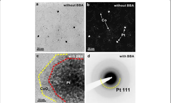
TEM/STEM images in Fig. 2 show the effect of a BBA additive on a synthesis of Pt/CoOxnanocomposites.
With-out BBA addition into the precursor solution, the synthe-sized Pt and Co are formed separately with forming a compound. (Fig.2)a Using the Z(atomic number)-contrast dark-field STEM imaging, 5 nm sized bright nanoparticles
© The Author(s). 2019 Open Access This article is distributed under the terms of the Creative Commons Attribution 4.0 International License (http://creativecommons.org/licenses/by/4.0/), which permits unrestricted use, distribution, and reproduction in any medium, provided you give appropriate credit to the original author(s) and the source, provide a link to the Creative Commons license, and indicate if changes were made.
* Correspondence:[email protected]
Department of Materials Science and Engineering, KAIST, Daejeon 305-701, South Korea
Applied Microscopy
Kim et al. Applied Microscopy (2019) 49:12 https://doi.org/10.1186/s42649-019-0016-2
are likely Pt while the rests with a size distribution of 0.96 ± 0.56 nm are Co. (Fig.2b, Additional file1: Figure S1) In the synthesis, the color of the reaction solution changes from yellow to black at around 140 °C when Pt(acac)2only
added in the solution. On the other hand, the color of the solution does not change at around 200 °C for 2 h when Co(acac)3 only added. However, in our study, the
reduction reaction of Co(acac)3 is observed at 200 °C
when two precursors are simultaneously added. This shows that pre-synthesized Pt nanoparticles act as cata-lysts to lower the reduction temperature of Co(acac)3 to
below 200 °C. However, the number of formed Pt nano-particles is not sufficient enough for Co reduction to grow Co nanoparticles, making Pt and Co form separately.
Fig. 1 Schematic illustration of the synthesis procedure for Pt/CoOxnanocomposite
Fig. 2 TEM image of Pt/CoOx nanocomposite (a) Bright-field and (b) dark-field STEM images of Pt and Co nanoparticles without using a BBA additive during synthesis. c TEM image of synthesized Pt/a-CoOxcore/shell nanocomposites when using a BBA additive. d Selected area electron
Thus, we recognize that Pt nanoparticles need to be re-duced at much lower than 140 °C in order to produce a composite of Pt and Co. Figure 2c shows the bright field TEM image of Pt/CoOxnanocomposites with the addition
of BBA. By adding BBA inside the solution, the color of the solution changes to black at 60 °C. This shows that re-duction temperature of Pt(acac)2is lowered below 60 °C.
This change makes Pt nanoparticles form inside the solu-tion much more than the one without BBA to ultimately have the reduced cobalt clusters increased. With BBA, the synthesized nanostructure forms a core-shell structure. Pt nanoparticles are well distributed with amorphous shells wrapped around them. In a selected area electron diffrac-tion (SAED) pattern of the nanocomposite, polycrystalline Pt is formed in a core with an amorphous shell formed outside. (Fig.2d).
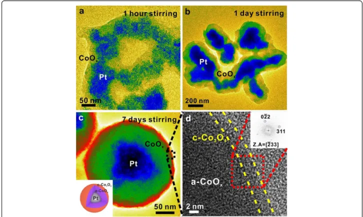
In order to change the morphology of nanocompos-ites, we further modify the synthesis of Pt/CoOx
nano-composites by adjusting the stirring time at 50 °C (Fig.3). Under 1 h stirring, Pt nanoparticles are found to spread widely while the shell structure is grown to sur-round them (Fig.3a). Under 1 day stirring, Pt nanoparti-cles coalesce to form a larger nanocomposite core than that with 1 h stirring (Fig. 3b). Finally, under 7 days stir-ring, Pt nanoparticles are aggregated to a size in between
50 and 100 nm, while an amorphous shell surrounds uni-formly to form a perfect spherical shape (Fig. 3c). The overall size of core/shell nanocomposites is 100~200 nm.
Although BBA originally reduces Pt precursors at 60 °C, stirring at 50 °C for a sufficiently long time after add-ing BBA produces agglomerated Pt seeds without growth due to low temperature. When the temperature is raised, the seeds grow and accumulate in the core. Further in-creasing the temperature reduces the Co precursor to form an oxide shell around the core. Figure 3d shows the high-resolution TEM image of the outer surface of the synthesized core/shell nanocomposites. It is clear that the shell is composed of an amorphous structure but with 3 nm crystalline CoOxformed on the outermost
surface. A corresponding fast Fourier transform (FFT) image identifies the structure to be Co3O4. This suggests
that the surface of the nanocomposites transforms from amorphous to crystalline by exposing it to air.
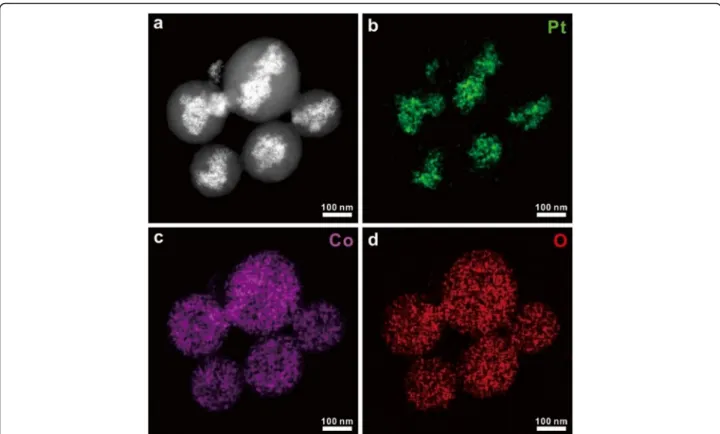
Figure 4 shows the STEM EDS mapping for compos-ition analysis of core/shell nanocomposites. At the nano-composite core, it is confirmed that the bright area in a STEM image in Fig. 4a consists of Pt while (Fig.4b) Co and O are shown at the shell (Fig.4c, d). The quantita-tive analysis clearly suggests that the shell structure is Co3O4-x (Additional file 1: Figure S2). In Fig. 4b, the
Fig. 3 Bright-field TEM images and high-resolution TEM analysis of the one-step synthesized Pt/CoOxnanocomposites under various stirring
conditions (a) 1 h, (b) 1 day and (c) 7 days. Inset: Schematic illustration of the synthesized Pt/CoOxcore/shell nanocomposite. d Zoomed in
high-resolution image of a square box in (c). Inset: fast Fourier transform (FFT) pattern of a red square in (d)
morphology of Pt is distributed like a band in a specific direction. However, in Fig. 4c and Fig. 4d, Co and O have a spherical shape. This result shows that the mag-netic stirring caused the aggregation of Pt seeds to be banded in a specific direction, and then the CoOx shell
was formed after temperature rises.
Conclusion
We synthesize the Pt/a-CoOxcore/shell nanocomposites
using one-step method. BBA is added to allow reduction of Pt and Co precursors to occur at low temperature. Low temperature stirring is performed to change the morphology of nanocomposites. After 7 days stirring, the core/shell nanocomposites are synthesized in which Pt nanoparticles are formed in the core with amorphous/ crystalline cobalt oxide formed at the shells.
Supplementary information
Supplementary information accompanies this paper athttps://doi.org/10. 1186/s42649-019-0016-2.
Additional file 1: Figure S1. Size distribution of cobalt nanoparticles in Fig.2b. average size of cobalt nanoparticles is 0.96 nm, and standard deviation of the sizes is 0.56 nm. Figure S2. Quantitative EDS graph of the entire particle in Fig.4. Atomic ratio of cobalt and oxygen is 45:55, seems very close to Co3O4.
Abbreviations
(S)TEM:(scanning) transmission electron microscopy; acac: acetylacetonate; BBA: Borane t-butylamine; BF: Bright-field; DF: Dark-field; EDS: Energy-dispersive X-ray spectroscopy; FFT: Fast fourier transform; SAED: Selected area diffraction pattern
Acknowledgements No applicable.
Authors’ contributions
DK has contributed to sample preparation, data analysis, and original data writing. SJK has contributed to TEM imaging. JMY has contributed for review and editing the manuscript. All authors read and approved the final manuscript.
Funding
This work supported by NRF grant funded by the Korea government (MSIP; Ministry of Science, ICT & Future Planning) (NRF2018R1C1B6002624) and Nano·Material Technology Development Program through the NRF funded by the Ministry of Science, ICT and Future Planning (2009–0082580).
Availability of data and materials
The datasets used and/or analyzed during the current study are available from the corresponding author on reasonable request.
Competing interests
There are no competing interests to declare.
Fig. 4 EDS mapping of Pt/CoOx nanocomposites (a) STEM dark-field image and (b-d) the corresponding EDS maps of Pt/CoOxnanocomposites
Received: 29 August 2019 Accepted: 25 October 2019
References
S. Alayoglu, A.U. Nilekar, M. Mavrikakis, B. Eichhorn, Ru–Pt core–shell nanoparticles for preferential oxidation of carbon monoxide in hydrogen. Nat. Mater. 7, 333 (2008)
H.A. Esfahani, L. Wang, Y. Nemoto, Y. Yamauchi, Synthesis of bimetallic Au@Pt nanoparticles with Au core and nanostructured Pt shell toward highly active electrocatalysts. Chem. Mater 22, 6310 (2010)
S.E. Habas, H. Lee, V. Radmilovic, G.A. Somorjai, P. Yang, Shaping binary metal nanocrystals through epitaxial seeded growth. Nat. Mater. 6, 692 (2007) Q. Li, L. Wu, G. Wu, D. Su, H. Lv, S. Zhang, W. Zhu, A. Casimir, H. Zhu, A.M. Garcia,
S. Sun, New approach to fully ordered fct-FePt nanoparticles for much enhanced electrocatalysis in acid. Nano Lett 15, 2468 (2015)
C. Liu, X. Wu, T. Klemmer, N. Shukla, D. Weller, Reduction of sintering during annealing of FePt nanoparticles coated with iron oxide. Chem. Mater. 17, 620 (2005)
S. Mostafa, F. Behafarid, J.R. Croy, L.K. Ono, L. Li, J.C. Yang, A.I. Frenkel, B.R. Cuenya, Shape-dependent catalytic properties of Pt nanoparticles. J. Am. Chem. Soc. 132, 15714 (2010)
Z. Peng, H. Yang, Desinger platinum nanoparticles: Control of shape, composition in alloy, nanostructure and electrocatalytic property. Nano Today 4, 143 (2009)
V.V. Pushkarev, N. Musselwhite, K. An, S. Alayoglu, G.A. Somorjai, High structure sensitivity of vapor-phase furfural decarbonylation/hydrogenation reaction network as a function of size and shape of Pt nanoparticles. Nano Lett. 12, 5196 (2012)
F. Tao, M.E. Grass, Y. Zhang, D.R. Butcher, J.R. Renzas, Z. Liu, J.Y. Chung, B.S. Mun, M. Salmeron, G.A. Somorjai, Reaction-driven restructuring of Rh-Pd and Pt-Pd core-shell nanoparticles. Science 322, 932 (2008)
X.W. Teng, D. Black, N.J. Watkins, Y.L. Gao, H. Yang, Platinum-maghemite core-shell nanoparticles using a sequential synthesis. Nano Lett. 3, 261 (2003) F.J. Vidal-lglesias, R.M. Aran-Ais, J.S. Gullon, E. Herrero, J.M. Feliu, Electrochemical
characterization of shape-controlled Pt nanoparticles in different supporting electrolytes. ACS Catal 2, 901 (2012)
D. Wang, H.L. Xin, R. Hovden, H. Wang, Y. Yu, D.A. Muller, F.J. DiSalvo, H.D. Abruna, Structurally ordered intermetallic platinum–cobalt core–shell nanoparticles with enhanced activity and stability as oxygen reduction electrocatalysts. Nat. Mater. 12, 81 (2013)
D. Wang, Y. Yu, J. Zhu, S. Liu, D.A. Muller, H.D. Abruna, Morphology and activity tuning of Cu3Pt/C ordered intermetallic nanoparticles by selective electrochemical dealloying. Nano Lett. 15, 1343 (2015)
G. Wang, H. Wu, D. Wexler, H. Liu, O. Savadogo, Ni@Pt core–shell nanoparticles with enhanced catalytic activity for oxygen reduction reaction. J. Alloy. Compd. 503, L1 (2010)
Y. Yin, R.M. Rioux, C.K. Erdonmez, S. Hughes, G.A. Somorjai, A.P. Alivisatos, Formation of hollow nanocrystals through the nanoscale kirkendall effect. Science 304, 711 (2004)
Y. Yu, W. Yang, X. Sun, W. Zhu, X.Z. Li, D.J. Sellmyer, S. Sun, Monodisperse MPt (M = Fe, Co, Ni, Cu, Zn) nanoparticles prepared from a facile oleylamine reduction of metal salts. Nano Lett 14, 2778 (2014)
H. Zeng, J. Li, Z.L. Wnag, J.P. Liu, S. Sun, Bimagnetic core/shell FePt/Fe3O4 nanoparticles. Nano Lett. 4, 187 (2004)
L. Zhang, R. Iyyamperumal, D.F. Yancey, R.M. Crooks, G. Henkelman, Design of Pt-shell nanoparticles with alloy cores for the oxygen reduction reaction. ACS Nano 7, 9168 (2013)
D. Zhao, B.Q. Xu, Enhancement of Pt utilization in electrocatalysts by using gold nanoparticles. Angew. Chem. Int. Ed. 45, 4955 (2006)
S.H. Zhou, B. Varughese, B. Eichhorn, G. Jackson, K. McIlwrath, Pt-Cu core-shell and alloy nanoparticles for heterogeneous NO(x) reduction: anomalous stability and reactivity of a core-shell nanostructure. Angew. Chem. Int. Ed. 44, 4539 (2005)
Publisher’s Note
Springer Nature remains neutral with regard to jurisdictional claims in published maps and institutional affiliations.