Copyright © 2013 Korean College of Helicobacter and Upper Gastrointestinal Research
The Korean Journal of Helicobacter and Upper Gastrointestinal Research is an Open-Access Journal. All articles are distributed under the terms of the Creative Commons Attribution Non-Commercial
License (http://creativecommons.org/licenses/by-nc/3.0) which permits unrestricted non-commercial use, distribution, and reproduction in any medium, provided the original work is properly cited.
CASE REPORT
The Korean Journal of Helicobacter and Upper Gastrointestinal Research, 2013;13(4):263-266ISSN 1738-3331, http://dx.doi.org/10.7704/kjhugr.2013.13.4.263Primary Adenocarcinoma of Duodenum Located in Third Portion
Cured by Wedge Resection
Chang Seok Bang, Jai Hoon Yoon, Sang Hyun Choi, Jeong Ho Eom, Yong Seop Lee, Yun Hyeong Lee, Sang Hak Han1
Departments of Internal Medicine and Pathology1, Hallym University College of Medicine, Chuncheon, Korea
Primary adenocarcinoma of duodenum is an uncommon neoplasm. Besides its rarity, vague symptoms and signs with the lack of physical findings can delay diagnosis and result in poor treatment outcome. Aggressive surgical managements including pan-creaticoduodenectomy was generally recommended for localized cancers despite high operational mortality. However, if early stage cancer is detected, wedge resection can be a therapeutic option. The authors encountered a 2.5×1.5 cm sized subepithelial tumor like mass with spontaneous bleeding and central dimpling located in the third portion of duodenum on esophagogastro-duodenoscopy. After repeated deep biopsy, the patient underwent wedge resection and regional lymph node dissection of the duodenum. Finally, the mass was proven as adenocarcinoma and the patient remains in good condition without recurrence for over 2 years. Due to it's low incidence and high mortality, meticulous endoscopic examination of duodenum is essential for early diag-nosis and limited operational methods may improve survival and quality of life of patients. (Korean J Helicobacter Up Gastrointest Res 2013;13:263-266)
Key Words: Duodenal neoplasms; Adenocarcinoma; Pancreaticoduodenectomy
Received: July 28, 2013 Accepted: September 5, 2013 Corresponding author: Jai Hoon Yoon
Department of Internal Medicine, Hallym University Chuncheon Sacred Heart Hospital, 77, Sakju-ro, Chuncheon 200-704, Korea
Tel: +82-33-240-5821, Fax: +82-33-241-8064, E-mail: [email protected]
INTRODUCTION
Primary adenocarcinoma of duodenum is a rare neo-plasm with an incidence of 0.35% of all gastrointestinal carcinomas.1,2 Adenocarcinoma accounts from 25% to 40% of small intestinal cancers and 57% to 65% cases are de-veloped in duodenum.1-8 Besides its rarity, vague symp-toms and signs with the lack of physical findings can make diagnosis delayed. Thus, cancers are found in-cidentally or seen in advanced stages complicating weight loss, abdominal pain, gastrointestinal bleeding or intestinal obstruction.9 At the time of the diagnosis, operation is considered for approximately 65% to 76% patients who don’t have distant metastasis.7,10 However, about half of these patients were reported to have lymph node metastasis.7,10 Pancreaticoduodenectomy was generally recommended for localized cancers despite high opera-tional morbidity and mortality. However, segmental re-section provides an equivalent survival outcomes
com-pared to more extensive method of pancreaticoduodenec-tomy for localized non-metastatic lesions in third or fourth portions of duodenum.11 Even this strategy is gen-erally accepted, wedge resection and regional lymph node dissection should be considered for cancers found in early stage if en-bloc resection is satisfied. We describe herein a case of primary adenocarcinoma resembling submucosal tumor located in third portion of duodenum treated by wedge resection.
CASE REPORT
A 64-year-old male presented to our hospital for the control of blood glucose levels for diabetes. The patient had been free of symptoms. Blood pressure was 130/80 mmHg, temperature 36.7oC, respiratory rate 20/min and pulse rate 70 beats/min. Complete blood counts revealed that white blood cells 9,220/mm3, hemoglobin 10.5 g/dL, and platelet counts 291,000/mm3. Other laboratory tests showed no specific findings including AST 50 IU/L, ALT 50 IU/L, and total bilirubin 0.46 mg/dL except fasting blood glucose 281 mg/dL. Endoscopic examination for the purpose of regular inspection revealed a 2.5×1.5 cm sized subepithelial tumor like mass located in third
por-264
Korean J Helicobacter Up Gastrointest Res: Vol 13, No 4, December 2013
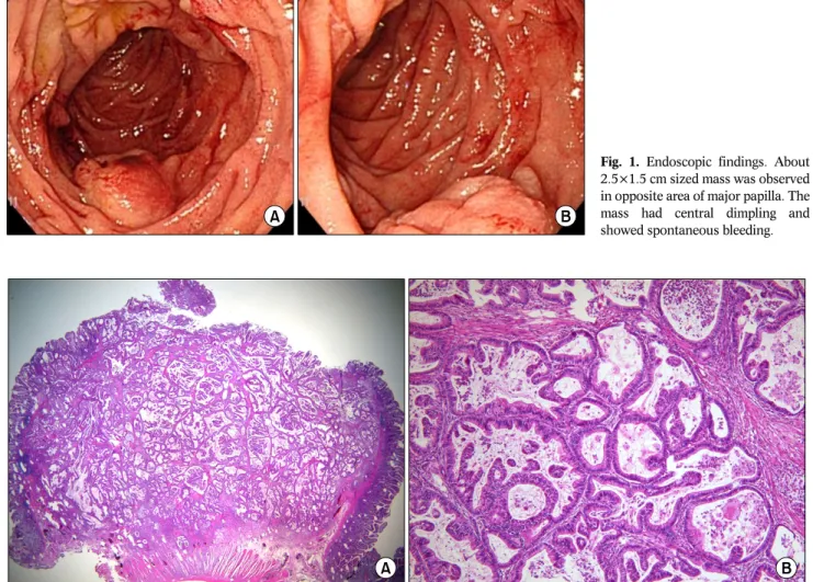
Fig. 1. Endoscopic findings. About
2.5×1.5 cm sized mass was observed in opposite area of major papilla. The mass had central dimpling and showed spontaneous bleeding.
Fig. 2. Histologic findings. Microscopic finding shows well-differentiated adenocarcinoma showing invasive neoplastic gland with involvement
of proper muscle layer without involvement of serosal layer (H&E; A, ×12.5; B, ×100).
tion of duodenum (Fig. 1). The mass was located in op-posite area of major papilla. The mass showed sponta-neous bleeding on the center pole and central dimpling. Hardness was felt on palpation by biopsy forcep. It was hard to differentiate between neoplastic epithelial tumor and subepithelial tumor. Repeated deep biopsy was per-formed for histologic confirmation of the mass. Besides this lesion, there was an ulcer on posterior wall of an-trum and biopsy was performed about that lesion. The mass proved adenocarcinoma and the ulcer proved ad-enoma with low grade dysplasia. There was no evidence of distant metastasis on ultrasonography (US), abdominal CT, 18FDG PET and colonoscopy. Only there were two gall bladder polyps of 10-mm and 11-mm size. Tumor
markers were as follows: AFP 4.0 ng/mL, CEA 1.4 ng/mL, CA 19-9 10.5 U/mL. The patient underwent surgical man-agements including wedge resection and regional lymph node dissection of duodenum, distal gastrectomy and cholecystectomy. Frozen section biopsy during operation revealed 2.5×1.5×0.9 cm sized well differentiated ad-enocarcinoma invading proper muscle layer of duodenum and resection margin was free. Also there was no cancer invasion in dissected lymph nodes around the mass. Finally, the mass was proven by adenocarcinoma of duo-denum located in third portion without lymph nodal, per-ineural and vascular invasions (Fig. 2). The stage of can-cer was pT2N0M0 (I) and the patient remains in good
Chang Seok Bang, et al: Adenocarcinoma of Duodenum
265
DISCUSSION
Primary adenocarcinoma of duodenum has been in-troduced as case reports or case series studies since Hamburger’s description.12 According to the data from National Cancer Information Center of Korea in 2011, 192,561 cancers were developed in 2009. Among the cancers, small bowel cancers were 578 and accounted for 0.3% of all cancers developed in 2009.13 In Surveillance, Epidemiology and End Results (SEER) program of US National Cancer Institute, adenocarcinoma accounted for 45% of small intestinal malignant tumors in 1987.14 In National Cancer Data Base (NCDB) from 1985 to 2005, adenocarcinoma also took up 33% of small intestinal ma-lignant tumors.10 According to the Korean Central Cancer Registry, 1.2 cases of small intestinal cancers are devel-oped per 100,000 population yearly and 2 cases per 100,000 population yearly in SEER program registry of US.13,14 Overall the incidence of small intestinal cancer appears to be increasing. This is because increased acces-sibility of endoscopic examination for the purpose of reg-ular health inspection and development of diagnostic tools like capsule or small intestinal endoscopy. There was male predominance (male 347, female 231) and age distribution was as follows: 60s 28.7%, 70s 24%, and 50s 16.8% according to the Korean Central Cancer Registry in 2009.13 In this case, patient is 64-year-old male typically. The distensibility and inaccessibility of small intestine can make diagnosis delayed hence many cases are found incidentally.9 The mass was also found incidentally on esophagogastroduodenoscopy for the purpose of regular health inspection in this case. Definite diagnostic methods are not established. Radiologic tests including CT, US and enteroclysis and endoscopic tests including capsule en-doscopy, push and double-balloon enteroscopy are usu-ally performed for the diagnosis. However, these tests cannot be performed without clear evidences or high clinical suspicion about small intestinal cancer. Thus, me-ticulous conventional endoscopic examination to the ac-cessible deep portion is needed although it has limitations for investigating duodenum. Regarding the cancer was detected at slight distal area from major papilla, minute observation of lumen seen beyond major papilla is also
important not to overlook malignant lesions. According to the retrospective observational study of Korea, 20% of malignant duodenal tumors were not diagnosed by initial endoscopy. Among them, 69.2% were not seen on first endoscopic examination while 30.7% were observed, however estimated by benign lesions. This study im-plicates importance of examination beyond the duodenal bulb and careful biopsy of suspicious lesions.15 In this case, subepithelial tumor like mass was observed. Hardness was felt on palpation by biopsy forcep and the mass showed spontaneous bleeding and central dimpling demanding histologic confirmation. Endoscopic features of adenocarcinoma of duodenum were reported as mucosal color change, mass with nodularity, ulcer on the pole not different to cancers of other gastrointestinal locations.15 Serum tumor markers have limitation in its role of duode-nal adenocarcinoma. Serum CEA and CA 19-9 of this case was also normal. The majority of small intestinal ad-enocarcinomas are positive for CEA by immunohisto-chemistry, however is doesn’t have diagnostic value.6,16 Immunohistochemical stain with CK 20 and CK 7 has limited value that approximately half of adenocarcinoma of duodenum are positive for CK 20 and CK 7.17
Because complete resection is achieved best in this methods, aggressive surgical managements including pan-creaticoduodenectomy was generally recommended for localized cancers despite high operational morbidity and mortality. However theoretically, lymphatics of distal por-tion of duodenum drains into small intestinal mesentery not into pancreaticoduodenal channel. Thus retrospective study of 68 patients revealed that segmental resection is appropriate for selected patients especially with the lesion of distal duodenum.11 Nevertheless the results above, the role of more minimal operations in duodenum were not established yet. Factors associated with decreased survival were reported as lymph node metastasis, advanced tumor stage, positive resection margins and advanced histologic grade.1,11,18 In this case, there was no factors predicting poor treatment outcomes and minimal surgical methods were considered even lacking of clinical experiences. Thus intraoperational assessments of lymph node meta-stasis and resection margin by frozen section biopsy were performed. And wedge resection with regional lymph
266
Korean J Helicobacter Up Gastrointest Res: Vol 13, No 4, December 2013
node dissection than segmental resection or pan-creaticoduodenectomy was decided promptly. If the can-cer proven to have lymph node metastasis, it is consid-ered to perform adjuvant chemotherapy or radiation therapy. However there is no study that prove the effect of this treatments.19 Five-year survivals by stage using NCDB from 1985 to 2005 were from stage I of 65% to stage IV of 4%.10
Regarding low incidence but high mortality of ad-enocarcinoma of duodenum, meticulous endoscopic ex-amination of duodenum and biopsy of suspicious lesion are essential for early diagnosis. And limited surgery with low operational mortality should be considered when it satisfies en bloc resection with no poor prognostic factors for the quality of life of patients.
REFERENCES
1. Ryder NM, Ko CY, Hines OJ, Gloor B, Reber HA. Primary duode-nal adenocarcinoma: a 40-year experience. Arch Surg 2000; 135:1070-1074.
2. Zollinger RM Jr. Primary neoplasms of the small intestine. Am J Surg 1986;151:654-658.
3. DiSario JA, Burt RW, Vargas H, McWhorter WP. Small bowel cancer: epidemiological and clinical characteristics from a population-based registry. Am J Gastroenterol 1994;89: 699-701.
4. Hatzaras I, Palesty JA, Abir F, et al. Small-bowel tumors: epi-demiologic and clinical characteristics of 1260 cases from the connecticut tumor registry. Arch Surg 2007;142:229-235. 5. Lepage C, Bouvier AM, Manfredi S, Dancourt V, Faivre J.
Incidence and management of primary malignant small bowel cancers: a well-defined French population study. Am J Gastroenterol 2006;101:2826-2832.
6. Talamonti MS, Goetz LH, Rao S, Joehl RJ. Primary cancers of the
small bowel: analysis of prognostic factors and results of surgi-cal management. Arch Surg 2002;137:564-570.
7. Dabaja BS, Suki D, Pro B, Bonnen M, Ajani J. Adenocarcinoma of the small bowel: presentation, prognostic factors, and outcome of 217 patients. Cancer 2004;101:518-526.
8. Halfdanarson TR, McWilliams RR, Donohue JH, Quevedo JF. A single-institution experience with 491 cases of small bowel adenocarcinoma. Am J Surg 2010;199:797-803.
9. Ciresi DL, Scholten DJ. The continuing clinical dilemma of pri-mary tumors of the small intestine. Am Surg 1995;61:698-702. 10. Bilimoria KY, Bentrem DJ, Wayne JD, Ko CY, Bennett CL,
Talamonti MS. Small bowel cancer in the United States: changes in epidemiology, treatment, and survival over the last 20 years. Ann Surg 2009;249:63-71.
11. Bakaeen FG, Murr MM, Sarr MG, et al. What prognostic factors are important in duodenal adenocarcinoma? Arch Surg 2000; 135:635-641.
12. Spira IA, Ghazi A, Wolff WI. Primary adenocarcinoma of the duodenum. Cancer 1977;39:1721-1726.
13. The Korea Central Cancer Registry; National Cancer Center. Annual report of cancer statistics in Korea in 2009. Seoul: Ministry of Health and Welfare, 2011.
14. Weiss NS, Yang CP. Incidence of histologic types of cancer of the small intestine. J Natl Cancer Inst 1987;78:653-656. 15. Sinn DH, Lee JH, Kang TW, et al. Primary duodenal malignant
tumors missed at initial upper endoscopy. Korean J Gastrointest Endosc 2005;31:211-215.
16. Zhu L, Kim K, Domenico DR, Appert HE, Howard JM. Adenocarcinoma of duodenum and ampulla of Vater: clin-icopathology study and expression of p53, c-neu, TGF-alpha, CEA, and EMA. J Surg Oncol 1996;61:100-105.
17. Lee MJ, Lee HS, Kim WH, Choi Y, Yang M. Expression of mucins and cytokeratins in primary carcinomas of the digestive system. Mod Pathol 2003;16:403-410.
18. Lee HG, You DD, Paik KY, Heo JS, Choi SH, Choi DW. Prognostic factors for primary duodenal adenocarcinoma. World J Surg 2008;32:2246-2252.
19. Singhal N, Singhal D. Adjuvant chemotherapy for small intestine adenocarcinoma. Cochrane Database Syst Rev 2007;(3): CD005202.