Long-term Patency of Nitinol Stent
Placement in Dysfunctional Hemodialysis
By
Tayhee Kim
Major in Medicine
Department of Medical Sciences
The Graduate School, Ajou University
Long-term Patency of Nitinol Stent
Placement in Dysfunctional Hemodialysis
By
Tayhee Kim
A Dissertation Submitted to The Graduate School of
Ajou University in Partial Fulfillment of The Requirements
for The Degree of Master of Medicine
Supervised by
Je Hwan Won, M.D.
Major in Medicine
Department of Medical Sciences
The Graduate School, Ajou University
This certifies that the dissertation
of Tayhee Kim is approved.
SUPERVISORY COMMITTE
_____________________
Je Hwan Won
_____________________
Doo Kyoung Kang
_____________________
Chang-Kwon Oh
The Graduate School, Ajou University
December 20th, 2011
i - ABSTRACT–
Long-term Patency of Nitinol Stent Placement in
Dysfunctional Hemodialysis
Purpose: To evaluate the long-term patency of stents placement on dysfunctional
hemodialysis shunt after failed balloon angioplasty and analysis the factors affecting the patency of nitinol stent.
Materials and methods: Eighty-three patients with dysfunctional dialysis shunts underwent
bail-out stenting to restore vascular access. The indications included 1) residual stenosis more than 50% after percutaneous transluminal antioplasty (PTA) 2) venous rupture or dissection 3) early restenosis less than 3months after PTA. Overall cumulative primary rates at 3 months, 6months, 1-, 2-, 3-, and 4-year and secondary patency rates at 3 months, 6months, 1-, 2-, 3-, 4-, 5-, and 6-year were calculated by Kaplan-Meier survival analysis. The primary and secondary patency rates according to age and gender of the patient, history of diabetes mellitus and hypertension, diameter and length of the stent, location of the stent (central, upper arm, cubital, forearm), type of the hemodialysis shunt (native arteriovenous fistula [AVF] or arteriovenous graft [AVG]), and presence of stent fracture, were compared by Kaplan-Meier log-rank test.
Results: The stents were placed in 26 central veins (31.3%), 40 upper arm veins (48.2%), 11
cubital veins (13.3%), and 6 forearm veins (7.2%) with 100% technical and clinical success rates. The 3months, 6months, 1-, 2-, 3-, and 4-year primary patency rates were 81%, 66.7%, 43.1%, 28.8%, 25.2%, and 25.2%, respectively. The 3months, 6months, 1-, 2-, 3-, 4-, 5-, and 6-year secondary patency rates were 91.6%, 88%, 82.9%, 73.9%, 62.6%, 59.1%, 59.1%, and 29.6%, respectively. During follow up, 12 stent fractures (14.5%) were occurred in 11 peripheral veins (4 cubital veins, 7 forearm veins) and 1 central vein. There was a one case of stent migration, which located in central vein. The primary patency rates did not statistically differ according to age and gender of the patient, history of diabetes mellitus and hypertension, diameter and length of the stent, location of the stent, and type of the hemodialysis shunt. However, stent fracture group showed significantly decreased primary
ii
patency rate in comparison with non-fracture group (p=0.0004), but there was no statistical difference in secondary patency rates between two groups (p=0.58). The secondary patency rates did not statistically differ according to gender of the patient, history of diabetes mellitus and hypertension, diameter and length of the stent, location of the stent, type of the hemodialysis shunt, and stent fracture. Only older age group (≥ 65 years) showed significantly decreased secondary patency rate in comparison with younger age group (p=0.045).
Conclusion : In conclusion, nitinol stent placement is effective method to prolong dialysis
access not only the central veins but also the peripheral veins including cubital veins. Stent fracture may shorten the primary patency, but not secondary patency.
Keywords: nitinol stent, patency, cubital vein, stent fracture, stent migration
iii
TABLE OF CONTENTS
ABSTRACT ···i
TABLE OF CONTENTS ··· iii
LIST OF FIGURES ··· iv
LIST OF TABLES ··· v
I. INTRODUCTION ··· 1
II. MATERIALS AND METHODS ··· 2
A. PATIENTS··· 2
B. INDICATIONS FOR STENT PLACEMENT ··· 2
C. BALLOON ANGIOPLASTY PROCEDURES ··· 2
D. SEDATION, ANALGESIA, AND PATIENT MONITORING ··· 3
E. STENT PLACEMENT ··· 3
F. STUDY DEFINITIONS AND STATISTICS ··· 4
G. FOLLOW-UP ··· 4 III. RESULTS ··· 6 IV. DISCUSSION··· 21 V. CONCLUSION ··· 25 REFERENCES ··· 26 국문요약 ··· 29
iv
LIST OF FIGURES
Fig. 1. Kaplan-Meier analysis of nitinol stent patency ··· 9
Fig. 2. Primary and secondary patency rates according to four stent locations ··· 9
Fig. 3. Primary and secondary patency rates according to two stent locations ··· 10
Fig. 4. Primary and secondary patency rates between types of hemodialysis shunts ··· 10
Fig. 5. Primary and secondary patency rates according to presence of diabetes mellitus · 11 Fig. 6. Primary and secondary patency rates between fracture and non-fracture group ··· 12
Fig. 7. Primary and secondary patency rates between age groups ··· 12
Fig. 8. A case of stent fracture ··· 17
v
LIST OF TABLES
Table 1. Nitinol stent fracture classification··· 7
Table 2. Clinical characteristics of eighty-three study patients ··· 8
Table 3. Factors affecting the primary patency ··· 14
1
I. INTRODUCTION
Vascular access stenosis is a common complication that may lead to significant morbidity and dysfunction of hemodialysis and increased cost of medical care. Recent studies show feasibility of endovascular stents which could be used in treatment of central as well as peripheral stenotic lesions (Beathard, 1993; Quinn et al., 1995; Hoffer et al., 1997; Chen et al., 2003; Maskova et al., 2003; Sprouse et al., 2004; Vogel and Parise, 2004; Rajan et al., 2007; Chan et al., 2008; Vesely, 2008). With recent technological advances, nitinol stents has become available and Vogel et al. reported that nitinol stent is safe and effective for treating dialysis-access venous stenoses that are resistant to standard angioplasty (Vogel and Parise, 2004, 2005). However, long-term patency of nitinol stent in large study population has not been reported and there are only few reports of complications associated with nitinol stent including fracture or migration in hemodialysis access (Maleux et al., 1998; Vogel and Parise, 2004; Brewster et al., 2006; Wada et al., 2007).
The aim of this study was to evaluate the long-term patency of stents placement on dysfunctional hemodialysis shunt after failed balloon angioplasty and analysis the factors affecting the patency of nitinol stents.
2
II. MATERIALS AND METHODS
A. Patients
From January 2005 to March 2011, 86 consecutive patients with dysfunctional dialysis shunts who underwent bail-out stenting to restore vascular access were retrospectively reviewed. After excluding three patients with follow up loss, ultimately 83 patients (47 men, 36 women; mean age, 63.24 ± 12.55 years) were enrolled in this study. All patients underwent hemodialysis access surgery at same institution (Ajou university hospital) by 15 year experienced vascular surgeon. Patients underwent hemodialysis at our dialysis center or our dialysis branches connected on the medical network. Consecutive medical records and radiographic images of all patients were reviewed.
B. Indications for stent placement
According to DOQI guidelines (1997), all angioplasties were performed on patients exhibiting >50% stenosis and clinical/physiologic abnormalities. The indications of stent placement included 1) residual stenosis more than 50% after percutaneous transluminal angioplasty (PTA), 2) venous rupture or dissection, 3) early restenosis less than 3months after PTA.
C. Balloon angioplasty procedures
All procedures were accomplished on outpatient basis at our hospital. Patients were monitored using pulse oximetry, blood pressure measurements, and electrocardiography. Fistulography was performed immediately before balloon angioplasty to measure vessel diameter. Thereafter, if a lesion was identified as requiring dilatation (>50% stenosis in diameter), a 6-8 Fr sheath introducer was positioned and balloon angioplasty proceeded. PTA balloon diameters ranged from 6-16mm with burst pressures ranged from 16 to 30 atm. The following balloons were used : Ultra Thin diamond, Blue Max20, XXL (Boston Scientific, MA, USA), and Conquest (Bard Peripheral vascular, Inc. AZ, USA). The required balloon diameters were 1-2mm larger than the diameter of the adjacent normal vein as measured on fistulography. After 2003, cutting balloons (Boston Scientific, Natick, MA, USA) with
3
larger-diameter conventional balloons were used in PTA-resistant lesions when they were available. We determined whether or not a continous palpable thrill was converted immediately after balloon angioplasty. Fistulography was performed after balloon angioplasty to confirm whether sufficient restoration of access flow could be obtained. The entire access circuit from the arterial anastomosis to the superior vena cava was evaluated by digital subtraction angiography during the procedure. All procedures were performed in the angiography room by two interventional radiologists (10 year- and 15 year experienced).
D. Sedation, analgesia, and patient monitoring
Intravenous remifentanil was used for procedural sedation and analgesia (Ultiva;GlaxoSmithKline, Zeist, Netherlands). Remifentanil was prepared as a 0.01mg/mL solution (1mg remifentanil mixed with 100mL normal saline) and was administered via an infusion pump at a rate of 0.1 g/kg/minute as the loading dose, 0.05 g/kg/minute as the maintenance dose, and 0.15 g/kg/minute as the maximal dose, through exclusive intravenous access made on the back of the contralateral hand (Wilhelm and Kreuer, 2008). Infusion was started with the loading dose, approximately 5 minutes before the balloon angioplasty, and the infusion rate was adjusted according to development of pain during the procedure. Patients were monitored by a designated nurse, who used non-invasive measurement of blood pressure and arterial oxygen saturation, checked venous lines, reactions and emotional status, and provided support. Following the procedure, patients were transferred to the recovery room and were monitored at 5-minute intervals by a nurse for at least 30 minutes, until they had fully recovered from the sedation. No anesthesiologist was present during the procedures.
E. Stent placement
All stents were bare metallic nitinol stents (S.M.A.R.T Control stent; Cordis, Miami, USA) ranging from 6-14 mm in diameter and 40-150mm in length. On follow-up, we also used PTA as the choice of treatment of restenosis or obstruction within the stents. Additional stenting was reserved for PTA-resistant lesions such as elastic recoil, dissection or rupture. After stent insertion, fistulography was performed to evaluate the access flow. All patients
4
who had stent placement received 100 mg Aspirin (Astrix cap, Boryung pharmaceutical co., Seoul, Korea) and 75mg Clopidogrel (Plavix tab, Handok, Seoul, Korea) once a day for 1month.
F. Study Definitions and Statistics
Technical success of the procedure was defined as restoration of flow combined with a maximal residual diameter stenosis of less than 30%. Clinical success was defined as the resumption of normal hemodialysis via the treated shunts for at least one session after the procedure. Patency estimates were calculated using the Kaplan-Meier life-table analysis and patency rates were measured from the time of percutaneous intervention. Primary patency was defined as the interval following intervention until the next access thrombosis or repeated intervention, and secondary patency as the interval after interventions until the access was surgically revised or abandoned due to various factors. Kidney transplantation and death with patent fistula were regarded as the end of the study. Complications were investigated and classified into major and minor complications according to definitions established by the Society of Interventional Radiology (Sacks et al., 1997; Gray et al., 2003). Primary and secondary patency rates were calculated using the Kaplan-Meier method. Patency rates were calculated according to age and gender of the patient, history of diabetes mellitus and hypertension, diameter and length of the stent, location of the stent (central, upper arm, cubital, forearm), type of the hemodialysis shunts (native arteriovenous fistula [AVF] or arteriovenous graft [AVG]), and stent fracture during follow up by Kaplan-Meier log-rank test to compare the probability of patency among these groups. Cox’s proportional hazard regression model was used separately in each sub groups to identify the factors affecting stent patency and to evaluate the relative risk rates. A p-value of 0.05 or less was considered significant. Statistical analyses were performed using SPSS 15.0 software (Statistical Package for the Social Sciences SPSS Inc., Chicago, IL, USA).
G. Follow-up
Follow-up was performed by review of patients’ records. Fistulography was performed for assessment of patency when restenosis or occlusion of the fistula was suspected during
5
physical examination or hemodialysis. In addition, some of the patients were also contacted by telephone and interviewed on the last date of follow up.
6
III. RESULTS
Eighty-three patients with dysfunctional dialysis shunts underwent bail-out stenting to restore vascular access. The stents were placed in 26 central veins (31.3%), 40 upper arm veins (48.2%), 11 cubital veins (13.3%), and 6 forearm veins (7.2%). There were 43 native arteriovenous fistulas (51.8%) and 40 arteriovenous grafts (48.2%). Patient characteristics are summarized in Table 2.
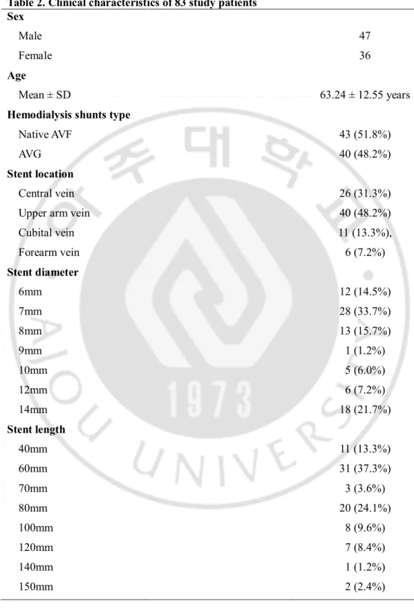
Technical success was achieved in 100% of procedures and clinical success was achieved in 100% of procedures. The mean duration of follow-up (±standard deviation) was 23.8 months ± 16.2 (range; 2 days to 6.3year) and the mean number of reintervention to maintain secondary patency after stent placement was 1.79 ± 2.5 (range; 0-17). The mean primary patency time (±standard deviation) was 19.88 months ± 2.691 (95% CI; 14.607 to 25.154) and the mean secondary patency time was 49.53 months ± 4.859 (95% CI; 40.001 to 59.049). The median primary and secondary patency times were 9.5 months and 63.9 months, respectively. The 3months, 6months, 1-, 2-, 3-, and 4-year primary patency rates were 81%, 66.7%, 43.1%, 28.8%, 25.2%, and 25.2%, respectively (Fig. 1A). The 3months, 6months, 1-, 2-, 3-, 4-, 5-, and 6-year secondary patency rates were 91.6%, 88%, 82.9%, 73.9%, 62.6%, 59.1%, 59.1%, and 29.6%, respectively (Fig. 1B).
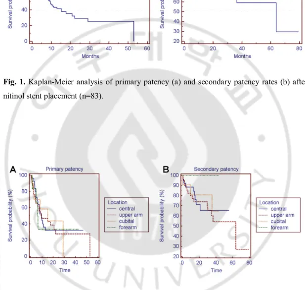
The primary and secondary patency rates did not statistically differ between stent location of four subgroups (central, upper arm, cubital, and forearm) by Kaplan-Meier log-rank test (p=0.99, 0.54, respectively, Fig. 2).
The mean primary patency times (±standard deviation) were 19.63 months ± 3.808 (95% CI; 12.170 to 27.097) in stents located at central veins and 20.35 months ± 3.288 (95% CI; 13.904 to 26.793) in stents located at peripheral veins, including upper arm, cubital, and forearm veins (p=0.87). The mean secondary patency times were 40.5 months ± 4.458 (95%CI; 31.767 to 49.242) in stents located at central veins and 50.04 months ± 5.349 (95%CI; 39.558 to 60.525) in stents located at peripheral veins (p=0.83). The median primary patency times were 9.5 months in stents located at central veins and 9.23 months in stents located at peripheral veins. The median secondary patency time in stents located at central veins cannot be computed in Kaplan-Meiere analysis due to survival curve does not fall to 50% (Fig. 3B). The median secondary patency time in stents located at peripheral
7
veins was 63.9 months.
For the central stents, the 3months, 6months, 1-, 2-, 3-, and 4-year primary patency rates were 92.1%, 68.1%, 36.1%, 36.1%, 32.1%, and 32.1%, respectively. For the peripheral stents including upper arm, cubital and forearm, the 3months, 6months, 1-, 2-, 3-, and 4-year primary patency rates were 75.9%, 66.2%, 47.4%, 29.8%, 25.6%, and 25.6%, respectively (Fig. 3A).There was no significant difference in primary patency between two subgroups (p=0.87) and relative risk for survival failure in peripheral stents, compared with central stents was 0.954 (95% CI; 0.530 to 1.715).
For the central stents, the 3months, 6months, 1-, 2-, 3-, 4-, 5-, and 6-year secondary patency rates were 92.3%, 88.5%, 88.5%, 65.5%, 65.5%, 65.5%, 65.5%, and 65.5%, respectively. For the peripheral stents including upper arm, cubital, and forearm, the 3months, 6months, 1-, 2-, 3-, 4-, 5-, and 6-year secondary patency rates were 91.2%, 87.7%, 80.3%, 78.2%, 63.6%, 59.1%, 59.1%, and 29.5, respectively (Fig. 3B). There was no significant difference in secondary patency between two subgroups (p=0.83) and relative risk for survival failure in peripheral stents, compared with central stents was 0.914 (95% CI; 0.391 to 2.135).
During follow up period, 12 stent fractures (12/83, 14.5%) were occurred. A total of 11 (91.7%) occurred in the peripheral veins (cubital vein 4, forearm vein 7), compared to 1 (8.3%) in the central vein. According to the Cardiovascular Institute of South (CIS) nitinol stent fracture classification system (David, 2004) (Table 1), there were 1/12 (8.3%) type I, 5/12 (41.7%) type II, 2/12 (16.7%) type III, and 4/12 (33.3%) type IV fractures.
Table 1. Nitinol stent fracture classification
Type 1 A single strut fracture only
Type 2 Multiple single nitinol stent fractures that can occur at different sites Type 3 Multiple nitinol stent fractures resulting in complete transverse linear
fracture but without stent displacement
8
Table 2. Clinical characteristics of 83 study patients Sex
Male 47
Female 36
Age
Mean ± SD 63.24 ± 12.55 years
Hemodialysis shunts type
Native AVF 43 (51.8%)
AVG 40 (48.2%)
Stent location
Central vein 26 (31.3%) Upper arm vein 40 (48.2%) Cubital vein 11 (13.3%), Forearm vein 6 (7.2%) Stent diameter 6mm 12 (14.5%) 7mm 28 (33.7%) 8mm 13 (15.7%) 9mm 1 (1.2%) 10mm 5 (6.0%) 12mm 6 (7.2%) 14mm 18 (21.7%) Stent length 40mm 11 (13.3%) 60mm 31 (37.3%) 70mm 3 (3.6%) 80mm 20 (24.1%) 100mm 8 (9.6%) 120mm 7 (8.4%) 140mm 1 (1.2%) 150mm 2 (2.4%)
9
Fig. 1. Kaplan-Meier analysis of primary patency (a) and secondary patency rates (b) after
nitinol stent placement (n=83).
Fig. 2. Primary and secondary patency rates did not statistically differ between the stent location of four subgroups by Kaplan Meier log rank test and p-values were 0.99 (a) and 0.54 (b), respectively.
10
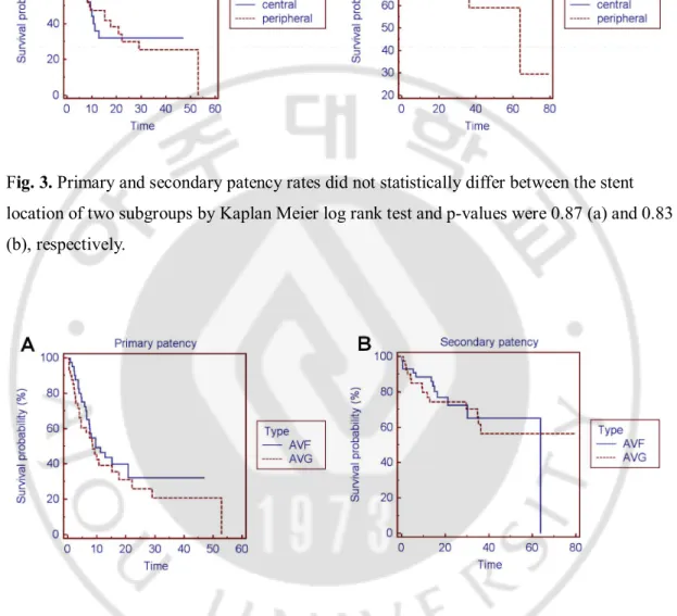
Fig. 3. Primary and secondary patency rates did not statistically differ between the stent location of two subgroups by Kaplan Meier log rank test and p-values were 0.87 (a) and 0.83 (b), respectively.
Fig. 4. Primary and secondary patency rates did not statistically differ between the type of
hemodialysis shunts by Kaplan Meier log rank test and p-values were 0.37 (a) and 0.81 (b), respectively.
The type of hemodialysis shunts did not significantly affect the primary patency (p=0.37) and relative risk for survival failure in AVG, compared with AVF was 1.278 (95% CI ; 0.737 to 2.214). There was also no significant difference in secondary patency between
11
two groups (p=0.81) and relative risk for survival failure in AVG, compared with AVF was 1.097 (95% CI ; 0.509 to 2.368) (Fig. 4).
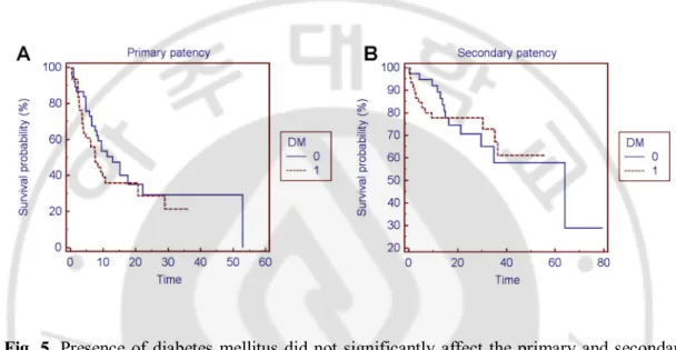
Presence of comorbity of diabetes mellitus did not significantly affect the primary and secondary patency (p=0.31, 0.92, respectively) and relative risk ratios for survival failure were 1.321 (95% CI; 0.762 to 2.292) and 0.964 (95% CI; 0.447 to 2.080), respectively (Fig. 5).
Fig. 5. Presence of diabetes mellitus did not significantly affect the primary and secondary
patency rates by Kaplan Meier log rank test and p-values were 0.31 (a) and 0.92 (b), respectively.
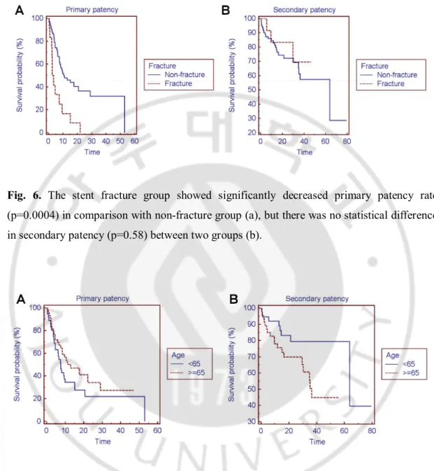
However, stent fracture group showed significantly decreased primary patency rate in comparison with non-fracture group (p=0.0004) and relative risk for survival failure was 2.998 (95% CI; 1.166 to 7.709) , but there was no statistical difference in secondary patency between two groups (p=0.58) and relative risk was 0.715 (95% CI; 0.247 to 2.073) (Fig.6, Fig.8).
In patients with same or more than 65 years old, the primary patency rate did not statistically differ comparison with less than 65 years old group (p=0.163, relative risk=0.682 with 95% CI; 0.392 to 1.186), however, there was significant difference between two groups in secondary patency (p=0.045) and relative risk of survival failure in older age group was 2.247 (95% CI : 1.042 to 4.846), compared with younger age group (Fig. 7).
12
Fig. 6. The stent fracture group showed significantly decreased primary patency rate
(p=0.0004) in comparison with non-fracture group (a), but there was no statistical difference in secondary patency (p=0.58) between two groups (b).
Fig. 7. Primary patency rate did not statistically differ between two groups (p=0.163) (a), but
older age group of same or more than 65 years old showed significantly decreased secondary patency rate (p=0.045) in comparison with younger age group by Kaplan Meier log rank test (b).
13
Only stent fracture significantly affect the primary patency and the other factors including age and gender of the patient, history of diabetes mellitus and hypertension, diameter and length of the stent, location of the stent, and type of the hemodialysis shunt did not significantly affect the primary patency by Kaplan Meier log rank test. P-values and hazard ratios are summarized in table 3.
Only patient’s age significantly affect the secondary patency and the other factors including gender of the patient, history of diabetes mellitus and hypertension, diameter and length of the stent, location of the stent, type of the hemodialysis shunt, and stent fracture did not significantly affect the secondary patency by Kaplan Meier log rank test. P-values and hazard ratios are summarized in table 4.
14
Table 3. Factors affecting the primary patency by log-rank test and Cox regression
P-value* Hazard ratio 95% CI lower level 95% CI upper level Agea 0.163 0.682 0.392 1.186 Sexb 0.443 0.810 0.464 1.413 Diabetes mellitus 0.312 1.321 0.762 2.292 Hypertension 0.790 1.077 0.619 1.876 Stent diameterc 0.714 0.901 0.514 1.579 Stent lengthd 0.845 1.055 0.609 1.828 Stent locatione 0.872 0.954 0.530 1.715 Shunt typef 0.374 1.278 0.737 2.214 Stent fracture 0.0004 2.998 1.166 7.709
a Classified into two subgroups of ≥65 years old and <65 years old. Hazard ratio means the
relative risk of older age group compared with younger age group.
b Hazard ratio means the relative risk of male group compared with female group.
c Classified into two subgroups according to the median value of 8 mm, >8mm and ≤8mm.
Hazard ratio means the relative risk of larger stent diameter group compared with smaller diameter group.
d Classified into two subgroups according to the median value of 60 mm, >60mm and
≤60mm. Hazard ratio means the relative risk of longer stent length group compared with shorter length group.
e Classified into two subgroups of central and peripheral location and Hazard ratio means the
relative risk of peripheral stent group compared with central stent group.
f Hazard ratio means the relative risk of AVG group compared with AVF group.
15
Table 4. Factors affecting the secondary patency by log-rank test and Cox regression
P-value* Hazard ratio 95% CI lower level 95% CI upper level Agea 0.045 2.247 1.042 4.846 Sexb 0.502 1.292 0.599 2.788 Diabetes mellitus 0.924 0.964 0.447 2.080 Hypertension 0.885 0.944 0.432 2.063 Stent diameterc 0.847 0.924 0.416 2.054 Stent lengthd 0.722 0.873 0.405 1.883 Stent locatione 0.830 0.914 0.391 2.135 Shunt typef 0.811 1.097 0.509 2.368 Stent fracture 0.581 0.715 0.247 2.073
a Classified into two subgroups of ≥65 years old and <65 years old. Hazard ratio means the
relative risk of older age group compared with younger age group.
b Hazard ratio means the relative risk of male group compared with female group.
c Classified into two subgroups according to the median value of 8 mm, >8mm and ≤8mm.
Hazard ratio means the relative risk of larger stent diameter group compared with smaller diameter group.
d Classified into two subgroups according to the median value of 60 mm, >60mm and
≤60mm. Hazard ratio means the relative risk of longer stent length group compared with shorter length group.
e Classified into two subgroups of central and peripheral location and Hazard ratio means the
relative risk of peripheral stent group compared with central stent group.
f Hazard ratio means the relative risk of AVG group compared with AVF group.
16
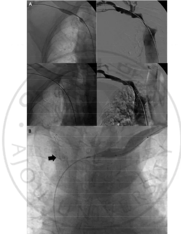
The complication rate was 7.2% (6/83) with four major complications and two minor complications. Three cases of major complications were wound infection at the surgical site. Another major complication was a case of stent migration, located in the central vein. After 17 days after the procedure, the patient had a progressive right arm swelling. The follow up venography showed that stent migrated to the SVC, so the graft abandoned (Fig. 9). Minor complications included one case of local infection which did not require hospitalization and one case of stent insertion site discomfort which located in cubital vein.
18
Fig. 8. 78-year old female with right brachio-brachial AVG. There was residual stenosis
more the 50% after balloon angioplasty at venous limb. The stent inserted and the patient achieved technical and clinical success (a). 26 days after stent insertion, the patient had follow up venography due to insufficient flow. There was type 4 nitinol stent fracture and significant in-stent restenosis. After balloon angioplasty, blood flow restored (b). The secondary patency was 3year 7months and four sessions of reintervention were performed to maintain secondary patency. Last venography which performed due to insufficient shunt flow shows significant stenosis at upper arm vein, but intact stent patency in spite of fracture (c).
19
Fig. 9. 75-year old male with right brachiocephalic AVF. Initial fistulography showed near
total occlusion of right brachiocephalic vein. There was residual stenosis more than 50% after balloon angioplasty. The stent inserted and the patient achieved technical and clinical
20
success (a). After 17days, the patient had follow up venography due to progressive right arm swelling. There was a migration of stent (arrow) to the SVC, so the graft abandoned.
21
IV. DISCUSSION
Most of previous study reported experience of endovascular treatment with the Wallstent (Boston Sicentific, Natick, MA, USA). However, recently Vogel et al. (Vogel and Parise, 2004) used nitinol stent for hemodialysis access in 64 patients and the mean patency durations were 14.9 months in stents located at central veins and 8.9 months in stents located at peripheral veins. In study of Vogel et al. (Vogel and Parise, 2004), the 3-, 6-, 12- months primary patency rates were 81%, 74, and 67% for central stents and 77%, 51%, and 20% for peripheral stents. The same author followed prospective study (Vogel and Parise, 2005) of comparing nitinol stent placement with PTA in 60 patients and all patients were followed for 1-year. The mean primary patency durations were 5.6months after PTA and 8.2 months after stent treatment and the 3-, 6-, 12-months primary patency rates of nitinol stents were 88%, 67%, and 41%.
The mean primary and secondary patency durations of nitinol stents in our center were 19.88 months and 49.53 months, respectively. The mean primary patency durations were 19.63 months in stents located at central veins and 20.35 months in stents located at peripheral veins and there was no significant difference in patency between central and peripheral located stents. In our study, the overall 3-, 6-, 12-months primary patency rates were 81%, 66.7%, 43.1% for overall, 92.1%, 68.1%, 36.1% for central stents and 75.9%, 66.2%, 47.4% for peripheral stents.
We subclassified peripheral veins to upper arm vein, cubital vein across the elbow joint, and lower arm vein. Side with previous study of Vogel et al. (Vogel and Parise, 2004, 2005), 11 of 83 (13.3%) stents located in cubital vein and there was no significant difference in patency between these patients and those who had stents placed at other peripheral veins. Consequently, our study shows that nitinol stent across the elbow joint could be feasible treatment and provide similar patency compared with stents placed at other peripheral veins.
In study of Vogel et al. (Vogel and Parise, 2004), there was only one stent-related complication, which was venous dissection in a patient with a stent across the elbow joint, and none of patients had stent fracture or migration. In another prospective study of Vogel et
al. (Vogel and Parise, 2005), all patients were followed for 1-year and there was only one
22
joint on follow-up. However, our study showed more frequent stent fractures (12/83, 14.5%), and among 11 patients with stents placed in cubital vein, there were 4 (36.4%) stent fractures during follow up. This result may be due to longer follow up period of our study, compared with two studies of Vogel et al. (Vogel and Parise, 2004, 2005). By Kaplan-Meier log-rank test, stent fracture significantly shortens the primary patency, but not secondary patency. There are several reports of nitinol stent fractures at lower extremity angioplasty, especially superficial femoral veins (Duda et al., 2002; David, 2004; Schlager et al., 2005), but only few reports exists at hemodialysis access. Tsuji et al. (Wada et al., 2007) described self-expanding stent fracture in the left brachiocephalic vein and a Brewster et al. (Brewster et al., 2006) described fracture of two overlapping stents, but long-term follow up data with large study population is not reported. Our study shows that the true incidence of nitinol stent fracture in hemodialysis access is more prevalent than suspected (14.5%) and may imply that stents fracture may lead to in-stent restenosis and shortens the primary patency. However, no significant difference was identified in secondary patency between fracture and non-fracture group. The etiology of stent fracture is not evaluated in this study, but more frequent fracture was identified at peripheral veins (91.7%, 4 cubital veins, 7 forearm veins) compared to 1 (8.3%) in the central vein. This finding could be explained by more biomechanical forces exerted on peripheral vein than central vein, but further investigations are needed.
Covered stent as they are known, lead to occasionally stent shortening and migration (Verstandig et al., 2003). However, to our knowledge, nitinol stent migration has not been reported in the literature. During follow up, one case of stent migration was occurred, which was located in the cephalic vein and migrated to the SVC. Verstandig et al. (Verstandig et al., 2003) explained the Wallstent (Schneider, Bulach, Switzerland) migration in central veins by shortening-lengthening-shortening mechanism. The normal expansion and contraction of the central vein during respiration, which when combined with the geometry of the stent, can lead to a sequence of shortening-lengthening-shortening of the stent, and this sequence can lead to stent movement and migration. However, unlike covered stent, bare-metal nitinol stent may provide more deployment in lumen and until now no report of nitinol stent shortening or migration exists. So not only the “shortening-lengthening-shortening” of the stent in central vein, but also the repeated extrinsic biomechanical forces (i.e. arm abduction and adduction) on the stents at thoracic outlet level, could be the possible mechanism of
23
nitinol stent migration.
We evaluated factors affecting stent patency, and except stent fracture, no other factors significantly affect the primary patency of stent. Also only the age of patients significantly affects the secondary patency of stent. Kolakowski et al. (Kolakowski et al., 2003) performed prospective, nonrandomized study on 61 subjects comparing the use of stents in forearm versus the upper arm, and primary patency was significantly decreased in forearm, but no significant difference was noted in secondary patency. Unlike Kolakowski et al. (Kolakowski et al., 2003), our study shows no significant difference in primary and secondary patency according to stent location between central, upper arm, forearm, and even more cubital veins. Other investigators (Rajan et al., 2007) have endeavored to determine the difference in access patency between AVF and AVG in central vein stenosis, and intervention primary patency remained significantly longer for fistulas than grafts. However, there was no significant difference in stent patency between and AVF and AVG group in our study.
The age of the patients was the only one variable affecting the secondary patency. The end point of this study includes the death with patent access, and that may lead to shortening of secondary patency in old age.
The limitations of our study include the restrospective study design and the absence of comparative control of angioplasty group. We defined the end of the primary patency to repeated intervention anywhere within the access circuit, like Vogel et al. (Vogel and Parise, 2004). However in one study (Vesely, 2008), patency of the access circuit was shorter than the patency of the stent per se (target lesion). Other areas of stenoses are the likely culprits in ultimate demise of patency of the access circuit. Vogel et al. (Vogel and Parise, 2004) also observed non-significant, but higher patency rate among patients with isolated central lesions compared with patients with other peripheral stenoses in addition to central stenosis. The additive effects of stenoses anywhere along the hemodialysis circuit will confer an additional risk to loss of patency. That will be the limitation of our study, since we did not retrospectively analysis the other additive stenoses anywhere along the access circuit. Finally, we cannot use the recent spotlighted expanded polytetrafluoroethylene self-expanding stent graft. Recently, Haskal et al. (Haskal et al., 2010) performed prospective randomized study of comparing expanded polytetrafluoroethylene self-expanding stent graft placement with PTA in 190 patients and showed that the stent graft provide longer-term and superior patency
24
than standard balloon angioplasty. However, expanded polytetrafluoroethylene self-expanding stent graft is not yet available in our country and expensiveness of graft could be the problem.
25
V. CONCLUSION
In conclusion, nitinol SMART stent can be effectively used to salvage suboptimal angioplasty results in hemodialysis access venous stenosis not only the central veins but also the peripheral veins including cubital veins. Long-term incidence of nitinol stent fractures in hemodialysis access was more prevalent than suspected (14.5%), especially which were located in peripheral veins. Stent fracture may lead to in-stent restenosis and shorten the primary patency, but not secondary patency.
26
REFERENCES
1. Beathard GA: Gianturco self-expanding stent in the treatment of stenosis in dialysis access grafts. Kidney Int 43: 872-877, 1993
2. Brewster UC, Mojibian HR, Aruny JE, Perazella MA: Access stenosis and stent fracture. Am J Kidney Dis 47: A45, e35-46, 2006
3. Chan MR, Bedi S, Sanchez RJ, Young HN, Becker YT, Kellerman PS, Yevzlin AS: Stent placement versus angioplasty improves patency of arteriovenous grafts and blood flow of arteriovenous fistulae. Clin J Am Soc Nephrol 3: 699-705, 2008 4. Chen CY, Liang HL, Pan HB, Chung HM, Chen WL, Fang HC, Lo A, Chen CK, Lai
PH, Yang CF: Metallic stenting for treatment of central venous obstruction in hemodialysis patients. J Chin Med Assoc 66: 166-172, 2003
5. David E. Allie CJH, Craig M.Walker: Nitinol stent fracture in the SFA.
Endovascular today 7: 22-34, 2004
6. Duda SH, Pusich B, Richter G, Landwehr P, Oliva VL, Tielbeek A, Wiesinger B, Hak JB, Tielemans H, Ziemer G, Cristea E, Lansky A, Beregi JP: Sirolimus-eluting stents for the treatment of obstructive superficial femoral artery disease: six-month results. Circulation 106: 1505-1509, 2002
7. Gray RJ, Sacks D, Martin LG, Trerotola SO: Reporting standards for percutaneous interventions in dialysis access. J Vasc Interv Radiol 14: S433-442, 2003
8. Haskal ZJ, Trerotola S, Dolmatch B, Schuman E, Altman S, Mietling S, Berman S, McLennan G, Trimmer C, Ross J, Vesely T: Stent graft versus balloon angioplasty for failing dialysis-access grafts. N Engl J Med 362: 494-503, 2010
9. Hoffer EK, Sultan S, Herskowitz MM, Daniels ID, Sclafani SJ: Prospective randomized trial of a metallic intravascular stent in hemodialysis graft maintenance.
J Vasc Interv Radiol 8: 965-973, 1997
10. Kolakowski S, Jr., Dougherty MJ, Calligaro KD: Salvaging prosthetic dialysis fistulas with stents: forearm versus upper arm grafts. J Vasc Surg 38: 719-723, 2003 11. Maleux G, Rousseau H, Otal P, Joffre F: Collapsed Palmaz stent after deployment
for hemodialysis access-related venous stenosis. J Vasc Interv Radiol 9: 169-171, 1998
27
12. Maskova J, Komarkova J, Kivanek J, Danes J, Slavikova M: Endovascular treatment of central vein stenoses and/or occlusions in hemodialysis patients. Cardiovasc
Intervent Radiol 26: 27-30, 2003
13. NKF-DOQI clinical practice guidelines for vascular access. National Kidney Foundation-Dialysis Outcomes Quality Initiative. Am J Kidney Dis 30: S150-191, 1997
14. Quinn SF, Schuman ES, Demlow TA, Standage BA, Ragsdale JW, Green GS, Sheley RC: Percutaneous transluminal angioplasty versus endovascular stent placement in the treatment of venous stenoses in patients undergoing hemodialysis: intermediate results. J Vasc Interv Radiol 6: 851-855, 1995
15. Rajan DK, Chennepragada SM, Lok CE, Beecroft JR, Tan KT, Hayeems E, Kachura JR, Sniderman KW, Simons ME: Patency of endovascular treatment for central venous stenosis: is there a difference between dialysis fistulas and grafts? J Vasc
Interv Radiol 18: 353-359, 2007
16. Sacks D, Marinelli DL, Martin LG, Spies JB: Reporting standards for clinical evaluation of new peripheral arterial revascularization devices. Technology Assessment Committee. J Vasc Interv Radiol 8: 137-149, 1997
17. Schlager O, Dick P, Sabeti S, Amighi J, Mlekusch W, Minar E, Schillinger M: Long-segment SFA stenting--the dark sides: in-stent restenosis, clinical deterioration, and stent fractures. J Endovasc Ther 12: 676-684, 2005
18. Sprouse LR, 2nd, Lesar CJ, Meier GH, 3rd, Parent FN, Demasi RJ, Gayle RG, Marcinzyck MJ, Glickman MH, Shah RM, McEnroe CS, Fogle MA, Stokes GK, Colonna JO: Percutaneous treatment of symptomatic central venous stenosis [corrected]. J Vasc Surg 39: 578-582, 2004
19. Verstandig AG, Bloom AI, Sasson T, Haviv YS, Rubinger D: Shortening and migration of Wallstents after stenting of central venous stenoses in hemodialysis patients. Cardiovasc Intervent Radiol 26: 58-64, 2003
20. Vesely T PT, Amin MZ: Use of stents and stent grafts to salvage angioplasty failures in patients with hemodialysis. Grafts Seminars in Dialysis: 100-104, 2008
21. Vogel PM, Parise C: SMART stent for salvage of hemodialysis access grafts. J Vasc
28
22. Vogel PM, Parise C: Comparison of SMART stent placement for arteriovenous graft salvage versus successful graft PTA. J Vasc Interv Radiol 16: 1619-1626, 2005 23. Wada M, Yamamoto M, Shiba M, Tsuji T, Iijima R, Nakajima R, Yoshitama T, Hara
H, Tsunoda T, Nakamura M: Stent fracture in the left brachiocephalic vein.
Cardiovasc Revasc Med 8: 103-106, 2007
24. Wilhelm W, Kreuer S: The place for short-acting opioids: special emphasis on remifentanil. Crit Care 12 Suppl 3: S5, 2008
29 - 국문요약 -
기능부전
혈액투석 동정맥루 치료법으로서 Nitinol 스텐트 설치
술의
장기 개통률 평가
아주대학교 대학원 의학과 김 태 희 (지도교수: 원 제 환) 연구목적: 기능부전 혈액투석동정맥루 치료로서의 Nitinol 스텐트 설치술의 장기 개통률과 이에 영향을 미치는 요소를 알아보고자 하였다. 재료 및 방법: 기능부전 혈액투석동정맥루를 가진 83명의 환자가 스텐트 설치술 을 시행받았고, 적응증으로는 첫째 경피경관혈관성형술 이후에도 50% 이상의 잔 여협착이 남았을 경우, 둘째 정맥의 파열 혹은 박리, 셋째 경피경관혈관성형술을 시행받은 3개월 이내에 조기 재협착이 생긴 경우로 하였다. 일차, 이차 개통률은 Kaplan-Meier 생존분석을 하였고, 환자의 성별과 나이, 당뇨와 고혈압의 유무, 스 텐트의 직경 및 길이, 스텐트가 설치된 위치 (중심정맥, 위팔, 팔꿈치, 아래팔), 혈액투석동정맥루의 종류 (자가정맥, 이식정맥), 스텐트 골절의 유무 에 따른 개 통률 차이를 Kaplan-Meier log-rank test로 분석하였다.결과: 스텐트는 26개의 중심정맥(31.3%), 40개의 위팔정맥 (48.2%), 11 개의 팔꿈치 정맥 (13.3%), 6개의 아래팔정맥 (7.2%)에 설치되었고, 기술적 그리고 임상적 성공 률은 모두 100% 였다. 3개월, 6개월, 1년, 2년, 3년, 4년 일차 개통률은 각각 81%, 66.7%, 43.1%, 28.8%, 25.2%, 25.2% 였다. 3개월, 6개월, 1년, 2년, 3년, 4년, 5년, 6년 이 차 개통률은 각각 91.6%, 88%, 82.9%, 73.9%, 62.6%, 59.1%, 59.1%, 29.6% 였다. 추적 관찰 기간 중에 12개의 스텐트 골절 (14.5%)가 발생하였고 11개는 말초정맥에 (팔꿈치정맥4, 아래팔정맥7), 1개는 중심정맥에 발생하였다. 중심정맥에 위치했던 한 개의 스텐트 이동이 있었다. Kaplan-Meier log-rank test로 분석하였을 때, 일차
30 개통률은 환자의 성별과 나이, 당뇨와 고혈압의 유무, 스텐트의 직경 및 길이, 스 텐트가 설치된 위치, 혈액투석동정맥루의 종류에 따른 차이가 없었다. 하지만 스 텐트 골절이 있는 집단은 통계학적으로 유의하게 스텐트 골절이 없는 집단보다 일차 개통률이 감소하였다 (p=0.0004). 하지만 이차 개통률는 두 그룹간에 차이가 없었다 (p=0.58). 이차 개통률은 환자의 성별, 당뇨와 고혈압의 유무, 스텐트의 직 경 및 길이, 스텐트가 설치된 위치, 혈액투석동정맥루의 종류, 스텐트 골절에 따 른 차이가 없었다. 하지만 유일하게 65세 이상의 환자 집단이 65세 미만보다 이 차 개통률이 감소하였다 (p=0.045). 결론: Nitinol 스텐트는 동정맥루 협착이 동반된 혈액투석 환자에서 중심정맥뿐 아 니라 말초정맥에도 효과적인 치료법이며 팔꿈치 정맥에서도 다른 위치의 스텐트 와 비교하여 개통률의 유의한 차이가 없었다. 장기 추적 관찰 시에 스텐트 골절 은 알려진 보고들 보다 많이 발생하였고 (14.5%), 한 개의 스텐트 이동이 있었다. 하지만 스텐트 골절은 일차 개통률을 유의하게 감소시켰으나, 이차 개통률에는 유의한 영향을 주지 않았다. 핵심어: Nitinol 스텐트, 개통률, 팔꿈치정맥, 스텐트 골절, 스텐트 이동