INTRODUCTION
Since Schaldenbrand and Appelman applied the term
‘‘stro-mal tumor’’ to collectively refer to a group of mesenchy‘‘stro-mal neoplasms of gastrointestinal tract in 1984 (1), there was con-siderable confusion with regards to the classification of the
853
Mee-Yon Cho1,26, Jin Hee Sohn2,26, Joon Mee Kim3,26, Kyoung-Mee Kim2,26, Young Su Park4,26, Woo Ho Kim5,26, Jin Sook Jung6,26, Eun Sun Jung7,26, So-Young Jin8,26, Dae Young Kang9,26, Jae Bok Park10,26, Ho Sung Park11,26, You Duck Choi12, Sun Hee Sung13, Young-Bae Kim14,26, Hogeun Kim15,26, Young-Kyung Bae16,26, Miseon Kang17,26, Hee Jin Chang18,26, Yang Seok Chae19,26, Hee Eun Lee5, Do Youn Park20,26, Youn Soo Lee7, Yun Kyung Kang18,26, Hye Kyung Kim8, Hee-Kyung Chang21,26, Soon Won Hong15, Young Hee Choi22,26, Okran Shin7, MiJin Gu23,26, Youn Wha Kim24,26, Gwang Il Kim25,26, and Sei Jin Chang27 Department of Pathology, Wonju College of Medicine Yonsei University1, Wonju; Sungkyunkwan University2, Seoul; Inha University3, Inchon; University of Ulsan4, Seoul; Seoul National University5, Seoul and Seongnam; Dong-A University6, Busan; The Catholic University of Korea7, Seoul and Uijeongbu; Soonchunhyang University8, Seoul; Chungnam National University9, Daejon; Daegu Catholic University10, Daegu; Chonbuk National University11, Chonju; Chonnam University12, Gwangju; Ewha University13, Seoul; Ajou Univerisity14, Suwon; Yonsei Univeristy15, Seoul; Youngnam Univeristy16, Daegu; Inje University17, Seoul and Busan; National Cancer Center18, Goyang; Korea University19, Seoul; Busan University20, Busan; Kosin University21, Busan; Hallym University22, Chunchon; Daegu Patima Hospital23, Daegu; Kyung Hee University24, Seoul; CHA Medical School25, Seongnam; The Gastrointestinal Pathology Study Group of Korean Society of Pathologists26; Department of Preventive Medicine, Yonsei University27, Wonju College of Medicine, Wonju, Korea
Address for Correspondence Jin Hee Sohn, M.D.
Department of Pathology, Kangbuk Samsung Medical Center, Sungkyunkwan University School of Medicine, 78 Saemunan-gil, Jongno-gu, Seoul 100-634, Korea Tel : +82.2-2001-2391, Fax : +82.2-2001-2398 E-mail : [email protected]
This study was supported by a grant from Novartis Korea Ltd.
Current Trends in the Epidemiological and Pathological Characteristics
of Gastrointestinal Stromal Tumors in Korea, 2003-2004
Despite remarkable progress in understanding and treating gastrointestinal stromal tumors (GISTs) during the past two decades, the pathological characteristics of GISTs have not been made clear yet. Furthermore, concrete diagnostic criteria of malig-nant GISTs are still uncertain. We collected pathology reports of 1,227 GISTs from 38 hospitals in Korea between 2003 and 2004 and evaluated the efficacy of the NIH and AFIP classification schemes as well as the prognostic factors among pathologic findings. The incidence of GISTs in Korea is about 1.6 to 2.2 patients per 100,000. Extra-gastrointestinal GISTs (10.1%) are more common in Korea than in Western countries. In univariate analysis, gender, age, tumor location, size, mitosis, tumor necrosis, vascular and mucosal invasions, histologic type, CD34 and s-100 protein expression, and classifications by the NIH and AFIP criteria were found to be signif-icantly correlated with patient’s survival. However, the primary tumor location, stage and classification of the AFIP criteria were prognostically significant in predicting patient’s survival in multivariate analysis. The GIST classification based on original tumor location, size, and mitosis is more efficient than the NIH criteria in predicting patient’s survival, but the mechanism still needs to be clarified through future studies. Key Words : Gastrointestinal Stromal Tumors; Epidemiology; Pathology; Prognosis
Received : 18 September 2009 Accepted : 21 December 2009
ⓒ 2010 The Korean Academy of Medical Sciences.
This is an Open Access article distributed under the terms of the Creative Commons Attribution Non-Commercial License (http://creativecommons.org/licenses/by-nc/3.0) which permits unrestricted non-commercial use, distribution, and reproduction in any medium, provided the original work is properly cited.
tumors. However, during the past two decades this confusion was eliminated, and clarification and restructuring became reliable with the introduction of the KIT protein expression in gastrointestinal stromal tumors (GISTs). Because most
GISTs have an activating mutation in the c-kit
proto-onco-gene that leads to expression of KIT protein, the immunohis-tochemical stain for the KIT (CD117) is now used by pathol-ogists to distinguish GISTs from non-GISTs mesenchymal tumors in gastrointestinal tract (2, 3). This was the chief rea-son why epidemiologic studies of GISTs in Korea were unre-liable before the 2000s. Based on the biological characteris-tics of this tumor, treatment with KIT/PDGFRatyrosine kinase inhibitors, such as imatinib, can lead to complete or partial remission when the tumor is unresectable or in a meta-static setting (4). Therefore, accurate pathological diagnosis is mandatory for proper treatment of patients with mesenchy-mal tumors of gastrointestinal tract. Recently, protein kinase C theta (PKC-q) (5) and DOG1 (6) over-expression in addi-tion to mutaaddi-tion analysis of the c-kit and PDGFRa(7) were described as diagnostic markers for GISTs.
Despite remarkable progress in understanding and treat-ing GISTs, pathologists still have a difficulty in classifytreat-ing GISTs because of a lack of concrete diagnostic evidence to predict patient’s prognosis. The incidence of GIST varies as reported in different studies from different countries (8-11). Gender, race, and location distribution of GISTs are also uncer-tain. Furthermore, there is no agreement on prognostic fac-tors of GISTs. In 2002, National Institute of Health (NIH) reported a consensus approach in diagnosing GISTs (2). The efficacy of the NIH criteria had been discussed, although it is commonly used in clinical setting. Recently, new diagnos-tic criteria in predicting prognosis of GISTs have been pro-posed by Miettinen et al in the Armed Forces Institutes of Pathology (AFIP) (12, 13). It has caused some confusion with regards to the terminology used in cancer registration, and it became difficult to identify the actual incidence of malig-nant GISTs.
To create informative and standardized pathological reports of GIST, we need to evaluate contents of pathology reports collected nationwide and to identify significant factors that predict the prognosis. We previously analyzed pathological characteristics of GISTs and described the pitfalls in interpre-tation of KIT expression as well as pathological diagnosis of GISTs (14) through a nationwide study of GISTs in Korea from 2001 to 2002 (14). One outstanding phenomena found in previous research was that small intestinal and esophageal GISTs were more common in Korea than in Western coun-tries, but the incidence and prognostic factors of GISTs in Korea have not been made clear yet.
In this study, we firstly described the population based inci-dence of GIST in Korea. Then, we identified pathological characteristics that should be mentioned in pathology reports of GISTs to predict patient’s prognosis and evaluated the effi-cacy of the NIH and AFIP classification criteria. Data obtained
from this nationwide multi-institutional study may provide an insight in the epidemiological characteristics and prognos-tic stratification of GISTs in Korea.
MATERIALS AND METHODS
Population based incidence of GISTs in Korea: Review the data of the National Cancer Institute Registry
To identify the population based incidence and trends in epidemiology of malignant GISTs in Korea, we reviewed all data collected by the Korean National Cancer Institute (NCI) and correlated the data with the population information of the Korea National Statistical Office. All tumors in the NCI Registry were identified by the International Classification of Disease for Oncology (ICD-O), 3rd edition, from the World Health Organization (WHO).
Reviewing pathology reports from 38 hospitals in Korea, 2003-2004
For survival analysis, we retrospectively collected nation-wide multi-institutional data of pathology reports diagnosed as GISTs not only in gastrointestinal tract but also in extra-gastrointestinal areas from 2003 to 2004.
The list of participant hospitals were as follows: Sungkyun-kwan University Seoul Samsung Medical Center and Kang-buk Samsung Medical Center, Asan Medical Center, Seoul National University Hospital and Bundang Hospital and Boramae Hospital, Yonsei University Severance Hospital and Kangnam Severance Hospital and Wonju Christian Hospi-tal, Chonnam University HospiHospi-tal, Ewha Woman’s sity Hospital, Ajou University Hospital, Yeungnam Univer-sity Hospital, Inje UniverUniver-sity Seoul Paik Hospital, Busan Paik Hospital and Ilsan Paik Hospital, The Catholic University of Korea Seoul St. Mary’s Hospital, Uijeongbu St. Mary’s Hos-pital and St. Vincent HosHos-pital, National Cancer Center, Dong-A University Hospital, Korea University Seoul Hospital and Ansan Hospital, Choongnam University Hospital, Daegu Catholic University Hospital, Soonchunhyang University Seoul Hospital and Bucheon Hospital, Inha University pital, Busan University Hospital, Choongbuk University pital, Eulji University Hospital, Chonbuk University pital, Gosin University Hospital, Hallym University Hos-pital, Daegu Fatima HosHos-pital, Kyunghee University Hospi-tal, CHA Medical School HospiHospi-tal, and Busan Baptist Hos-pital. Institutional Review Board of Sungkyunkwan Univer-sity Kangbuk Samsung Medical Center gave a permission for this research (C 0847).
Data collected from pathology reports
tumor size, mitotic count, diagnosis, stage, and immunohis-tochemical findings (c-kit, desmin, actin, s-100, CD34) in patients, if they were described. Then, we evaluated the con-tents of pathology reports. GIST locations were categorized into esophagus, stomach, small intestine, large intestine, and extra-gastrointestinal areas. Follow-up data were taken from the Korea National Statistical Office. All the data were em-ployed to perform an anonymous and aggregate statistical analysis.
To estimate prognostic significance of the classification schemes of GISTs, each tumor was re-classified the basis of descriptions in the pathology reports. The used diagnostic criteria were those proposed by the National Institutes of Health (NIH) GIST Workshop (2001) and the AFIP (2002). The NIH criteria includes four risk groups; very low risk (<2 cm and <5 mitoses/50 HPF), low risk (2-5 cm and <5 mito-ses/50 HPF), intermediate risk (<5 cm and 6-10 mitomito-ses/50 HPF or 5-10 cm and <5 mitoses/50 HPF), and high risk (>5 cm and >5 mitoses/50 HPF or >10 cm regardless of mitot-ic activity or >10 mitoses/50 HPF regardless of the tumor size). On the other hand, the AFIP criteria (2002) includes three groups; benign (stomach ≤5 cm and <5/50 HPF; intes-tine ≤2 cm and <5/50 HPF), uncertain or low malignant potential (stomach >5 cm and ≤10 cm and <5/50 HPF; intestine >2 cm but ≤5 cm and <5/50 HPF), and malig-nant (stomach >10 cm or ≥5/50 HPF; intestine >5 cm or ≥5/50 HPF).
Statistical analysis
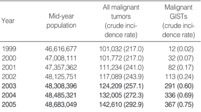
Data were presented as numbers (%) for categorical vari-ables. To estimate the association between eligible variables and mean survival time, the Kaplan-Meier test was applied together with the log-rank test to compare various groups. We conducted the Cox proportional hazard regression anal-ysis to estimate hazard risk ratios and 95% confidence inter-vals (CI) of the possible risk factors for survival after adjust-ment in age and sex. The SPSS (version 13.0) was used for statistical analyses. A P value of <0.05 was considered statis-tically significant. Year Mid-year population All malignant tumors (crude inci-dence rate) Malignant GISTs (crude inci-dence rate) 1999 46,616,677 101,032 (217.0) 12 (0.02) 2000 47,008,111 101,772 (217.0) 32 (0.07) 2001 47,357,362 111,234 (241.0) 82 (0.17) 2002 48,125,751 117,089 (243.9) 113 (0.24) 2003 48,308,396 124,209 (257.1) 291 (0.60) 2004 48,485,321 132,005 (272.3) 336 (0.69) 2005 48,683,049 142,610 (292.9) 367 (0.75) Table 1. Changes in incidence of malignant GISTs compared with all malignant tumors from 1999 to 2005: Data of Korean National Cancer Registry
NA, Not available; Number in parenthesis is percentage. Number of
subjected (%) Subgroups
Variables
Age 1,227 (100.0) <60 (43.7) ≥60 (56.3) 11-86 (57.85±
12.62 yr old) Gender 1,227 (100.0) Male (52.3) Female (47.7)
Size (cm) 1,129 (92.0) <2 (23.9) 2-4.9 (34.9) 5-9.9 (26.7) ≥10 (14.5)
Location 1,223 (99.7) Esophagus Stomach Small intestine Large intestine
Extra-gastro-(1.2) (59.9) (23.2) (5.8) intestinal (10.1)
Mitosis 1,125 (91.7) <5 (57.9) ≥5 (42.1)
Stage 1,122 (91.4) Confined to organs Invasion into adjacent Distant metastasis Recurrences
(76.7) organs (4.3) (9.0) (1.5)
Diagnosis by 1,140 (92.9) Benign (36.6) Uncertain Malignancy (40.9) NA (7.1)
AFIP (2002) malignancy (15.4)
Diagnosis by NIH 1,125 (91.7) Very low (16.1) Low (27.7) Intermediate (17.4) High (27.4) Metastatic, recurrences (3.1)
Necrosis 264 (21.5) - (54.5) + (45.5)
Vessel Invasion 104 ( 8.5) - (91.3) + (8.7) Mucosal Invasion 145 (11.8) - (60.7) + (39.3)
Histologic type 805 (65.6) Spindle (81.7) Epithelioid (6.3) Mixed (7.6) Other (4.3)
C-Kit 1,112 (90.6) - (3.8) + (96.2)
CD34 971 (79.1) - (18.8) + (81.2)
Actin 1,013 (82.6) - (64.2) + (35.8)
S-100 988 (80.5) - (83.7) + (16.3)
RESULTS
Population based incidence of malignant GISTs in Korea
There were 1,233 cases of malignant GISTs in the Nation-al Cancer Institute (NCI) Registry from 1999 to 2005 (Table 1). Table 1 shows the incidence of malignant GISTs in com-parison with all malignant tumors registered. There was a big shift in GISTs incidences between 1999 and 2002 and also from 2003 to 2005, while the incidence of all malignant tumors was almost the same during the same time. Extra-gas-trointestinal GISTs (8.3-8.7%) showed to be more common than esophageal GISTs (0.7-1.1%).
Characteristics of GISTs: nationwide collection from 38 hospitals in Korea during 2003-2004
We collected 1,227 pathology reports from the 38 partic-ipating hospitals from January 1, 2003 to December 31, 2004. The characteristics of GISTs and contents of pathology reports collected nationwide are shown in Table 2. The range of
pati-ent age was from 11 to 86 yr old (mean 57.83±12.62); 5
(0.4%) was in their 1st decade, 20 (1.6%) in 2nd, 87 (7.1%) in 3rd, 200 (16.3%) in 4th, 302 (24.6%) in 5th, 382 (31.1%) in 6th, 210 (17.1%) in 7th, and 21 (1.7%) in 8th. Male to female ratio was 1 to 1.7. The most common location of tumor was stomach. Extra-gastrointestinal locations were omentum and mesentery (45.1%). Then, pelvis (9.8%), intra-abdomi-nal (34.3%), retroperitoneum (3.9%), abdomiintra-abdomi-nal wall (3.9%), and pancreas (3%) were found to be occurring sites of extra-gastrointestinal GISTs. Liver (46), lymph node (9), bone, lung, spleen, diaphragm, and so on were found to be metastatic sites
of GISTs. One hundred cases (8.2%: 3 in the esophagus, 85 in the stomach, 8 in the small intestine, 1 in the large intes-tine and 1 in extra-gastrointestinal) were incidentally found during operations for other diseases. More than 90% of the tumors showed the result of c-kit immunostain in patholo-gy reports in contrast with CD34, actin and s-100 protein in which about 80% were found to have. Almost all GISTs, which provided the results of immunostains, were c-kit posi-tive (96.2%). The relationship between tumor location and malignancy, defined by variable criteria are presented in Tables 3-5.
The incidence of gastric GIST was slightly increased in ages older than 60 yr, in contrast to decreases in the incidence of the small intestinal GISTs. However, the location of tumor was not significantly related with patient’s age (P=0.083). In general, it showed male preponderance. Esophageal, large intestinal, and extra-gastrointestinal GISTs were more com-mon in males than in females, but gastric GISTs were more common in females (P=0.027).
In the collected pathology reports, GISTs were classified by the NIH criteria (55.4%), the AFIP criteria (41.7%), and other (2.9%). To compare the prognostic value of the diag-nostic criteria, we re-classified each GIST by the NIH crite-ria as well as the AFIP critecrite-ria based on the descriptions in the pathology reports, including tumor, size, location, and mitosis (Tables 3, 4). About half of the GISTs in our study were malignant according to the AFIP criteria. Benign tumors were common in stomach in contrast to malignant tumors which were more common in intestinal and extra-gastroin-testinal areas (P=0.000). High risk tumors, as classified by the NIH criteria, were also more common in the small and large intestinal and extra-gastrointestinal locations than in
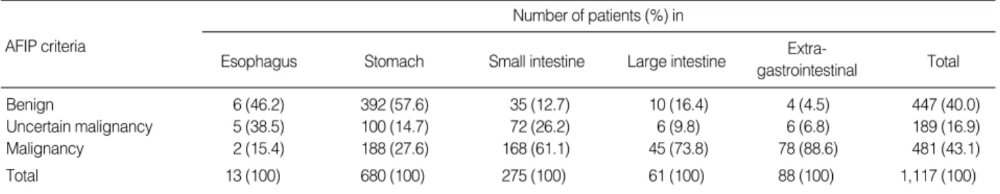
AFIP criteria Number of patients (%) in Extra- gastrointestinal Total Large intestine Small intestine Stomach Esophagus Benign 6 (46.2) 392 (57.6) 35 (12.7) 10 (16.4) 4 (4.5) 447 (40.0) Uncertain malignancy 5 (38.5) 100 (14.7) 72 (26.2) 6 (9.8) 6 (6.8) 189 (16.9) Malignancy 2 (15.4) 188 (27.6) 168 (61.1) 45 (73.8) 78 (88.6) 481 (43.1) Total 13 (100) 680 (100) 275 (100) 61 (100) 88 (100) 1,117 (100)
Table 3. The relationship between tumor location and malignancy, defined by the AFIP criteria
NIH criteria Number of patients (%) in Extra- gastrointestinal Total Large intestine Small intestine Stomach Esophagus
Very low risk 6 (46.2) 158 (23.5) 22 (8.1) 7 (11.9) 3 (3.5) 196 (17.8)
Low risk 0 (0.0) 242 (36.0) 83 (30.7) 8 (13.6) 4 (4.7) 337 (30.6)
Intermediate risk 5 (38.5) 125 (18.6) 56 (20.7) 11 (18.6) 17 (19.8) 214 (19.5)
High risk 2 (15.4) 147 (21.9) 109 (40.4) 33 (55.9) 62 (72.1) 353 (32.1)
Total 13 (100) 672 (100) 270 (100) 59 (100) 86 (100) 1,100 (100)
stomach (P=0.001). Most of extra-gastrointestinal tumors were malignant (88.6%) or high risk (72.1%). We also re-classified GISTs into eight groups based on tumor size and mitosis according to a new classification by Miettinen and Lastosa 2006 (15) (Table 5). However, statistical analysis was not available due to the small number of tumors in some cat-egories. As shown in Table 5, tumor size was correlated with mitosis. In the group with mitotically active GISTs, gastric tumors tend to be small in size compared with tumors in intes-tine, extra-gastrointestinal areas, and esophagus. The descrip-tion of tumor necrosis, vessel and mucosal invasions, and his-tologic type were found only in 21.5%, 8.5%, 11.8%, and 65.6% of pathology reports collected for this study,
respec-tively.
Survival analysis
During the follow-up study period, we found that 102 patients died as a result of GISTs. Most GISTs, which caused mortality, were malignant (80.4%) or high risk (66.7%). How-ever, 1.8% of benign or uncertain malignant tumors and 6.9% of very low risk, low risk or intermediate tumors led to mor-tality. In the same risk group by the NIH criteria, the inci-dence of patients, who died of the diseases, was higher in small intestinal and extra-gastrointestinal GISTs than gastric ones. The malignant GISTs by the AFIP criteria also
demonstrat-Number of patients (%) in tumor size of 2-4.9 cm <2 cm 5-9.9 cm ≥10 cm Total Mitosis <5 Eosphagus 7 (70.0) 1 (10.0) 2 (20.0) 0 (0.0) 10 (100) Stomach 169 (36.0) 190 (40.4) 87 (18.5) 24 (5.1) 470 (100) Small intestine 18 (12.9) 59 (42.1) 45 (32.1) 18 (12.9) 140 (100) Large intestine 13 (39.4) 9 (27.3) 4 (12.1) 7 (21.2) 33 (100) Extra-gastrointestinal 17 (37.0) 4 (8.7) 16 (34.8) 9 (19.6) 46 (100) Total 224 (32.0) 263 (37.6) 154 (22.0) 58 (8.3) 699 (100) Mitosis ≥5 Esophagus 0 (0.0) 2 (40.0) 2 (40.0) 1 (20.0) 5 (100) Stomach 23 (8.8) 112 (42.7) 77 (29.4) 50 (19.1) 262 (100) Small intestine 11 (7.6) 34 (23.4) 62 (42.8) 38 (26.2) 145 (100) Large intestine 7 (18.4) 9 (23.7) 16 (42.1) 6 (15.8) 38 (100) Extra-gastrointestinal 9 (16.1) 7 (12.5) 16 (28.6) 24 (42.9) 56 (100) Total 50 (9.9) 164 (32.4) 173 (34.2) 119 (23.5) 506 (100)
Table 5. Size distribution of all GISTs based on mitosis and location according to the new Miettinen criteria (2006)
Cumulative survival (%) 100 80 60 40 20 0 0 24 48 72 96 Time (months) A Cumulative survival (%) 100 80 60 40 20 0 0 24 48 72 96 Time (months) B Cumulative survival (%) 100 80 60 40 20 0 0 24 48 72 96 Time (months) D Cumulative survival (%) 100 80 60 40 20 0 0 24 48 72 96 Time (months) E Cumulative survival (%) 100 80 60 40 20 0 0 24 48 72 96 Time (months) C Female (n=565) Male (n=611) <2 cm (n=280) 2-9.9 cm (n=728) >10 cm (n=169) 5/50 HPF (n=685) 5/50 HPF (n=492) <60 yr (n=457) >=60 yr (n=601) Stomach (n=703) Small intestin (n=270) Large intestin (n=68) Extragastroinsestine (n=99)
Fig. 1. Univariate survival analyses of varia-bles: gender (A, P=0.026), age (B, P=0.006), location (C, P=0.000), tumor size (D, P= 0.000) and mitosis (E, P=0.000) are signifi-cantly correlated with patient’s survival.
ed a higher mortality rate.
All the variables except for smooth muscle actin immuno-expression that was significantly correlated with the survival were analyzed in this study with univariate analysis (Figs. 1-4). In ages older than 60 yr, male gender, small intestinal and extra-gastrointestinal location, larger than 10 cm in size, and mitosis more than 5/50 HPF pointed to poor survival rate. All histopathological variables analyzed in the study: mucos-al and vessel invasions and necrosis were significantly related with the survival (Fig. 2, P=0.000, P=0.000, and P=0.000, respectively). Spindle cell type tumors showed a better
prog-nosis than mixed, epithelioid, and pleomorphic cell types (Fig. 2, P=0.016). In the classification of GISTs by the NIH criteria, there was no prognostically significant difference between very low and low risk versus intermediate risk tumors, but high-risk tumors pointed significantly to a shorter sur-vival time (Fig. 4, P=0.000). According to the AFIP criteria, the median survival time of malignant tumors was signifi-cantly shorter than those of benign and uncertain malignan-cy (Fig. 4, P=0.000).
In multivariate analysis, tumor location, patient’s age, malig-nant GISTs by the AFIP criteria, and stages of tumors showed
Fig. 2. Univariate analysis of variables of microscopic findings: Tumor necrosis (A), vessel invasion (B), mucosal invasion (C) and histo-logic type (D) are significantly correlated with patient’s survival (P=0.000, 0.000, 0.000, 0.016, respectively).
Cumulative survival (%) 100 80 60 40 20 0 0 24 48 72 Time (months) Cumulative survival (%) 100 80 60 40 20 0 0 24 48 72 Time (months) Cumulative survival (%) 100 80 60 40 20 0 0 24 48 72 Time (months) Cumulative survival (%) 100 80 60 40 20 0 0 24 48 72 96 Time (months) Spindle (n=643) Epithelioid (n=50) Negative (n=84) Positive (n=55) Negative (n=138) Positive (n=115) Negative (n=84) Positive (n=8) Mixed (n=60)
Other (Pieomerphic. etc) (n=31)
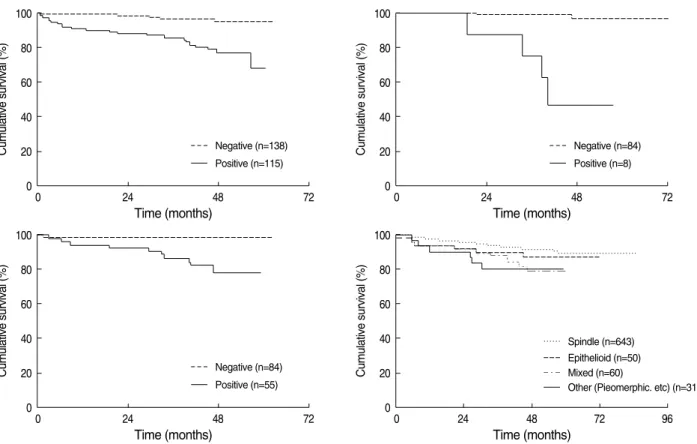
Fig. 3. Univariate analysis of variables of immunohistochemical findings: CD34 expression (A) inversely relates with a patient’s survival (P=0.001) in contrast to poor survival of GISTs with S-100 protein expression (B) (P=0.000). However a-smooth muscle actin expression (C) is not correlated with patient’s survival (P=0.573).
Cumulative survival (%) 100 80 60 40 20 0 0 24 48 72 96 Time (months) A Negative (n=174) Positive (n=756) Cumulative survival (%)
100 80 60 40 20 0 0 24 48 72 96 Time (months) B Negative (n=791) Positive (n=156) Cumulative survival (%)
100 80 60 40 20 0 0 24 48 72 96 Time (months) C Negative (n=620) Positive (n=351)
significant correlation with the prognosis (Table 6). People with tumor sizes more than 2 cm were more likely at an in-creased risk of death compared to those with tumor sizes less than 2 cm, but this data was statistically not significant (P= 0.247). In terms of tumor location, the risk was 1.985 and 2.423 times higher in small intestines and extra-gastrointesti-nal than in stomachs (P=0.030 & 0.014), but the risk in large intestinal GISTs was not higher (P=0.602). Malignant GISTs by the AFIP criteria revealed a 6.211 times higher risk of mortality compared with benign and borderline ones (P= 0.027). The risk in aggressive behavior of GISTs by the NIH criteria slightly increased in intermediate tumors compared with very low and low risk GISTs (odds ratio=0.443 & 0.585),
but this data was not statistically significant (P=0.524). GISTs limited to organs revealed better survival of patients than tumors, which invaded adjacent organs or metastasis.
The immunoexpression of a-smooth muscle actin, CD34
and s-100 protein, necrosis, and mucosal and vascular inva-sions were not available for multivariate analysis because of the small number of results.
As a result of this study, the GIST classifications based on original tumor location together with size and mitosis is more efficient than the NIH criteria in predicting patient’s survival. However, the mechanism still needs to be clarified through future studies.
Fig. 4. Univariate survival analysis of variables of diagnostic criterias: Both of the NIH criteria (A) and the AFIP criteria (B) are significantly correlated with patient’s survival (P=0.000 & P=0.000).
A Cumulative survival (%) 100 80 60 40 20 0 0 24 48 72 96 Time (months) Intermediate risk (n=204) Very low risk/Low risk (n=517)
High risk (n=358) B Cumulative survival (%) 100 80 60 40 20 0 0 24 48 72 96 Time (months) Benign/Uncertain malignancy (n=615) Malignancy (n=480) *P<0.05.
Variables P value Relative risk 95% C.I.
Size: <2 cm vs. 0.247 <5 cm 0.899 0.937 0.344-2.522 5-10 cm 0.507 1.370 0.540-3.478 >10 cm 0.139 2.043 0.793-5.261 Mitosis: <5 vs. ≥5 0.428 0.775 0.413-1.455 Location*: Stomach vs. Small Intestine* 0.030* 1.958 1.066-3.595 Large intestine 0.602 1.313 0.472-3.657 Extra-gastrointestinal* 0.014* 2.423 1.199-4.897 Diagnosis by AFIP*
Benign & borderline vs. malignant 0.027* 6.211 1.210-29.603
Diagnosis by NIH: Very low & low risk vs. 0.524
Intermediate risk 0.325 0.443 0.088-2.240
High risk 0.548 0.585 0.102-3.366
Stage*: Confined to organs vs.
Invasion to adjacent organs 0.000* 4.133 2.062-8.281
Distant metastasis 0.001* 2.872 1.512-5.458
Recurrence 0.055 2.911 0.976-8.661
Gender 0.214 1.352 0.840-2.175
Age <60 vs. ≥60* 0.001* 2.340 1.385-3.952
Table 6. Multivariate analysis of eligible GISTs risk factors such as: size, mitosis, tumor location, diagnosis by AFIP criteira, diagnosis by NIH criteria, gender and age
DISCUSSION
This is the first description of a population based on the incidence of GISTs in Korea. In the review of the Korean NCI Registry, we found a huge change regarding the incidences of malignant GISTs between 1999-2002 and 2003-2005. For example, the incidence of malignant GISTs in 2003 (0.69 per 100,000 population) was a double that of 2002 (0.24 per 100,000), even though there were no significant changes in the incidences of all malignant tumors during the same time. In regard with the change in incidences of GISTs, several his-torical backgrounds may be related with them. The main reasons of the incidence increase of GISTs during 2002-2005 in Korea is related to an improved understanding of patho-biology, the treatment of GISTs, the introduction of schemat-ic diagnostschemat-ic criteria proposed by the NIH consensus work-shop together with improved convenience of immunohisto-chemical staining for c-KIT. Changes in the GIST incidences similar to those in our study also has been described by Goettsch et al. (16) and Steigen and Eide (17).
Since the NIH publication, which was unfortunately based on a consensus opinion rather than actual follow-up data (2), numerous additional pathologic and biologic variables have been evaluated as prognostic factors. In 2002 Miettien et al. (18) analyzed a large AFIP series of GISTs coupled with long term follow-up data and then proposed guidelines for the evaluation of GISTs malignancies. Taking the Korean NCI Registry in consideration, and including only tumors coded as having malignant behavior, the incidence of all GISTs in Korea can be estimated as 1.6 per 100,000 by the AFIP cri-teria. The proportion of malignant GISTs by the AFIP crite-ria was 43.1%. However, by the NIH critecrite-ria, the proportion of high-risk GISTs was 32.1%, and the incidence of GISTs was estimated 2.2 per 100,000. The incidence of GISTs have been previously reported at 1-2 per 100,000 population by Miettinen and Lastosa (3), 1.85-2.2 per 100,000 population in the United States by Fletcher et al. (2), 2 per 100,000 pop-ulation in southwestern Sweden by Nilsson et al. (9), and 1.1 per 100,000 population in Iceland by Tryggvason et al. (10). The incidence of GIST in Korea did not appear to be differ-ent from theirs.
In the review of pathology reports, we found that most hos-pitals in Korea used either the NIH criteria or the AFIP crite-ria to predict the prognosis of GISTs. The results of our study, different diagnostic criteria and terminology in two diagnos-tic schemes, may have created some confusion in classifying and estimating the incidence of GISTs. However, the iden-tification of GISTs with poor prognosis is very important in the choice of treatment modality to improve the survival. We found a prognostic significance of the AFIP criteria in uni-variate as well as multiuni-variate analyses. In our previous report on guideline for cancer registration, the intermediate and high risk tumors were regarded as malignancy. However, the guide-line is based on a consensus opinion rather than an actual
fol-low-up data (19). In this study, the prognosis of intermedi-ate risk groups of GIST by the NIH criteria was not differ-ent from those of the very low and low risk tumor groups when follow-up data were examined. Previously published reports also described no prognostic differences in interme-diate risk GISTs and the very low and low risk ones (20). In multivariate analysis of our study, the patients with interme-diate and high risk GISTs were more likely to increase the risk of mortality compared with very low and low risk GISTs, but it was statistically not significant (P=0.325 and 0.548, respectively). It appeared to be appropriate that only high-risk tumors were regarded as malignant GISTs. However, a few patients with low and intermediate risk tumors died of diseases in our study. The prognosis of low and intermediate risk GISTs by the NIH criteria remains to be clarified through long term follow-up. The prognostic efficacy of the NIH cri-teria has been discussed by several researchers (9, 21), and the modification of the NIH criteria (20-22) was published recent-ly to improve the risk-based stratification of GISTs. The orig-inal NIH criteria adopted only tumor size and mitosis but the significance of tumor size and location was considered on AFIP criteria (12, 18). The impact of the location of GISTs on the patient’s survival is still controversial. Some investiga-tors demonstrated a more aggressive behavior in intestinal GISTs than in gastric GISTs (9-12, 21, 23, 24), while oth-ers found no differences in the patient’s survival (25). In the results of our study, the AFIP criteria as well as the original tumor location were prognostically significant in multivari-ate analysis. There seems to be several explanations for nostic significances of the tumor location. Firstly, the prog-nostic significance of the tumor location may be related to early detection of gastric GISTs. This is supported by the fact that gastric GISTs were significantly smaller than the non-gastric ones (10). The results of our study demonstrated that the malignant GISTs by the AFIP criteria or high risk by the NIH criteria were far more common in the intestine and extra-gastrointestinal than in the stomach at the time of diagno-sis. Secondly, Miettinen et al. recently reported the true bio-logic differences between gastric and non-gastric GISTs (26). The mutation in exon 9 of the c-kit gene, which is related to more aggressive tumors, was reported to be more frequently found in small intestinal GISTs (24). Although Park et al. (25) described that the tumor location was significant to predict the overall survival of patients in univariate analysis of 93 high-risk GISTs, but c-kit exon 9 mutations were not related with poor prognosis in Korean patients. In the data of patients, who died of the disease in our study, we found that the pro-portion of intestinal and extra-gastrointestinal GISTs were higher than gastric GISTs among the same categories by the NIH criteria. This means that the anatomical tumor location has to be related to a patient’s survival but it remains to be clarified as to why. Furthermore, we found that three benign and two uncertain malignant GISTs by the AFIP criteria had mortality in this study. Therefore, further efforts to improve
the diagnostic criteria to trace the malignant potential of GISTs are needed.
We found tumors with invasion or metastasis at the time of diagnosis in 13.7%. As expected, the tumors, which were confined to the original organs, revealed a better chance of survival than tumors with local invasion or metastasis. Any tumors showing metastatic lesion were classified into high risk or malignant GISTs in this study.
From the results of our study, we found several different epidemiologic characteristics of GISTs in Korea. Male gen-der was related with poor patient’s survival in univariate anal-ysis, but not in multivariate analysis. Male gender has been described as an independent adverse prognostic factor, (11) but not fully consistent. The average patient’s age was 57.83 yr, and results in patients older than 60 yr indicated signifi-cant poor survival rates. The patient’s age at the time of diag-nosis has been described as a GIST prognosticator (11, 17, 21, 27). Extra-gastrointestinal tumors were found to be more common than in previous reports. We found 8.3% of extra-gastrointestinal malignant GISTs from the Korean NCI Reg-istry and 10.1% of the data collected from 38 hospitals in this study. However, the proportion was reported to be 5% in previous reports (2). Extra-gastrointestinal GIST locations were omentum, mesentery, pelvis, intra-abdominal, retroperi-toneum, abdominal wall and pancreas in our study. All the GISTs had been regarded as tumors with some malignant potential, but this is being challenged by recent data reflect-ing clinically insignificant and incidental GISTs. Microscopic GISTs were detected during autopsies of the stomach in 23% of patients older than 50 yr (28) and 35% of patients with gastric carcinoma (29). Incidental GISTs were found during operations of various diseases, but, except for the neurofibro-matosis type 1 and Carney syndrome, no risk factors were identified. During the course of our study, we incidentally found GISTs in 8.2% and most commonly in the stomach during surgery for gastric carcinoma, but in patients with esophageal, colorectal, biliary tract, pancreatic, and hepato-cellular carcinomas, gastrinoma, and ischemic colitis, GISTs were rarely found.
We found the prognostic significance in the pathologic findings of tumor necrosis and mucosal and vascular inva-sions, but they were rarely described in the pathology reports. From the results of this research, we recommend to describe these prognostic factors in daily practice, even though it is negative. We have three major limitations with regards to data of this study. The first one is incomplete information of chemotherapy. We reviewed the pathology reports with infor-mation of surgery. The treatment modality was not consid-ered for survival analysis in this study. The second limitation is insufficient follow-up data. We analyzed the overall survival time but not the recurrence of diseases. The follow-up dura-tion in our study was less than 7 yr. The third one is from the retrospective analysis of pathology reports. We could not ran-domize the treatment modality. Even though these
limita-tions, we have concluded that the classification of GISTs based on original tumor location together with size and mitosis is more valuable to predict the prognosis. In addition, male gen-der, age and stage should be considered as factors for risk strat-ification of GISTs. Additionally, we recommend the descrip-tion of the immunohistochemical results of CD34 and s-100 protein as well as histologic parameters such as tumor necro-sis, mucosal and vascular invasions in pathology reports for daily practice.
Recently, many researchers focus on the significance of c-kit and PDGFRA mutation in predicting the response to tyro-sine kinase inhibitor therapy (30) and the correlation of res-ponse to the therapy with type of c-kit mutation in GISTs (13, 25). Because the biologic behavior of GISTs is different in different locations and prognosis of low and intermediate risk tumors is uncertain, the genetic analysis of GISTs might be valuable, when assessing the prognosis of GISTs as well as the efficacy of therapy.
REFERENCES
1. Schaldenbrand JD, Appelman HD. Solitary solid stromal
gastroin-testinal tumors in von Recklinghausen’s disease with minimal smooth muscle differentiation. Hum Pathol 1984; 15: 229-32.
2. Fletcher CD, Berman JJ, Corless C, Gorstein F, Lasota J, Longley BJ, Miettinen M, O'Leary TJ, Remotti H, Rubin BP, Shmookler B, Sobin LH, Weiss SW. Diagnosis of gastrointestinal stromal tumors:
a consensus approach. Hum Pathol 2002; 33: 459-65.
3. Miettinen M, Lasota J. Gastrointestinal stromal tumors--definition,
clinical, histological, immunohistochemical, and molecular genetic features and differential diagnosis. Virchows Arch 2001; 438: 1-12.
4. Dematteo RP, Heinrich MC, El-Rifai WM, Demetri G. Clinical
man-agement of gastrointestinal stromal tumors: before and after sti-571. Hum Pathol 2002; 33: 466-77.
5. Kim KM, Kang DW, Moon WS, Park JB, Park CK, Sohn JH, Jeong JS, Cho MY, Jin SY, Choi JS, Kang DY. Pkctheta expression in
gas-trointestinal stromal tumor. Mod Pathol 2006; 19: 1480-6.
6. Espinosa I, Lee CH, Kim MK, Rouse BT, Subramanian S, Montgo-mery K, Varma S, Corless CL, Heinrich MC, Smith KS, Wang Z, Rubin B, Nielsen TO, Seitz RS, Ross DT, West RB, Cleary ML, van de Rijn M. A novel monoclonal antibody against dog1 is a sensitive
and specific marker for gastrointestinal stromal tumors. Am J Surg Pathol 2008; 32: 210-8.
7. Heinrich MC, Corless CL, Duensing A, McGreevey L, Chen CJ, Joseph N, Singer S, Griffith DJ, Haley A, Town A, Demetri GD, Fletcher CD, Fletcher JA. Pdgfra activating mutations in
gastroin-testinal stromal tumors. Science 2003; 299: 708-10.
8. Miettinen M, Sobin LH, Sarlomo-Rikala M. Immunohistochemical
spectrum of gists at different sites and their differential diagnosis with a reference to cd117 (kit). Mod Pathol 2000; 13: 1134-42.
9. Nilsson B, Bumming P, Meis-Kindblom JM, Oden A, Dortok A, Gustavsson B, Sablinska K, Kindblom LG. Gastrointestinal stromal
prognostica-tion in the preimatinib mesylate era--a populaprognostica-tion-based study in western Sweden. Cancer 2005; 103: 821-9.
10. Tryggvason G, Gislason HG, Magnusson MK, Jonasson JG.
Gas-trointestinal stromal tumors in Iceland, 1990-2003: the Icelandic gist study, a population-based incidence and pathologic risk stratifi-cation study. Int J Cancer 2005; 117: 289-93.
11. Tran T, Davila JA, El-Serag HB. The epidemiology of malignant
gas-trointestinal stromal tumors: an analysis of 1,458 cases from 1992 to 2000. Am J Gastroenterol 2005; 100: 162-8.
12. Miettinen M, Lasota J. Gastrointestinal stromal tumors: pathology
and prognosis at different sites. Semin Diagn Pathol 2006; 23: 70-83.
13. Hornick JL, Fletcher CD. The role of kit in the management of
pati-ents with gastrointestinal stromal tumors. Hum Pathol 2007; 38: 679-87.
14. Kim KM, Kang DW, Moon WS, Park JB, Park CK, Sohn JH, Jeong JS, Cho MY, Jin SY, Choi JS, Kang DY. Gastrointestinal stromal
tumors in Koreans: it’s incidence and the clinical, pathologic and immunohistochemical findings. J Korean Med Sci 2005; 20: 977-84.
15. Miettinen M, Lasota J. Gastrointestinal stromal tumors: review on
morphology, molecular pathology, prognosis, and differential diag-nosis. Arch Pathol Lab Med 2006; 130: 1466-78.
16. Goettsch WG, Bos SD, Breekveldt-Postma N, Casparie M, Herings RM, Hogendoorn PC. Incidence of gastrointestinal stromal tumours
is underestimated: results of a nation-wide study. Eur J Cancer 2005; 41: 2868-72.
17. Steigen SE, Eide TJ. Trends in incidence and survival of
mesenchy-mal neoplasm of the digestive tract within a defined population of northern Norway. APMIS 2006; 114: 192-200.
18. Miettinen M, El-Rifai W, H L Sobin L, Lasota J. Evaluation of
malig-nancy and prognosis of gastrointestinal stromal tumors: a review. Hum Pathol 2002; 33: 478-83.
19. Cho MY, Kang YK, Kim KM, Chang HK, Chang HJ, Chang MS, Kim JM, Kang DY, Park C, Sohn JH. Porposal for creating a
guide-line for cancer registration of the gastrointestinal tumors (i). Kore-an J Pathol 2008; 42: 140-50.
20. Joensuu H. Risk stratification of patients diagnosed with
gastroin-testinal stromal tumor. Hum Pathol 2008; 39: 1411-9.
21. Hou YY, Lu SH, Zhou Y, Xu JF, Ji Y, Hou J, Qi WD, Shi Y, Tan YS, Zhu XZ. Predictive values of clinical and pathological
parame-ters for malignancy of gastrointestinal stromal tumors. Histol
Histo-pathol 2009; 24: 737-47.
22. Huang HY, Li CF, Huang WW, Hu TH, Lin CN, Uen YH, Hsiung CY, Lu D. A modification of NIH consensus criteria to better
distin-guish the highly lethal subset of primary localized gastrointestinal stromal tumors: a subdivision of the original high-risk group on the basis of outcome. Surgery 2007; 141: 748-56.
23. Lasota J, Kopczynski J, Sarlomo-Rikala M, Schneider-Stock R, Stachura T, Kordek R, Michal M, Boltze C, Roessner A, Stachura J, Miettinen M. Kit 1530ins6 mutation defines a subset of
predomi-nantly malignant gastrointestinal stromal tumors of intestinal ori-gin. Hum Pathol 2003; 34: 1306-12.
24. Antonescu CR, Sommer G, Sarran L, Tschernyavsky SJ, Riedel E, Woodruff JM, Robson M, Maki R, Brennan MF, Ladanyi M, DeMat-teo RP, Besmer P. Association of kit exon 9 mutations with
nongas-tric primary site and aggressive behavior: kit mutation analysis and clinical correlates of 120 gastrointestinal stromal tumors. Clin Can-cer Res 2003; 9: 3329-37.
25. Park CK, Lee EJ, Kim M, Lim HY, Choi DI, Noh JH, Sohn TS, Kim S, Kim MJ, Lee HK, Kim KM. Prognostic stratification of high-risk
gastrointestinal stromal tumors in the era of targeted therapy. Ann Surg 2008; 247: 1011-8.
26. Miettinen M, Sobin LH, Lasota J. Gastrointestinal stromal tumors
of the stomach: a clinicopathologic, immunohistochemical, and molecular genetic study of 1765 cases with long-term follow-up. Am J Surg Pathol 2005; 29: 52-68.
27. Woodall CE 3rd, Brock GN, Fan J, Byam JA, Scoggins CR, McMas-ters KM, Martin RC 2nd. An evaluation of 2537 gastrointestinal
stromal tumors for a proposed clinical staging system. Arch Surg 2009; 144: 670-8.
28. Agaimy A, Wunsch PH, Hofstaedter F, Blaszyk H, Rummele P, Gaumann A, Dietmaier W, Hartmann A. Minute gastric sclerosing
stromal tumors (gist tumorlets) are common in adults and frequent-ly show c-kit mutations. Am J Surg Pathol 2007; 31: 113-20.
29. Kawanowa K, Sakuma Y, Sakurai S, Hishima T, Iwasaki Y, Saito K, Hosoya Y, Nakajima T, Funata N. High incidence of
microscop-ic gastrointestinal stromal tumors in the stomach. Hum Pathol 2006; 37: 1527-35.
30. Steigen SE, Eide TJ, Wasag B, Lasota J, Miettinen M. Mutations in
gastrointestinal stromal tumors--a population-based study from North-ern Norway. APMIS 2007; 115: 289-98.