활액막 세포에서 기원하는 거대 세포종은 국소형과 미만형으로 분류되며 국소형 거대 세포종은 주로 지 관절의 활액막에서 호발 하는 것으로 알려져 있다.1,2) 그러나 이는 슬관절 내에서는 드물게 발생하는 병변으로 이를 보고하는 문헌도 제한되어 있으며, 후방 십자 인대에서 발생한 국내 보고도 2차례에 불과했다.3,4) 저자들은 슬관절 후방 십자 인대의 전연에 발생한 거대 세포종 1예를 관절경 하에서 절제 후 병리학적으로 확진하였으며 임상적 으로 좋은 결과를 보였기에 이를 문헌 고찰과 함께 보고하고자 한 다.
증례 보고
21세 여자 환자가 2개월 전 특별한 외상력 없이 걷던 중 발생한 좌 측 슬관절 전방의 통증 및 간헐적 잠김 증상이 발생하여 타 병원 에서 자기 공명 영상(MRI) 검사 후 반월상 연골판 파열 의심 하에 전원되었다. 내원 시 시행한 신체 검사에서 슬관절 운동 범위는 0-135도로 정상이었으며, 슬관절 전방 부위의 압통이 있었으며 슬관절 30도Case Report
J Korean Bone Joint Tumor Soc 2014; 20: 85-88 • http://dx.doi.org/10.5292/jkbjts.2014.20.2.85 www.kbjts.or.kr슬관절 후방 십자 인대에서 기원한 건막 거대 세포종: 1예
보고
A Tenosynovial Giant Cell Tumor Arising from Posterior Cruciate Ligament of Knee
Joint: A Case Report
김홍균 • 최창현 • 정국진 • 이영민 • 신미경* • 황지효
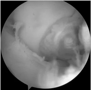
한림대학교 의과대학 강남성심병원 정형외과, *해부병리과 국소형 거대 세포종은 주로 지 관절의 활액막에서 호발하는 것으로 알려져 있으며 슬관절 내에서는 드물게 발생하는 것으로 알려져 있다. 병 리학적으로 다핵의 거대 세포를 특징적으로 가지는 질환으로 완전 절제 시 재발율은 낮다. 슬관절 내에 발생하는 경우 무증상에서 간헐적 잠 김 증상까지 다양하게 나타날 수 있으며, 관절경적으로 완전 절제가 가능하나 불완전 절제 시 45%까지 재발하는 것으로 보고되고 있다. 저자 들은 후방 십자 인대의 전연에 발생한 거대 세포종 1예를 관절경 하에서 절제 후 병리학적으로 확진하여 이를 보고하고자 한다. 색인단어: 거대 세포종, 후방 십자 인대, 슬관절 굴곡 시 전방부 동통을 호소하였다. 이밖의 다른 이학적 검사에서 는 특이 소견은 관찰되지 않았다. 단순 방사선 검사에서 슬관절 부위의 연부 조직의 부종이 약간 관찰되었으나 골 파괴 소견은 관찰되지 않았으며 관절 간격도 정 상이었다. 외부 자기 공명 영상의 시상면 proton density 영상에서 저신호 강도의 9×6 mm의 주위 조직과 경계가 명확한 종괴가 관 찰되었는데, 이는 T2 강조 영상에서 저신호 강도의 소견을 보였다 (Fig. 1). 관절경 하에서 황색의 타원형 종물이 관찰되었는데, 종물은 후 방 십자 인대 전연(anterior margin)과 연결되어 있었으며 이를 관 절경 하에서 완전 절제하였다(Fig. 2). 종물이 인대의 전연 활액막 에 부착되어 있어서 후방 십자 인대에는 손상 없이 종물의 완전 절제가 가능하였다. 해부 병리 검사에서 다핵의 거대 세포들이 섞여있는 소견이 관 찰되었으며 거대 세포종으로 진단되었다(Fig. 3). 관절경 하 종물 의 제거 후 증상의 소실과 함께 수술 후 1년 9개월 지난 후 외래 방 문하여 시행한 이학적 검사에서 증상 재발 없이 일상 생활이 가능 한 상태이다.고 찰
슬관절 내에 조직구(histiocyte) 기원의 종물로는 건막 거대 세포 종(tendon sheath giant cell tumor)과 색소 융모 결절성 활액막염 접수일 2014년 9월 17일 심사수정일 2014년 11월 24일 게재확정일 2014년 11월 26일 교신저자 황지효 서울시 영등포구 대림1동 948-1, 한림대학교 의과대학 강남성심병원 정형외과 TEL 02-829-5165, FAX 02-834-1728 E-mail [email protected]Copyrights © 2014 by The Korean Bone and Joint Tumor Society
“This is an Open Access article distributed under the terms of the Creative Commons Attribution Non-Commercial License (http://creativecommons.org/licenses/by-nc/3.0/) which permits unrestricted non-commercial use, distribution, and reproduction in any medium, provided the original work is properly cited.”
대한골관절종양학회지:제20권 제2호 2014
86
김홍균·최창현·정국진 외 3인
(pigmented villonodular synovitis)이 보고되어 있다. 이 두 질환은 유사한 조직학적 특징을 가지고 있으나, 활액막 증식의 형태가 서 로 다르다.1,2) 국소형(localized type) 건막 거대 세포종은 주로 수부 및 족부에 호발하고, 경계가 명확한 결절형 또는 폴립형의 종물 로 발견되며, 관절 내 또는 관절 외에 발생한다. 이들은 건막에 부 착되어 있으며 간혹 수부에 발생 시 신경을 압박할 수 있으며, 다 핵의 거대 세포를 특징적으로 가지는 질환으로 완전 절제 시 재발 율은 낮다.5) 미만형(diffuse type)은 증식형 활액막염으로도 불리우 며, 불규칙하고 침윤하는 양상을 띠며, 주로 관절 외에 발생한다. 색소형 융모 결절성 활액막염은 관절 내 활액막 대부분을 침범하 며 특히 슬관절의 침범이 많다.6-8) 조직학적으로 건막 거대 세포종과 색소성 융모 결절성 활액막 염은 구별이 어려우나, 국소형 거대 세포종은 세포의 침윤이 국소 적이고 경(pedicle)에 의해 활액막과 연결되어 있다. 미만형은 침 윤성의 불규칙한 소견은 보이나, 융모성의 증식은 없다. 색소성 융모 결절성 활액막염은 관절 내의 활액막의 전반적인 융모성 증 식 소견을 보인다. 국소형 건막 거대 세포종의 재발율은 7-23%로 보고되고 있으며, 미만형 건막 거대 세포종은 40-50%로 보고되고 있다. 색소성 융모 결절성 활액막염의 재발은 매우 높으며 활액막 제거를 통한 완전 절제가 필요하다.2,7,8) 건막 거대 세포종의 증상으로는 무증상에서부터 종창, 간헐적 잠김 등 다양한 증상을 유발하게 되어 임상적으로는 진단 및 색소 성 융모 결절성 활액막염과의 감별 진단이 어렵다. 또한 자기 공 명 영상에서도 T1 및 T2 강조 영상에서 중등도 또는 저신호 강도 로 나타나게되어 자기 공명 영상으로도 색소성 융모 결절성 활액 막염과 유사한 소견을 보이게 되어 더욱 감별하기 힘들다.6,9) 저자들의 경우 환자의 임상 증상 및 외부 자기 공명 영상 소견 에서 슬관절 내 반월상연골 손상이나 유리체를 예상하여 관절경 검사를 시행하였으나, 후방 십자 인대의 전연에 경계가 명확한 종 괴를 발견해 내고 완전 절제하였다. 수술 전 검사에서 외부 자기 공명 영상이라는 한계점이 있지만 이전의 증례 보고들과 다르게 명확한 임상 증상이 나타나지 않았으며, 후방 십자 인대의 전연에 종괴를 형성하고 있다는 차이점이 있었다. 관절경 검사 시 관절 내 병변에 대해 정확히 관찰할 수 있으며, 특히 국소형의 경우 간단하게 제거가 가능하다. 또한 감염이나 관 절 강직증 등의 합병증 가능성이 있으나 관혈적 제거에 비해 합병 증 발생율이 낮고 덜 침습적이어서 빠른 기능적 회복이 가능하다. 그러나 후방에 국소적으로 침범한 경우나 미만형의 경우 관절경 하 접근이 용이하지 않을 경우 관혈적 접근이 근치적 절제에 필요 할 수 있다. 따라서 건막 거대 세포종의 진단에 있어서 면밀한 이학적 검진 및 자기 공명 영상을 통한 의심이 필요하지만, 무엇보다 관절경 검사를 통한 완전 절제 및 종괴의 제거 시 조직학적으로 확진하는 것이 바람직하다. 또한, 불완전한 절제 시 슬관절의 경우 45%까지 재발하는 경향이 있으므로 지속적인 경과 관찰이 필요하리라 사
Figure 1. (A) On proton density coronal image, a 9×6 mm sized mass (white arrow) with well circumscribed intermediate signal intensity was seen lateral side of knee joint. (B) On T2 sagittal image, a 9×6 mm sized mass (white arrow) with well circumscribed intermediate signal intensity was seen just anterior margin of PCL.
A B
Figure 2. The mass was visible at the anterolateral area of the PCL by arthroscope.
87
슬관절 후방 십자 인대에서 기원한 건막 거대 세포종
료된다.5)
참고문헌
1. Enzinger FM, Weiss SW. Benign tumors and tumor like le-sions of synovial tissue. In: Enzinger FM, Weiss SW, ed. Soft tissue tumors. 3rd ed. St. Louis: Mosby; 1995. 412-41.
2. Rosenberg AE. Soft tissue tumors and tumor-like lesions. In: Cotran RS, Kumar V, Robbins SL, ed. Robbins pathologic basis of disease. 5th ed. Philadelphia: Saunders; 1994. 1260-1. 3. Chung WY, Kim YC, Jo SK. Localized form of tenosynovial
gi-ant cell tumor arising from the posterior Cruciate Ligament of the knee. 2 case report. J Korean Arthroscopy Soc. 2003;7:87-91.
4. Kim KT, Kang MS. Localized tenosynovial giant cell tumor involving the posterior cruciate ligament of the Knee. 1 case
report. J Korean Arthroscopy Soc. 2011;15:113-6.
5. Insall JN, Scott WN. Surgery of the knee. 4th ed. Philadelphia: Elsevier; 2006. 1026-9.
6. Koo BS, Kim KC, Lee HJ. Localized giant cell tumor of tendon sheath arising from the anterior cruciate ligament of the knee. 2 case report. J Korean Arthroscopy Soc. 1999;3:146-9. 7. Lee GW, Lee KS, Song SH, Kim MK, Yun SH. Snow-man
shaped nodular tenosynovitis in the knee. case report. J Ko-rean Arthroscopy Soc. 1999;3:44-7.
8. Rao AS, Vigorita VJ. Pigmented villonodular synovitis (giant-cell tumor of the tendon sheath and synovial membrane). A review of eighty-one cases. J Bone Joint Surg Am. 1984;66:76-94.
9. Jelinek JS, Kransdorf MJ, Shmookler BM, Aboulafia AA, Mala-wer MM. Giant cell tumor of the tendon sheath: MR findings in nine cases. AJR Am J Roentgenol. 1994;162:919-22.
Figure 3. (A) Well defined mass with capsule shows closely packed cells (H&E, ×10). (B) Relatively closely packed medium sized polyhedral cells with giant cells is shown (H&E, ×100).
A Tenosynovial Giant Cell Tumor Arising from Posterior Cruciate
Ligament of Knee Joint: A Case Report
Hong Kyun Kim, Chang Hyun Choi, Kook Jin Chung, Young Min Lee,
Mi Kyung Shin*, and Ji Hyo Hwang
Departments of Orthopedics, *Pathology, Kangnam Sacred Heart Hospital, College of Medicine, Hallym University, Seoul, Korea
Localized forms of giant cell tumor are known to arise commonly in the synovial membrane of the finger joints. Multinucleated giant cells are its characteristic pathology finding, giant cell tumor shows a low rate of recurrence after complete excision. When occurring at the knee joints, giant cell tumor manifests a wide form of symptoms, from no symptom at all, to intermittent locking. Complete excision is possible by arthroscopy, but if done incompletely, it is reported to recur in 45% of cases. We present here a case of giant cell tumor that has arisen from the anterior portion of the posterior cruciate ligament, excised by arthroscopy and followed by patho-logic confirmation.
Key words: giant cell tumor (GCT), posterior cruciate ligament (PCL), knee joint
Received September 17, 2014 Revised November 24, 2014 Accepted November 26, 2014 Correspondence to: Ji Hyo Hwang
Department of Orthopedic Surgery, Kangnam Sacred Heart Hospital, Hallym University College of Medicine, 948-1, Daerim-1 dong, Yeongdeungpo-gu, Seoul 150-950, Korea
TEL: +82-2-829-5165 FAX: +82-2-834-1728 E-mail: [email protected]
Case Report
J Korean Bone Joint Tumor Soc 2014; 20: 85-88 • http://dx.doi.org/10.5292/jkbjts.2014.20.2.85 www.kbjts.or.krCopyrights © 2014 by The Korean Bone and Joint Tumor Society
“This is an Open Access article distributed under the terms of the Creative Commons Attribution Non-Commercial License (http://creativecommons.org/licenses/by-nc/3.0/) which permits unrestricted non-commercial use, distribution, and reproduction in any medium, provided the original work is properly cited.”
The Journal of the Korean Bone and Joint Tumor Society Vol. 20, No. 2 (December 2014)