저작자표시-비영리-변경금지 2.0 대한민국 이용자는 아래의 조건을 따르는 경우에 한하여 자유롭게 l 이 저작물을 복제, 배포, 전송, 전시, 공연 및 방송할 수 있습니다. 다음과 같은 조건을 따라야 합니다: l 귀하는, 이 저작물의 재이용이나 배포의 경우, 이 저작물에 적용된 이용허락조건 을 명확하게 나타내어야 합니다. l 저작권자로부터 별도의 허가를 받으면 이러한 조건들은 적용되지 않습니다. 저작권법에 따른 이용자의 권리는 위의 내용에 의하여 영향을 받지 않습니다. 이것은 이용허락규약(Legal Code)을 이해하기 쉽게 요약한 것입니다. Disclaimer 저작자표시. 귀하는 원저작자를 표시하여야 합니다. 비영리. 귀하는 이 저작물을 영리 목적으로 이용할 수 없습니다. 변경금지. 귀하는 이 저작물을 개작, 변형 또는 가공할 수 없습니다.
Surgical treatment of cavernous malformation in spinal
cord
by
Chul-kyu Lee
Major in Medicine
Department of Medical Sciences
The Graduate School, Ajou University
2
Surgical treatment of cavernous malformation in spinal
cord
by
Chul-kyu Lee
A Dissertation Submitted to The Graduate School of
Ajou University in Partial Fulfillment of the Requirements for the
Degree of Master of Medicine
Supervised by
Soo Han Yoon, M.D., Ph.D.
Major in Medicine
Department of Medical Sciences
The Graduate School, Ajou University
3
This certifies that the dissertation
of Chul-kyu Lee is approved.
SUPERVISORY COMMITTEE
Soo Han Yoon
Ki Hong Cho
Se-hyuk Kim
The Graduate School, Ajou University
December, 20rd, 2013
4 - ABSTRACT -
Surgical treatment of carvenous malformation in spinal cord
Intramedullary CMs accounts for approximately 3-5% of all CMs of the CNS and 5%–12% of all spinal cord vascular lesions. The decision between conservative management and surgical treatment of intramedullary CMs is discussed controversially and the opinions diverge. The choice of microsurgical treatment of symptomatic intramedullary CMs is usually made on a case-by-case basis, considering the neurological complaints, general condition and the level of suffering of the patients as well as the surgical accessibility.
Patients with spinal intramedullary CM were enrolled who treated with surgical resection in the department of neurosurgery at our single institute between March 2007 and March 2012, retrospectively. Clinical presentation was determined using the Ogilvy classification and symptoms were evaluated on the McCormick scale.
Laminoplastic laminotomies were performed for 7 patients and a one level corpectomy was performed for 1 patient. Six patients (75%) had dorsally located lesions, whereas 2 patients (25%) had ventrally located lesions. Complete microsurgical resections were done in all patients. In terms of outcome as assessed according to the McCormick classification, 4 patients (50%) had stable outcome, 3 patients (37.5%) had improved outcome, and 1 patient (12.5%) had worsened outcome.
Clinical presentation based on Ogilvy’s classification at presentation showed 3 cases manifested as acute episodes of stepwise neurological deterioration (class 1), 1 case as slow progression of neurological deterioration (class 2), and 4 cases as acute onset of neurological deterioration with rapid decline (class 3). Four patients had history of acute bleeding within 1 month before surgery and 3 patients (75%) among those 4 patients clinically presented Ogilvy class 3.
In Group I (early resection), the average McCormick classification at follow-up was 3.25 0.5, which was clinically improved compared with 3.0 0.8 before surgery. However, this was not statistically significant (p > 0.05). In Group II (delayed resection), the average McCormick classification at follow-up was 2.5 0.5, which was statistically improved compared with 1.5 0.5 before surgery (p < 0.05).
The average preoperative VAS score for pain was 7.8 range 5-9). VAS score was improved up to 2.5
1.3 right after the surgery and well maintained until final follow
-up (2.6 1.4). The average VAS scores
at immediate postoperative and follow-up were statistically improved compared preoperative VAS score (p < 0.05).
The principal feasibility of surgical treatment has been accepted to complete resection because recurrent episodes of bleeding raise the risk for neurological deterioration and surgery for satisfaction of long-term results. Nevertheless limitation of small number of cases, our study had reviewed considerations of surgery for CM. We suggest that a full understanding of clinical manifestations, optimal surgical timing and complete resection of intramedullary CM can guide to make a favorable clinical outcome.
5
TABLE OF CONTENTS
ABSTRACT ………..4 TABLE OF CONTENTS ………..5 LIST OF FIGURES ………...6 LIST OF TABLES ………....7 Ⅰ. INTRODUCTION……….……….. 8 Ⅱ. METHODS………..……….………8A. Study design and data collection………...8
B. Evaluation method……….9 C. Surgical technique………...….10 Ⅲ. RESULTS………...11 Ⅳ. DISCUSSION ………12 Ⅴ. CONCLUSION ……….15 REFERENCES……….16 국문요약………..19
6
List of Figure
Fig. 1 Delayed operation ………..10 Fig.2 Early sesection ………. …………..15
7
List of Table
Table 1. Ogilvy classification……….9
Table 2. McCormick Grade……….9 Table 3. Summary of 8 patients with intraspinal CM………...12
8
Introduction
Cavernous malformations (CMs) are angiographically occult, well-circumscribed lesions that consist of closely packed, capillary-like vessels, without intervening brain or spinal tissue.9, 11, 13, 22, 30) Spinal CMs can occur along the neuraxis and are predominantly located intramedullary. However, exophytic growth and extradural CMs are reported as well .23) Incidence of CM was found 0.4~0.53% by prospective autopsy series in central nerve system (CNS).1-2, 5, 17, 22, 28) Intramedullary CMs accounts for approximately 3-5% of all CMs of the CNS and 5%–12% of all spinal cord vascular lesions.11, 13, 15, 17, 26, 30) Clinically, their natural history and risk of hemorrhage remain unclear.30) After the application of Magnetic resonance image (MRI), intramedullary CMs are increasingly being diagnosed in patients with varying spinal cord related symptoms or pain syndromes.19, 22) Spinal intramedullary CMs are clinically more aggressive than their intracranial counterparts.11) Due to space occupying growth, recurrent micro-bleeding or significant hemorrhage spinal CMs can lead to severe neurological deteriorations, such as progressive myelopathy and right up to paraplegia, unlike their intracranial counterparts. The latter typically presents with progressive focal neurological deficits and haemorrhage, whereas the risk of significant hemorrhage like other vascular malformations is considered to be low due to the low venous pressure.1-2, 5, 9, 11, 13, 17, 19, 22)
The decision between conservative management and surgical treatment of intramedullary CMs is discussed controversially and the opinions diverge. The choice of microsurgical treatment of symptomatic intramedullary CMs is usually made on a case-by-case basis, considering the neurological complaints, general condition and the level of suffering of the patients as well as the surgical accessibility.19)
The aim of this study was to present data of microsurgically treated patients with symptomatic intramedullary CMs in our department and to discuss about the optimal surgical timing according to clinical manifestations.
Methods
Study design and data collection
Patients with spinal intramedullary CM were enrolled who treated with surgical resection in the department of neurosurgery at our single institute between March 2007 and March 2012. The medical records, radiographic images, intraoperative photographs, neurologic examinations, preoperative and postoperative clinical findings of all patients were reviewed retrospectively. The diagnosis was made in all patients after the onset of clinical symptoms. All patients had a single, monofocal intramedullary pathology and no family history. We include the patients with acute episode of hemorrhage and neurological deteriorations. We did not include asymptomatic lesions diagnosed in patients with multiple cavernomas.
9
Clinical presentation was determined using the Ogilvy classification and symptoms were evaluated on the McCormick scale. Ogilvy classification distinguishes 4 clinical presentation, as follows : 1. acute episode of stepwise neurological deterioration; 2. Slow progression of neurological deterioration; 3. Acute onset of neurological deterioration with rapid decline; 4. Acute onset of mild symptoms of neurological deterioration with gradual decline over weeks to months.26) (Table 1) The McCormick classification distinguishes 4 functional stages, as follows: Grade 1 denotes neurologically normal, mild focal deficit not significantly affecting function of involved limb, and normal gait; Grade 2 a sensorimotor deficit affecting function of the involved limb, mild to moderate gait difficulty, although patient still can walk independently; Grade 3 designates more severe neurological deficiencies, patient requires a cane/brace for ambulation, and may or may not be independent; Grade 4, signifies severe deficit, patient requires wheelchair or has bilateral upper-extremity impairment, and usually is not independent.10) (Table 2)
We evaluated clinical outcomes and postoperative complications at 1 month, 2 month, 6 month and every 6 month after the surgery. Preoperative and final follow-up Visual Analog Scales (VAS) for pain were prospectively collected and used in this analysis. The measurement was assessed using a 10-point VAS with endpoint anchors of no pain (0 point) and severe pain (10 point).
We divided the patients into two groups according to the surgical timing of intramedullary CMs consider Group I (early resection) included the patients who underwent surgical resection of the lesion within 4 weeks from the presentation of symptoms. Group II (delayed resection) included the patients who underwent surgical resection of the lesion 4 weeks later after the presentation of symptoms. Statistical analysis was performed using the Student t-test method, with p < 0.05 deemed as significant.
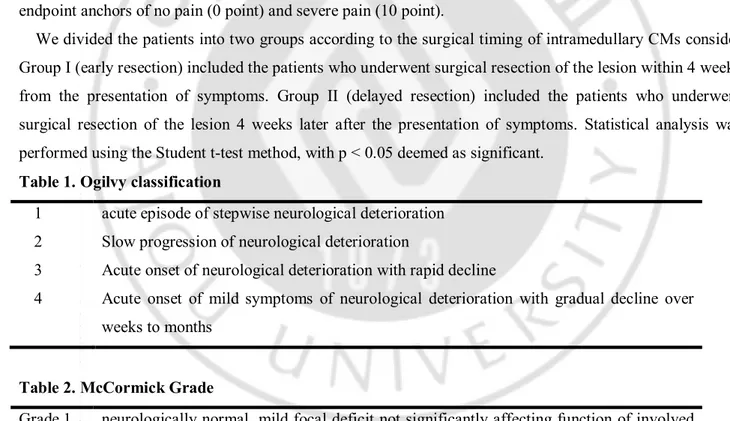
Table 1. Ogilvy classification
1 acute episode of stepwise neurological deterioration 2 Slow progression of neurological deterioration
3 Acute onset of neurological deterioration with rapid decline
4 Acute onset of mild symptoms of neurological deterioration with gradual decline over weeks to months
Table 2. McCormick Grade
Grade 1 neurologically normal, mild focal deficit not significantly affecting function of involved limb, and normal gait
Grade 2 sensorimotor deficit affecting function of the involved limb, mild to moderate gait difficulty, although patient still can walk independently
Grade 3 more severe neurological deficiencies, patient requires a cane/brace for ambulation, and may or may not be independent
Grade 4 severe deficit, patient requires wheelchair or has bilateral upper-extremity impairment, and usually is not independent
10
Surgical technique
Seven patients with dorsal or ventral lesions underwent posterior approach followed by laminoplastic laminotomy and 1 patient with ventral lesions underwent anterior approach followed by corpectomy.
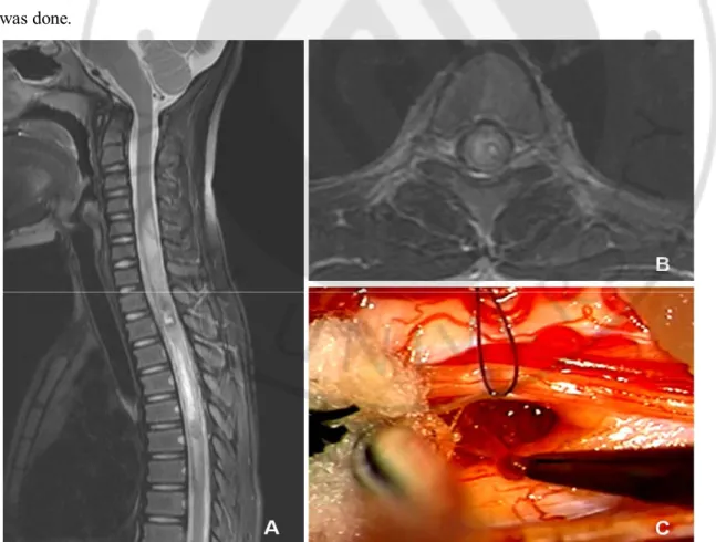
For the posterior approach, (Figure 1) the patient was placed prone on the operating room table without the head fixation and electrophysiological monitoring was used in all most patients. Midline incision was made spanning the affected levels. Laminotomy was performed overlying the area of interest and extending a single spinal level above and below the lesion for lateral and ventral lesions. With microscopic assistance, Small myelotomy with sufficient opening of the pia over the suspected area was done to access the lesion directly after midline dural incisions and resection of lesion was done. For ventrolateral lesion, division of dentate ligament allowing gentle rotation of the spinal cord with of 6-0 silk sutures holding. For lesions that come to the surface, incisions were made overlying the lesion by bi-polor coagulation. Micorbrunt-hook was used to separate the CM from the surrounding tissue. For lesions with a hematoma component, the hematoma was drained to decompress the spinal cord at this time. The CM was pulled into the center of the cavity and separated from the surrounding spinal cord. After inspection for residual malformation, the gliotic, hemosiderin-stained walls were left. The lesion bed was carefully examined after resection to avoid residual caverns, a common cause of recurrence. The dura mater was closed by automatic stamp and laminoplasty was done.
Fig. 1. Delayed resection A : Sagittal T2-weighted MR image showed intralesional hemorrhage with cord
swelling in cervical segment. B : Axial MR image showed the lesion with hemorrhage located in the left lateral-dorsal portion of spinal cord. C : intraoperative photograph showed removing a part of the lesion
11
For the anterior approach, the patient was placed supine and 1 level corpectomy was done. With microscopic assistance, Small myelotomy over the suspected area was done to access the lesion directly after midline dural incisions and resection of lesion was done as mentioned above.
Result
Between March 2007 and March 2012, 8 patients were performed resection of CM at our institute were pathologically proven to CM. The patient profiles are presented in Table 3. Four patients were men and 4 patients were women with a mean age of 39.8 years (range 7-81 years) at presentation. Male/ female ratio was equal. The average follow-up period was 39.7 months (range 15-71 months).
The most common presenting symptoms were motor weakness (62.5%), sensory deficit (37.5%) and dysesthesia (12.5%). All patients suffered from progressive clinical symptoms and signs. Average duration of clinical symptoms was 8.6 months (range 1-18 months). The lesions were located in the thoracic region in 50% of the patients, in the cervical region in 40% and in the cervicothoracic region in 10%. All lesions involved a single level and the average lesion size was 8.1mm (range 0.7–1.1 mm).
Laminoplastic laminotomies were performed for 7 patients and a one level corpectomy was performed for 1 patient. Six patients (75%) had dorsally located lesions, whereas 2 patients (25%) had ventrally located lesions. Complete microsurgical resections were done in all patients. In terms of outcome as assessed according to the McCormick classification, 4 patients (50%) had stable outcome, 3 patients (37.5%) had improved outcome, and 1 patient (12.5%) had worsened outcome.
Clinical presentation based on Ogilvy’s classification at presentation showed 3 cases manifested as acute episodes of stepwise neurological deterioration (class 1), 1 case as slow progression of neurological deterioration (class 2), and 4 cases as acute onset of neurological deterioration with rapid decline (class 3). Four patients had history of acute bleeding within 1 month before surgery and 3 patients (75%) among those 4 patients clinically presented Ogilvy class 3.
The average McCormick classification at follow-up was 2.30 ± 1.0, which was clinically improved compared with 3.0 ± 0.7 before surgery. However, this was not statistically significant (p > 0.05). There was a slight worsening of the McCormick grade at postoperative assessment (3.0 ± 1.0), this did not reach significance compared with the preoperative state (p > 0.05). The McCormick grade got worse in 1 patient who was paraplegic postoperatively.
In Group I (early resection), the average McCormick classification at follow-up was 3.0 ± 0.8, which was clinically improved compared with 3.25 ± 0.5 before surgery. However, this was not statistically significant (p > 0.05). In Group II (delayed resection), the average McCormick classification at follow-up was 1.5 ± 0.5, which was statistically improved compared with 2.5 ± 0.5 before surgery (p < 0.05).
The average preoperative VAS score for pain was 7.8 ± 1.5 (range 5-9). VAS score was improved up to 2.5 ± 1.3 right after the surgery and well maintained until final follow-up (2.6 ± 1.4). The average VAS
12
scores at immediate postoperative and follow-up were statistically improved compared preoperative VAS score (p < 0.05).
Number of bleeding episode was not statistically correlated with clinical manifestations and outcomes (p > 0.05). There was no complication and kyphosis in all patients during the follow-up period.
In Takeshi et al., the average McCormick classification at follow-up was 1.85 ± 0.5, which was clinically improved compared with 2.30 ± 0.5 before surgery.36) However, this was not statistically significant (p > 0.05). In our study, the average McCormick classification at follow-up was 2.30 ± 1.0, which was statistically improved compared with 3.0 ± 0.7 before surgery (p < 0.05).
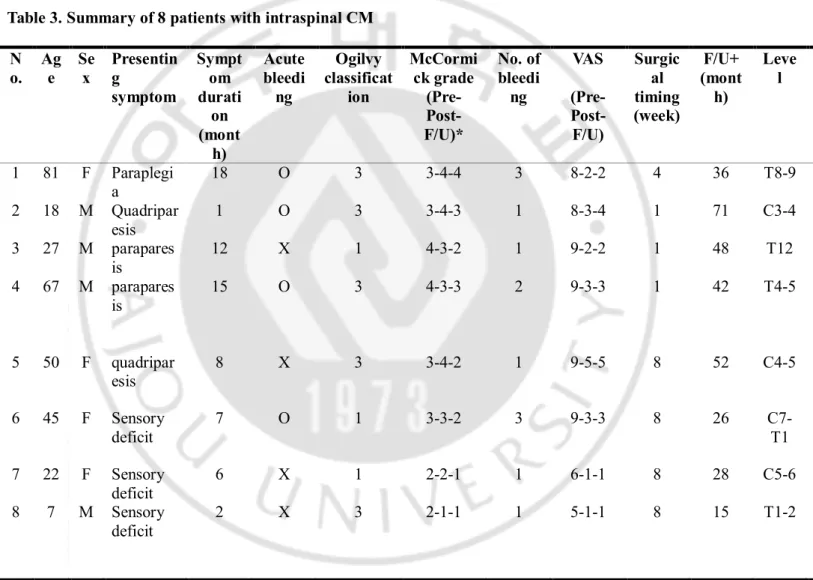
Table 3. Summary of 8 patients with intraspinal CM N o. Ag e Se x Presentin g symptom Sympt om durati on (mont h) Acute bleedi ng Ogilvy classificat ion McCormi ck grade (Pre- Post-F/U)* No. of bleedi ng VAS (Pre- Post-F/U) Surgic al timing (week) F/U+ (mont h) Leve l 1 81 F Paraplegi a 18 O 3 3-4-4 3 8-2-2 4 36 T8-9 2 18 M Quadripar esis 1 O 3 3-4-3 1 8-3-4 1 71 C3-4 3 27 M parapares is 12 X 1 4-3-2 1 9-2-2 1 48 T12 4 67 M parapares is 15 O 3 4-3-3 2 9-3-3 1 42 T4-5 5 50 F quadripar esis 8 X 3 3-4-2 1 9-5-5 8 52 C4-5 6 45 F Sensory deficit 7 O 1 3-3-2 3 9-3-3 8 26 C7-T1 7 22 F Sensory deficit 6 X 1 2-2-1 1 6-1-1 8 28 C5-6 8 7 M Sensory deficit 2 X 3 2-1-1 1 5-1-1 8 15 T1-2
No. : number, M : male, F : female, Pre-post- F/U : preoperative-postoperative-follow-up, CR : complete microsurgical resection
Discussion
CMs can occur throughout the central nervous system, but are most commonly located in the supratentorial compartment.11) The intramedullary location is relatively rare, but recent MR imaging studies
13
have demonstrated that lesions in this region are more common than originally thought.7, 19, 22) It is likely that the myelopathy previously diagnosed as idiopathic was caused by these intraspinal CMs, because these lesions were difficult to identify on computed tomography and myelography studies.6) Intraspinal spinal cord CMs are rare entities and account for approximately 5% of all CMs. .34) Patients without a family history of intraspinal CMs are more likely to present at an earlier age of onset than those with a family history.18, 36) Although a prevalence of females has been described in the literature with a female/male ratio of 2:1,27, 33, 41) Deutsch and coworkers described a female/male ratio of 1:1.5) There was no female predominance and female/male ratio was 1:1 in our study. Although spinal CMs have been identified in all age groups, symptomatic cases tend to be diagnosed in the 3rd to 4th decades of life.24) A mean age at presentation was 39.8 years in our study. Our result was consisted with what the recent literatures said.
Patients may have a diagnosis of asymptomatic intramedullary CM, although the classic presentation is either acute or chronic progressive clinical symptoms. Pathological studies have shown that CMs are composed of an aberrant ultrastructure and are characterized by thin-walled sinusoidal vascular channels that are dilated and contiguous.29, 35, 38) Repetitive intra- or paralesional microhemorrhages usually lead to slow growth over years and intermittent or chronic progressive symptoms are the normal course of the disease.8, 25, 32, 42) Thrombotic venous occlusion, compression of draining vessels, or sudden rupture of the amuscular wall with extralesional hematoma can lead to an acute increase in volume and sudden onset of clinical symptoms. Nevertheless, rapid clinical deterioration including sudden paraparesis or tetraparesis generally is a rare event and usually caused by massive space occupying bleeding in about 70% and acute decompensation due to mass effect, with previous minor. 3, 12, 14, 20) However, some patients have a combined presentation in which an acute deterioration is followed by neurological improvement. Subsequently, a gradual decline in function and worsening myelopathy occurs, which may be punctuated with recurrent acute hemorrhages. Although frequency of bleeding can deteriorate the clinical presentations, this is not statistically correlated with clinical outcomes and prognosis in our study.
CMs must remain in the differential diagnosis of intradural intramedullary lesions. The deposition of hemosiderin resulting in a hypointense rim around a mixed signal intensity leads to the pathognomonic appearance of CMs on T2-weighted MR imaging.6, 37) Infrequently, they may take a more homogeneous hyper- or hypointense appearance.37) In 24 (34%) of 70 patients (range 0–94% per series) in 6 series, venous malformations were observed.1, 4, 31, 39-40) Distinguishing CMs from multiple sclerosis on MR imaging can be difficult because inactive lesions may not enhance and can demonstrate mixed or hyperintense signal intensity. Clinical histories of progressive or recurrent episodes of the neurological decline seen with CMs also mimic multiple sclerosis; however, a distinguishing feature for these lesions in contrast to multiple sclerosis is a symptomatology that is referable to only 1 location of the neuraxis.9) In addition, demyelinating lesions will more consistently respond to steroids and tend to have more rapid changes in appearance on serial imaging. The broader differential includes spinal ependymomas, astrocytomas, metastatic disease, hemangioblastomas, spinal arteriovenous malformations (AVMs), and transverse myelitis. A lack of
14
enhancement can help distinguish CMs from most of these lesions, and cranial MR imaging demonstrating additional CMs can also help elucidate the diagnosis. Angiography will facilitate the diagnosis of a spinal AVM or hemangioblastoma.9)
Gross et al. recently suggested management algorithm for spinal intramedullary CMs according to the modes of clinical presentations and MR findings.9) Once spinal CM is diagnosed, the decision for microsurgical removal of spinal CMs of the slow progressive type is made individually due to variance of clinical constellations and the level of suffering. Possible postoperative morbidity and long-term prognosis should be considered exactly, when patients are advised and prepared for surgery. In contrast to this, surgical procedure in the cases of acute neurological deterioration is clearly indicated in the early stage.
The principal feasibility of surgical treatment has been accepted to complete resection. Because recurrent episodes of bleeding raise the risk for neurological deterioration and surgery for satisfaction of long-term results.26, 31) This hemorrhage rate for symptomatic intramedullary CMs has been reported as 1.4 to 4.5% per year.30, 42) 16)CMs have a significant rebleeding rate associated with further neurological decline. These vascular malformations lead to a progressive myelopathy that can be arrested by surgery.39) The neurological decline secondary to chronic myelopathy is not as reversible as the acute presentation, although others have reported general improvement in neurological function in all patients.2, 11, 21, 30, 34, 41) It may be that the recurrent hemorrhages irritate the normal spinal cord, so that it becomes gliotic and edematous. There are no alternatives in the treatment of CMs; surgery is the mainstay treatment
Optimal time for surgery is another critical factor to make a favorable clinical outcome. There are some argues for a posthemorrhagic effect that induces dysesthesia that is probably associated with perturbation to the spinothalamic tract, and may suggest that early surgical intervention may be important to prevent this pain syndrome, contrary to a previous study in which an optimal time for surgery of 4–6 weeks after hemorrhage was suggested.18, 42) In our series, massive intramedullary hemorrhages with cord swelling were seen in 2 patient. (Figure 2) Early surgical interventions were needed because they presented rapid neurologic deterioration. However, early surgical intervention has several risk features. The lesion is poorly demarcated from normal neural tissue. Surgical dissection and retraction are not easy to achieve because of extensive intramedullary hemorrhage and surrounding cord edema. During surgery total resection should be done to prevent recurrence and re-bleeding under preservation of the venous anomaly to avoid postoperative edema and thereby associated neurological deterioration. Incomplete surgical resection and additional injuries to spinal cord can be happened because of those conditions and good clinical outcome can not be expected. Our study showed statistical improvement of neurological status in Group II (delayed resection). This emphasizes that optimal surgical timing is important to make a favorable postoperative outcome. Possibly some patients can be treated conservatively, but prediction of re-bleeding and its result is difficult. Therefore we suggest that decision of operation was delayed for 4 weeks during conservative care.
15
Fig.2. Early resection A : Sagittal T2-weighted MR image showed massive intramedullary hemorrhage with
cord swelling in cervical segment. B, C: Axial MR images showed the lesion with hemorrhage located in the right lateral-dorsal portion of spinal cord. C : intraoperative photograph showed blood-tinged neural tissue with cavity which was creasted after removal of CM
Conclusion
The intramedullary CM is a relatively rare spinal lesion which has various clinical presentations. The principal feasibility of surgical treatment has been accepted to complete resection because recurrent episodes of bleeding raise the risk for neurological deterioration and surgery for satisfaction of long-term results. Nevertheless limitation of small number of cases, our study had reviewed considerations of surgery for CM. We suggest that a full understanding of clinical manifestations, optimal surgical timing and complete resection of intramedullary CM can guide to make a favorable clinical outcome.
16
Reference
1. Bian, Bertalanffy, Sun, Shen: Intramedullary cavernous malformations: clinical features and surgical technique via hemilaminectomy. Clin Neurol Neurosurg 111: 511-517, 2009.
2. Canavero, Pagni, Duca, Bradac: Spinal intramedullary cavernous angiomas: a literature meta-analysis.
Surg Neurol 41: 381-388, 1994.
3. Caruso, Galarza, Borghesi, Pozzati, Vitale: Acute presentation of spinal epidural cavernous angiomas: case report. Neurosurgery 60: E575-576; discussion E576, 2007.
4. Cosgrove, Bertrand, Fontaine, Robitaille, Melanson: Cavernous angiomas of the spinal cord. J
Neurosurg 68: 31-36, 1988.
5. Deutsch, Jallo, Faktorovich, Epstein: Spinal intramedullary cavernoma: clinical presentation and surgical outcome. J Neurosurg 93: 65-70, 2000.
6. Fontaine, Melanson, Cosgrove, Bertrand : Cavernous hemangiomas of the spinal cord: MR imaging.
Radiology 166: 839-841, 1988.
7. Gao, [Magnetic resonance imaging of occult intracranial malformations] Zhonghua Yi Xue Za Zhi 69: 375-377, 328, 1989.
8. Goyal, Singh, Gupta, Tatke: Spinal epidural cavernous haemangioma: a case report and review of literature. Spinal Cord 40: 200-202, 2002.
9. Gross, Du, Popp, Day: Intramedullary spinal cord cavernous malformations. Neurosurg Focus 29: E14, 2010.
10. Guirado, Taricco, Nobre, Couto Junior, Ribas, Meluzzi: Quality of life in adult intradural primary spinal tumors: 36-Item Short Form Health Survey correlation with McCormick and Aminoff-Logue scales. J
Neurosurg Spine: Oct 11, 2013.
11. Jallo, Freed, Zareck, Epstein, Kothbauer: Clinical presentation and optimal management for intramedullary cavernous malformations. Neurosurg Focus 21: e10, 2006.
12. Jo, Lee, Chung, Paeng, Kim, Yoon: Pure epidural cavernous hemangioma of the cervical spine that presented with an acute sensory deficit caused by hemorrhage. Yonsei Med J 47: 877-880, 2006.
13. Kharkar, Shuck, Conway, Rigamonti: The natural history of conservatively managed symptomatic intramedullary spinal cord cavernomas. Neurosurgery 60: 865-872; discussion 865-872, 2007.
14. Kivelev, Ramsey, Dashti, Porras, Tyyninen, Hernesniemi: Cervical intradural extramedullary cavernoma presenting with isolated intramedullary hemorrhage. J Neurosurg Spine 8: 88-91, 2008. 15. Kolias, Pal, Shivane, Ismail, Tyagi: Paediatric intramedullary spinal cord cavernous malformations:
case report and review of the literature. Clin Neurol Neurosurg 111: 784-788, 2009.
16. Kondziella, Brodersen, Laursen, Hansen: Cavernous hemangioma of the spinal cord - conservative or operative management? Acta Neurol Scand 114: 287-290, 2006.
17. Labauge, Bouly, Parker, Gallas, Emery, Loiseau: Outcome in 53 patients with spinal cord cavernomas.
17
18. Lu, Lawton: Clinical presentation and surgical management of intramedullary spinal cord cavernous malformations. Neurosurg Focus 29: E12, 2010.
19. Maslehaty, Barth, Petridis, Doukas, Mehdorn: Symptomatic spinal cavernous malformations: indication for microsurgical treatment and outcome. Eur Spine J 20: 1765-1770, 2011.
20. Mathews, Peck, Brant-Zawadzki: Brown-Sequard syndrome secondary to spontaneous bleed from postradiation cavernous angiomas. AJNR Am J Neuroradiol 29: 1989-1990, 2008.
21. McCormick, Michelsen, Post, Carmel, Stein: Cavernous malformations of the spinal cord.
Neurosurgery 23: 459-463, 1988.
22. Mehdorn, Stolke: Cervical intramedullary cavernous angioma with MRI-proven haemorrhages. J
Neurol 238: 420-426, 1991.
23. Minh: Cervicothoracic spinal epidural cavernous hemangioma: case report and review of the literature.
Surg Neurol 64: 83-85; discussion 85, 2005.
24. Mitha, Turner, Abla, Vishteh, Spetzler: Outcomes following resection of intramedullary spinal cord cavernous malformations: a 25-year experience. J Neurosurg Spine 14: 605-611, 2011.
25. Nagi, Megdiche, Bouzaidi, Haouet, Khouja, Douira: Imaging features of spinal epidural cavernous malformations. J Neuroradiol 31: 208-213, 2004.
26. Ogilvy, Louis, Ojemann: Intramedullary cavernous angiomas of the spinal cord: clinical presentation, pathological features, and surgical management. Neurosurgery 31: 219-229; discussion 229-230, 1992. 27. Pagni, Canavero, Forni: Report of a cavernoma of the cauda equina and review of the literature. Surg
Neurol 33: 124-131, 1990.
28. Robinson, Awad, Little: Natural history of the cavernous angioma. J Neurosurg 75: 709-714, 1991. 29. Robinson, Awad, Masaryk, Estes: Pathological heterogeneity of angiographically occult vascular
malformations of the brain. Neurosurgery 33: 547-554; discussion 554-545, 1993.
30. Sandalcioglu, Wiedemayer, Gasser, Asgari, Engelhorn, Stolke: Intramedullary spinal cord cavernous malformations: clinical features and risk of hemorrhage. Neurosurg Rev 26: 253-256, 2003.
31. Santoro, Piccirilli, Frati, Salvati, Innocenzi, Ricci: Intramedullary spinal cord cavernous malformations: report of ten new cases. Neurosurg Rev 27: 93-98, 2004.
32. Satpathy, Das, Das: Spinal epidural cavernous hemangioma with myelopathy: a rare lesion. Neurol
India 57: 88-90, 2009.
33. Scott, Barnes, Kupsky, Adelman: Cavernous angiomas of the central nervous system in children. J
Neurosurg 76: 38-46, 1992.
34. Spetzger, Gilsbach, Bertalanffy: Cavernous angiomas of the spinal cord clinical presentation, surgical strategy, and postoperative results. Acta Neurochir (Wien) 134: 200-206, 1995.
35. Tomlinson, Houser, Scheithauer, Sundt, Okazaki, Parisi: Angiographically occult vascular malformations: a correlative study of features on magnetic resonance imaging and histological examination. Neurosurgery 34: 792-799; discussion 799-800, 1994.
18
36. Tong, Deng, Li, Fu, Xu: Clinical presentation and surgical outcome of intramedullary spinal cord cavernous malformations. J Neurosurg Spine 16: 308-314, 2012.
37. Turjman, Joly, Monnet, Faure, Doyon, Froment: MRI of intramedullary cavernous haemangiomas.
Neuroradiology 37: 297-302, 1995.
38. Vanefsky, Cheng, Chang, Norbash, Snipe, Marks: Correlation of magnetic resonance characteristics and histopathological type of angiographically occult vascular malformations. Neurosurgery 44: 1174-1180; discussion 1180-1171, 1999.
39. Vishteh, Sankhla, Anson, Zabramski, Spetzler: Surgical resection of intramedullary spinal cord cavernous malformations: delayed complications, long-term outcomes, and association with cryptic venous malformations. Neurosurgery 41: 1094-1100; discussion 1100-1091, 1997.
40. Weinzierl, Krings, Korinth, Reinges, Gilsbach: MRI and intraoperative findings in cavernous haemangiomas of the spinal cord. Neuroradiology 46: 65-71, 2004.
41. Zentner, Hassler, Gawehn, Schroth: Intramedullary cavernous angiomas. Surg Neurol 31: 64-68, 1989. 42. Zevgaridis, Medele, Hamburger, Steiger, Reulen: Cavernous haemangiomas of the spinal cord. A
19 - 국문요약 -
척수내 해면 혈관종의 치료
아주대학교 대학원 의학과 이 철 규 (지도교수: 윤 수 한) 척수내 해면 혈관종은 중추 신경계에서 생기는 해면 혈관종의 약 3~5% 정도이며 전체 척수내 발생할 수 있는 혈관성 질병의 5~12% 정도이다. 척수내 해면 혈관종 치료 결정에 있어서 수술적 치료와 보존적 치료 사이에는 아직 논란의 여지가 남아 있는 실정이다. 신경학적인 증상을 나타내는 척수내 해면 혈관종의 수술적 치료는 수술 접근의 용이성, 환자의 전신 상태, 신경학적인 증상의 심각성 등을 고려해 환자에 따라 다르게 적용되고 있다. 2007 년 3 월부터 2012 년 3 월까지 본원에서 수술적 치료를 받은 척수내 해면 혈관종 환자를 대상으로 후향적 연구 방법으로 연구를 진행했다. 환자에 대한 병력 평가는 오길비 분류(Ogilvy classification)로 평가되었고 환자 증상에 대한 평가는 맥코믹 지수(Mccormick scale)로 평가되었다. 7 명의 환자는 후궁 성형술을 시행 받았고 한 명의 환자는 한 마디 척추체 제거술을 시행 받았다. 6 명의 환자(75%)는 척수의 배측(dorsal)에 종양이 있었고 2 명의 환자는 척수의 복측(ventral)에 종양이 위치해 있었다. 맥코믹 지수에 따라 4 명의 환자(50%)는 비슷한 결과를, 3 명의 환자는(37.5%) 호전된 결과를, 1 명의 환자는(12,5%) 악화된 결과를 나타냈다. 오길비 분류에 따라 3 명은 단계적으로 급격한 악화 양상을 보였고(class I), 1 명은 점진적인 악화를(class II), 4 명은 지속적인 급격한 악화 양상을 보였다.(class III)Group I (급성기에 수술한 환자군)에서는,평균 맥코믹 지수가 수술 전 3.0 ± 0.5 에서 외래 추적 관찰 기간 동안 3.25 ± 0.5 로 통계학적으로 유의한 차이가 없었다.(p > 0.05) Group II (아급성기에
20 수술한 환자군)에서는 평균 맥코믹 지수가 수술 전 2.5 ± 0.5 에서 외래 추적 관찰 기간 동안 1.5 ± 0.5 로 통계학적으로도 호전된 결과를 나타냈다. 평균 수술 전 통증 평가 점수(VAS)는 7.8 ± 1.5 (range 5-9)이었고 수술 직후 통증 평가 점수(VAS)는 2.5 ± 1.3 이었으며 외래 추적 관찰 기간 동안에는 2.6 ± 1.3 이었다. 평균 통증 평가 점수는 통계학적으로 유의한 호전된 결과를 나타냈다. (p < 0.05) 심각한 신경학적인 결손을 유발할 수 있는, 반복적인 척수내 해면 혈관종의 출혈은 장기적인 예후를 호전시키기 위해서 수술적 전적출술이 필요하며 수술 시기는 아급성기에 하는 것이 보다 적절할 것으로 사료된다. 환자 표본이 적음에도 불구하고 우리 연구에서는 척수내 해면 혈관종에 대한 이해 및 수술 시기, 전적출술의 필요성에 대해 긍정적인 결론을 가질 수 있었다.