Critical Role of Phospholipase C
␥1 in the Generation
of H
2
O
2
-evoked [Ca
2
ⴙ
]
i
Oscillations in Cultured Rat
Cortical Astrocytes
*
□SReceived for publication, February 23, 2006 Published, JBC Papers in Press, March 16, 2006, DOI 10.1074/jbc.M601726200
Jeong Hee Hong
‡, Seok Jun Moon
‡1, Hae Mi Byun
‡, Min Seuk Kim
‡, Hae Jo
‡, Yun Soo Bae
§, Syng-Ill Lee
‡,
Martin D. Bootman
¶2, H. Llewelyn Roderick
¶储3, Dong Min Shin
‡4, and Jeong Taeg Seo
‡5From the
‡Department of Oral Biology, Brain Korea 21 Project for Medical Science, Oral Science Research Center, Yonsei University
College of Dentistry, Seoul, 120-752, Korea,
§Division of Molecular Life Science, Center for Cell Signaling Research, Ewha Womans
University, Seoul 120-750, Korea,
¶Laboratory of Molecular Signalling, The Babraham Institute, Babraham, CB2 4AT Cambridge,
United Kingdom, and the
储Department of Pharmacology, University of Cambridge, CB2 1PD Cambridge, United Kingdom
Reactive oxygen species, such as the superoxide anion, H2O2, and
the hydroxyl radical, have been considered as cytotoxic by-products of cellular metabolism. However, recent studies have provided
evi-dence that H2O2serves as a signaling molecule modulating various
physiological functions. Here we investigated the effect of H2O2on
the regulation of intracellular Ca2ⴙsignaling in rat cortical
astro-cytes. H2O2triggered the generation of oscillations of intracellular
Ca2ⴙconcentration ([Ca2ⴙ]i) in a concentration-dependent manner
over the range 10 –100M. The H2O2-induced [Ca
2ⴙ]
ioscillations
per-sisted in the absence of extracellular Ca2ⴙand were prevented by
depletion of intracellular Ca2ⴙstores with thapsigargin. The H
2O2
-induced [Ca2ⴙ]
ioscillations were not inhibited by pretreatment with
ryanodine but were prevented by 2-aminoethoxydiphenyl borate and caffeine, known antagonists of inositol 1,4,5-trisphosphate
recep-tors. H2O2activated phospholipase C (PLC)␥1 in a dose-dependent
manner, and U73122, an inhibitor of PLC, completely abolished the
H2O2-induced [Ca
2ⴙ]
ioscillations. In addition, RNA interference
against PLC␥1 and the expression of the inositol
1,4,5-trisphos-phate-sequestering “sponge” prevented the generation of [Ca2ⴙ]
i
oscillations. H2O2-induced [Ca2ⴙ]
ioscillations and PLC␥1
phos-phorylation were inhibited by pretreatment with dithiothreitol, a
sulf-hydryl-reducing agent. Finally, epidermal growth factor induced H2O2
production, PLC␥1 activation, and [Ca2ⴙ]
i increases, which were
attenuated by N-acetylcysteine and diphenyleneiodonium and by the overexpression of peroxiredoxin type II. Therefore, we conclude
that low concentrations of exogenously applied H2O2 generate
[Ca2ⴙ]
ioscillations by activating PLC␥1 through sulfhydryl
oxi-dation-dependent mechanisms. Furthermore, we show that this mechanism underlies the modulatory effect of endogenously
pro-duced H2O2on epidermal growth factor-induced Ca2ⴙsignaling in
rat cortical astrocytes.
H2O2is a member of the reactive oxygen species (ROS),
6which cause
oxidative damage to cellular components such as lipids, nucleic acids,
and proteins. Therefore, H2O2has generally been considered to be
cyto-toxic and hazardous to living organisms. However, a growing body of
evidence suggests that H2O2serves as an intracellular signaling molecule
modulating various physiological functions (1). Cells possess mechanisms
that can rapidly synthesize and destroy H2O2in response to receptor
stimulation. For example, stimulation of membrane receptors of various growth factors, such as transforming growth factor-1, platelet-derived growth factor, and epidermal growth factor (EGF) triggers the rapid and
transient production of H2O2(2–5). H2O2generated in response to
receptor stimulation has been shown to play an important role in regu-lating various normal cell functions, such as cell proliferation, platelet aggregation, and vasodilation (6 – 8). In addition to this, exogenous
addition of H2O2at low concentrations affects the functions of various
ion channels and other proteins involved in signal transduction (8 –10).
Therefore, H2O2fulfills the prerequisites for being considered as a
gen-uine intracellular messenger.
Recently, a great deal of attention has focused on the sensitivity of the
mechanisms responsible for Ca2⫹mobilization in response to changes
in the cellular redox state. Ca2⫹plays a pivotal role in the regulation of a
diverse range of cellular functions, such as muscle contraction, secre-tion, synaptic plasticity, cell proliferasecre-tion, and cell death (11). Many
hormones and neurotransmitters increase intracellular Ca2⫹
concen-tration ([Ca2⫹]i) by mobilizing Ca2⫹from intracellular stores and by
inducing an influx from the extracellular space (12, 13). H2O2has been
shown to enhance the activity of L-type Ca2⫹channels (10). Peroxide
can also stimulate the mobilization of Ca2⫹in many cell types by
mod-ifying Ca2⫹release channels, such as TRPM2 (14), ryanodine receptors
(15), and inositol 1,4,5-trisphosphate (IP3)-dependent Ca2⫹channels
(16). In addition, H2O2can modify the activity of Ca2⫹pumps involved
in Ca2⫹homeostasis, such as the sarcoendoplasmic reticulum Ca2⫹
-ATPase (SERCA) (17) and plasma membrane Ca2⫹-ATPase (17).
Fur-thermore, enzymes involved in Ca2⫹signaling pathways, such as
phos-pholipase C (PLC)␥1 (18) and phospholipase D (19) are also targets.
However, most of the previous studies employed high concentrations of
H2O2, and it is questionable whether such diverse actions of H2O2on
*This work was supported in part by Korea Health 21 R & D Project, the Ministry of Health & Welfare, and Republic of Korea Grants 03-PJ1-PG3-21400-0005, 02-PJ1-PG3-20706-0001, and A050002. The costs of publication of this article were defrayed in part by the payment of page charges. This article must therefore be hereby marked
“advertise-ment” in accordance with 18 U.S.C. Section 1734 solely to indicate this fact.
□SThe on-line version of this article (available at http://www.jbc.org) contains
supple-mental Fig. S1.
1Present address: Dept. of Biological Chemistry, The Johns Hopkins University School of
Medicine, Baltimore, MD 21205-2196.
2Recipient of support from the Biotechnology and Biological Sciences Research Council. 3Recipient of a Royal Society for Research fellowship.
4To whom correspondence may be addressed. Tel.: 82-2-2228-3051; Fax:
82-2-364-1085; E-mail: dmshin@yumc.yonsei.ac.kr.
5To whom correspondence may be addressed: Tel.: 82-2-2228-3054; Fax:
82-2-364-1085; E-mail: jeong@yumc.yonsei.ac.kr.
6The abbreviations used are: ROS, reactive oxygen species; PLC␥1, phospholipase C␥1;
[Ca2⫹]i, intracellular Ca2⫹concentration; IP3, inositol 1,4,5-trisphosphate; EGF,
epi-dermal growth factor; SERCA, sarcoendoplasmic reticulum Ca2⫹ATPase; PMCA, plasma membrane Ca2⫹ATPase; PLC1, phospholipase C 1; DTT, dithiothreitol; DPI, diphenyleneiodonium; NAC, N-acetylcysteine; DCF, 2⬘,7⬘-dichlorofluorescein diac-etate; Prx II, peroxiredoxin type II; GFP, green fluorescent protein; siRNA, small inter-fering RNA; PSS, physiological salt solution; 2-APB, 2-aminoethoxydiphenyl borate; MEM, minimum essential medium; RNAi, RNA interference.
THE JOURNAL OF BIOLOGICAL CHEMISTRY VOL. 281, NO. 19, pp. 13057–13067, May 12, 2006 © 2006 by The American Society for Biochemistry and Molecular Biology, Inc. Printed in the U.S.A.
at Ewha Medical Library on July 5, 2016
http://www.jbc.org/
Downloaded from
at Ewha Medical Library on July 5, 2016
http://www.jbc.org/
Downloaded from
at Ewha Medical Library on July 5, 2016
http://www.jbc.org/
However, despite the lack of information about the physiological roles of ROS in astrocytes, NADPH oxidase was shown to be involved in the
generation of H2O2and cell survival in this cell type (25). Given the
widespread involvement of H2O2in modulating Ca2⫹signaling
cas-cades, it is tempting to speculate that astrocytes may also use redox
signaling to modify their Ca2⫹signaling.
Therefore, in the present study, we sought to investigate the roles of
H2O2in Ca2⫹signaling in cultured rat astrocytes. Our results indicate
that a low, physiologically relevant concentration of H2O2 (30 M)
induces [Ca2⫹]ioscillations in a PLC␥1- and IP3-dependent manner. In
addition, H2O2produced endogenously by EGF receptor stimulation is
involved in the modulation of Ca2⫹signaling in rat astrocytes.
EXPERIMENTAL PROCEDURES
Materials—H2O2, dithiothreitol (DTT), thapsigargin,
2-aminoe-thoxydiphenyl borate (2-APB), ryanodine, N-acetylcysteine (NAC), diphenyleneiodonium (DPI), caffeine, histamine, EGF, U73122, U73343, and 2⬘,7⬘-dichlorofluorescein diacetate (DCF) were purchased from Sigma. Minimum essential medium (MEM) containing 100 mg/liter sodium suc-cinate and 75 mg/liter succinic acid, trypsin-EDTA, Opti-MEM, penicillin, streptomycin, and fetal bovine serum were purchased from Invitrogen. Fura-2-acetoxymethyl ester was purchased from Teflabs (Austin, TX). All other chemicals were of reagent grade. The polyclonal antibody against PLC␥1 and monoclonal antibody against phosphotyrosine (PY783) of PLC␥1 were purchased from Upstate Biotechnology, Inc. (Lake Placid, NY). Anti-PLC1 polyclonal antibody was kindly provided by Dr. Shmuel Muallem (University of Texas Southwestern Medical Center, Dallas).
Cell Cultures—Primary cultures of cortical astrocytes were prepared from neonatal (12–24 h) Wistar rats. Briefly, cortices were dissected, and the tissues were minced and mechanically dissociated. Then the isolated cells were plated on 60-mm culture dishes and maintained at
37 °C in a humidified 5% CO2and 95% air for 2–3 weeks. For [Ca2⫹]i
measurements, cells were cultured on 0.01% poly-L-lysine-coated cover
glasses in 60-mm dishes for 7–10 days. The culture medium consisted of
MEM supplemented with 2 mMglutamine, 25 mMglucose, 100g/ml
penicillin, 25 ng/ml streptomycin, and 10% fetal bovine serum. The culture medium was replaced every 3 days. Cells were serum-starved for 2 days before each experiment.
Expression of IP3Sponge and Peroxiredoxin Type II (Prx
II)—Astro-cytes were transiently transfected with a green fluorescent protein
(GFP)-tagged high affinity (R441Q) or low affinity (K508A) IP3
-seques-tering sponge (26), or were cotransfected with Prx II (1g/ml; kindly
provided by Professor S. W. Kang, Ewha Womans University, Seoul,
Korea) and eGFP-N1 (1.2g/ml; Clontech) using Lipofectamine 2000
mMglucose (pH 7.4). Cells were then lysed at 4 °C in lysis buffer (150 mM
NaCl, 1% Triton X-100, 50 mMTris, 10 mMNaF, 1 mMEDTA, 1 mM
phenylmethylsulfonyl fluoride, 1g/ml aprotinin, 1 mMleupeptin, and
1 mMsodium orthovanadate) and centrifuged at 12,000 rpm for 10 min
at 4 °C. The samples were subjected to SDS-PAGE and subsequently transferred to nitrocellulose membranes. The membranes were incu-bated with specific antibodies against PLC␥1, phosphospecific tyrosine 783, PLC1, and actin, and the proteins were detected by ECL (Amer-sham Biosciences). The intensity of bands was quantified using Meta-Morph Analysis System (Universal Imaging Co., Downingtown, PA).
[Ca2⫹]
iMeasurements—For [Ca2⫹]imeasurements, attached cells
were loaded with fura-2 by incubation with 3.5M
fura-2-acetoxym-ethyl ester in PSS equilibrated with 100% O2for 40 min at room
temperature. The cells were then washed twice and rested for at least 20 min before use. The fura-2-loaded cells were mounted on the stage of an inverted microscope (Nikon, Tokyo, Japan) for imaging. The cells were superfused at a constant perfusion rate with the PSS.
In Ca2⫹-free solutions, CaCl2was omitted, and 1 mMEGTA was
added. The excitation wavelength was alternated between 340 and 380 nm, and the emission fluorescence was monitored at 510 nm with a CCD camera using MetaFluor system (Universal Imaging Co., Downingtown, PA). Fluorescence images were obtained at 4-s inter-vals. Background fluorescence was subtracted from the raw signals at
each excitation wavelength, and the values of [Ca2⫹]iwere calculated
using the equation described previously (28).
ROS Imaging—ROS levels were measured using the fluorescence probe DCF. In brief, cells were incubated for 5 min in the presence of 5g/ml DCF and washed in Hanks’ balanced salt solution. DCF fluores-cence was measured using a confocal laser-scanning microscope (Leica, Buffalo, NY) with an excitation wavelength at 488 nm and an emission at 525 nm. To avoid photo-oxidation of DCF, the fluorescence images were collected using a single rapid scan, and identical settings were used for all samples.
Data Analysis—The results are presented as mean⫾ S.E. Statistical analysis was performed by unpaired Student’s t test. p values lower than 0.05 were considered to be statistically significant.
RESULTS
H2O2Mobilizes Ca
2⫹in Cultured Rat Cortical Astrocytes in a
Con-centration-dependent Manner—The effect of H2O2on Ca2⫹
mobili-zation was examined in fura-2-loaded cultured rat astrocytes. Exposure
of the cells to H2O2at concentrations lower than 3Mfailed to increase
[Ca2⫹]iat least for 20 min (Fig. 1A). However, 10MH2O2was shown to
induce [Ca2⫹]ioscillations in 34.7⫾ 8.4% of the 18 tested cells, and the
at Ewha Medical Library on July 5, 2016
http://www.jbc.org/
number of the responding cells increased concentration-dependently
(60.9⫾ 5.8% for 30M, 84.9⫾ 10.2% for 100M, and 93.1⫾ 1.7% for
higher than 300MH2O2; Fig. 1D, n⫽ 18–20).
In general, as shown in Fig. 1, B and C, concentrations of H2O2less
than 30Minduced oscillations of [Ca2⫹]i, whereas doses higher than
300Mcaused spike and plateau type [Ca2⫹]iincreases. However, in
some cases, 30MH2O2also caused spike and plateau signals. The
average lag time between exposure to 10MH2O2and the generation of
Ca2⫹responses was 7.4⫾ 1.0 min, and it tended to decrease as the
concentration of H2O2was increased (5.1⫾ 0.5 min for 30M, 4.0⫾ 0.4
min for 100M, and 3.2⫾ 0.4 min for 300M). Because 30MH2O2did
not induce cell death (data not shown) and generally produced reliable
and reversible [Ca2⫹]ioscillations, we chose this concentration to
ana-lyze the mechanism by which peroxide-stimulated Ca2⫹ signaling
occurred in subsequent experiments.
Thapsigargin-releasable, IP3-sensitive Ca2⫹Stores Are Responsible for
H2O2-induced [Ca2⫹]iOscillations—To identify the source of the Ca2⫹
mobilization, Ca2⫹was removed from the bath solution and then 30M
H2O2was added. As shown in Fig. 2A, [Ca2⫹]ioscillations persisted in
the absence of extracellular Ca2⫹(n⫽ 18), suggesting that intracellular
Ca2⫹stores were the main source for H2O2-induced [Ca2⫹]i
oscilla-tions. Depletion of the intracellular Ca2⫹stores with thapsigargin, a
specific inhibitor of SERCA, prevented H2O2-induced [Ca2⫹]i
oscilla-tions (n⫽ 20), indicating that the intracellular Ca2⫹stores responsible
for the [Ca2⫹]ioscillations were thapsigargin-sensitive (Fig. 2B). To
examine whether the thapsigargin-sensitive intracellular Ca2⫹ store
was releasable by IP3 receptors or ryanodine receptors, cells were
exposed to 75M2-APB, 20 mMcaffeine (IP3receptor antagonists), or
100Mryanodine (a ryanodine receptor antagonist), prior to the
addi-tion of 30MH2O2. As shown in Fig. 2C and supplemental Fig. S1,
2-APB (n⫽ 17) and caffeine (n ⫽ 13) completely prevented the
gener-ation of [Ca2⫹]ioscillations evoked by H2O2. In contrast to this,
ryano-dine failed to inhibit the H2O2-induced [Ca2⫹]ioscillations (n⫽ 15),
although it effectively blocked the [Ca2⫹]iincreases induced by 500M
caffeine, a ryanodine receptor agonist (n⫽ 16; Fig. 2, D and E). These
results suggest that thapsigargin-releasable, IP3-sensitive Ca2⫹stores
are responsible for H2O2-induced Ca2⫹mobilization.
Activation of PLC␥1 Is Essential for the Generation of H2O2-induced
[Ca2⫹]
iOscillations—Because H2O2has been shown to activate PLC␥1
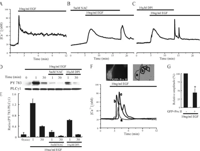
in some cell types (29, 30), we investigated whether PLC␥1 was phos-FIGURE 1. H2O2mobilizes Ca2ⴙin a dose-dependent manner in cultured rat cortical astrocytes. Cells were loaded with fura-2 as described under “Experimental Procedures,” and
changes in [Ca2⫹]iwere measured using ratiometric fluorescence imaging. Cells were exposed to 1M(A), 30M(B), or 1 mM(C) H2O2. For each panel, the trace is representative of
18 –20 cells in four or five independent experiments. D, the number of cells responding to H2O2is plotted as a function of the added H2O2concentrations (1, 3, 10, 30, 100, and 300
M). Data are expressed as the percentage of responding cells. Results are depicted as mean⫾ S.E.
Role of PLC
␥1 in H
2O
2-induced [Ca
2ⴙ]
i
Oscillations
at Ewha Medical Library on July 5, 2016
http://www.jbc.org/
phorylated following H2O2 stimulation of cultured rat astrocytes.
PLC␥1 possesses three tyrosine residues, 771, 783, and
Tyr-1254. Among them, Tyr-783 is known to be essential for IP3formation
(31). Therefore, a phosphospecific tyrosine 783 antibody was used to
detect the H2O2-induced phosphorylation of PLC␥1. As shown in Fig. 3,
Aand B, exposure of the astrocytes to various concentrations of H2O2
for 10 min induced PLC␥1 phosphorylation on tyrosine residue 783 in a
dose-dependent manner (n⫽ 6).
To clarify further the involvement of PLC␥1 in the generation of
[Ca2⫹]ioscillations, we used the PLC inhibitor U73122 and as control its
inactive analogue U73343. As shown in Fig. 3, C and D, 10MU73122, but
not 10MU73343, prevented the H2O2-evoked [Ca2⫹]ioscillations. The
frequencies of H2O2-induced [Ca2⫹]ioscillations were 6.5⫾ 1.2 peaks/20
min (n⫽14)inthepresenceofU73343and0.8⫾0.8peaks/20min(n⫽16)
in the presence of U73122. The effect of U73122 and U73343 on the H2O2
-induced PLC␥1 phosphorylation was also examined. As shown in Fig. 3, E
and F, the phosphorylation of PLC␥1 induced by H2O2was inhibited by 10
MU73122 but not by 10MU73343 (n⫽ 3).
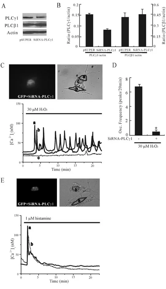
The involvement of PLC␥1 was further investigated by using RNA interference (RNAi). As shown in Fig. 4, A and B, transfection of siRNA-PLC␥1 suppressed the siRNA-PLC␥1 expression level, although that of PLC1
expression was not affected (n⫽ 5). To examine the functional
conse-quences of depletion of PLC␥1 by RNAi, cells were transfected with pSUPER (empty vector) or siRNA-PLC␥1 prior to measurement of FIGURE 2. H2O2mobilizes Ca2ⴙfrom thapsigargin-releasable, IP3-sensitive Ca2ⴙstores in cultured rat cortical astrocytes. A, cells were exposed to 30MH2O2in the absence
of extracellular Ca2⫹as indicated by the bars. B, cells were exposed to 1Mthapsigargin (Tg) followed by 30MH2O2in a nominally Ca2⫹-free solution. The absence of [Ca2⫹]iincrease
after H2O2application indicates that H2O2mobilizes Ca2⫹from a thapsigargin-sensitive Ca2⫹store. C and D, H2O2-induced [Ca2⫹]ioscillations were prevented by 75M2-APB but
not by 100Mryanodine. E, ryanodine blocked the [Ca2⫹]iincreases induced by 500Mcaffeine, a ryanodine receptor agonist. For each panel, the trace is representative of 15–20 cells
in three or four independent experiments.
at Ewha Medical Library on July 5, 2016
http://www.jbc.org/
[Ca2⫹]i. As shown in Fig. 4, C and D, transfection with GFP and
siRNA-PLC␥1 resulted in the prevention of Ca2⫹responses to H
2O2(n⫽ 10).
The frequencies of H2O2-induced [Ca2⫹]ioscillations were 6.8⫾ 0.26
peaks/20 min in GFP/PLC␥1-negative cells and 0.5 ⫾ 0.2 peaks/20 min in GFP/PLC␥1-positive cells. Transfection with GFP and pSUPER had
no effect on H2O2-induced [Ca2⫹]ioscillations (data not shown).
To confirm whether the transfection of siRNA-PLC␥1 had any effect
on the Ca2⫹response elicited by a PLC-activating agonist, we
stimu-lated the cells with histamine. As shown in Fig. 4, E and F, the
cotrans-fection of GFP and siRNA-PLC␥1 did not prevent the Ca2⫹responses to
histamine (n⫽ 6). These results indicate that PLC␥1 is necessary for the
generation of [Ca2⫹]ioscillations in response to H2O2.
In addition, we also observed that expression of the IP3sponge
com-pletely abrogated [Ca2⫹]ioscillations in response to H2O2(n⫽ 8; see
Fig. 5, A and B), although the expression of low affinity IP3sponge did
not prevent the H2O2-evoked [Ca2⫹]ioscillations (n⫽ 6; Fig. 5, C and
D). This result strongly suggests that IP3production through the
acti-vation of PLC␥1 is a critical step for the H2O2-induced generation of
[Ca2⫹]ioscillations.
Oxidation of a PLC␥1-associated Signaling Component Is Responsible for H2O2-induced [Ca
2⫹]
iOscillations—To examine whether the H2O2
-induced PLC␥1 phosphorylation and [Ca2⫹]
ioscillations were
attrib-uted to the sulfhydryl oxidation-dependent mechanisms, we treated
cells with 1 mMDTT, a sulfhydryl-reducing agent, 4 min prior to the
addition of H2O2. As shown in Fig. 6, A and B, the phosphorylation of
PLC␥1 by H2O2was prevented by pretreatment with 1 mMDTT (n⫽ 5).
Furthermore, the generation of [Ca2⫹]ioscillations by 30MH2O2was
also inhibited by 1 mMDTT (n⫽ 16; Fig. 6C). These data suggest that
FIGURE 3. Activation of PLC␥1 plays an essential role in H2O2-induced [Ca 2ⴙ]
ioscillations in cultured rat cortical astrocytes. A, cells were treated with the indicated
concen-trations of H2O2for 10 min, and lysates were subjected to immunoblot analysis with specific antibodies to phosphotyrosine (PY783) or PLC␥1. B, data from six experiments were
quantified, and the ratio (PY783/PLC1) was calculated. C, cells were exposed to 30 MH2O2in combination with U73343 or U73122 as indicated by the bars. D, quantitation of results
in C. Oscillation frequency (peaks/20 min) was counted (n⫽ 14–16). * indicates the difference of the oscillation frequencies between U73343- and U73122-treated groups ( p ⬍ 0.05).
E, cells were treated with the indicated concentrations of H2O2in combination with 10MU73343 or 10MU73122 for 10 min, and lysates were subjected to immunoblot analysis
with specific antibodies to phosphotyrosine (PY783) or PLC␥1. F, data from three experiments were quantified, and the ratio (PY783/PLC␥1) was calculated. Results are presented as mean⫾ S.E.
Role of PLC
␥1 in H
2O
2-induced [Ca
2ⴙ]
i
Oscillations
at Ewha Medical Library on July 5, 2016
http://www.jbc.org/
oxidation of a PLC␥1-associated signaling component is responsible for
the activation of PLC␥1 and generation of [Ca2⫹]
ioscillations by H2O2.
H2O2Might be Involved in the EGF-induced Activation of PLC␥1 and
Ca2⫹ Mobilization—Because it has been known that EGF elevates
[Ca2⫹]iand produces H2O2in fibroblasts (32), we investigated whether
H2O2was produced in response to EGF and if the endogenously
pro-duced H2O2was involved in PLC␥1 activation and [Ca2⫹]iincreases in
cultured rat cortical astrocytes. As shown in Fig. 7, A and B, EGF at a concentration of 10 ng/ml induced an increase in DCF fluorescence
inten-sity that was prevented by 5 mMNAC, indicating that EGF increased an
FIGURE 4. PLC␥1 RNA interference results in the
prevention of Ca2ⴙresponses to H 2O2in cul-tured rat cortical astrocytes. A, Western blots of
cell lysates transfected with pSUPER (empty vec-tor) or siRNA for PLC␥1 (siRNA-PLC␥1). Gels were transferred and blotted with specific antibodies to PLC␥1, PLC1, or actin. B, data from four experi-ments were quantified, and the ratios (PLC␥1/
Actin and PLC1/Actin) were calculated. C and E,
cells were cotransfected with GFP and siRNA-PLC␥1, and [Ca2⫹]
iwas measured. Note that
GFP-and siRNA-PLC␥1-transfected cells (designated as
c in C ) exhibited a lack of Ca2⫹response to 30M
H2O2(n⫽ 8; C), although GFP and siRNA-PLC␥1
transfection (designated as b in E) did not affect the Ca2⫹response to histamine (n⫽ 6; E ). D, the
oscillation frequency (peaks/20 min) in cells trans-fected with or without siRNA-PLC␥1 was counted during H2O2stimulation. Results are means⫾ S.E.
* indicates the difference of the oscillation fre-quencies between siRNA-PLC␥1-transfected and nontransfected groups ( p⬍ 0.05).
at Ewha Medical Library on July 5, 2016
http://www.jbc.org/
accumulation of ROS (n⫽ 5). The DCF fluorescence intensity was also
decreased by 10MDPI (an inhibitor of NADPH oxidase), suggesting that
NADPH oxidase, at least in part, participated in the EGF-triggered
gener-ation of ROS (n⫽ 5).
EGF induced a rapid transient peak increase in [Ca2⫹]ithat
subse-quently declined (n⫽ 15; Fig. 8A). However, pretreatment with 5 mM
NAC or 10MDPI attenuated the EGF-induced [Ca2⫹]iincreases, and
removal of NAC and DPI in the continued presence of EGF increased
[Ca2⫹]iagain (n⫽ 16–20; Fig. 8, B and C). The effect of NAC and DPI
on the EGF-induced activation of PLC␥1 was also investigated. As shown in Fig. 8, D and E, the immediate strong activation of PLC␥1 followed by a sustained weak activation was observed following EGF
stimulation, but in the presence of 5 mMNAC or 10MDPI the
activa-tion of PLC␥1 was greatly reduced (n ⫽ 3).
Because Prx II is a cellular peroxidase that eliminates endogenous
H2O2produced in response to growth factors such as EGF (33), we
examined whether the overexpression of Prx II also attenuated
EGF-induced [Ca2⫹]ioscillations. As shown in Fig. 8, F and G, overexpression
of Prx II decreased the amplitude of EGF-induced [Ca2⫹]iincrease by
about 57% (n⫽ 6). These data suggested that H2O2is generated by the
activation of NADPH oxidase and is subsequently involved in the
acti-vation of PLC and the eleacti-vation of [Ca2⫹]iduring EGF stimulation in
cultured rat astrocytes. DISCUSSION
In this study, we report that exogenous addition of low
concentra-tions of H2O2triggers [Ca2⫹]ioscillations through the activation of
PLC␥1 in cultured rat cortical astrocytes. H2O2-mediated elevation of
cytosolic Ca2⫹levels has been shown previously in various cell types
(32–36). However, in many cases, [Ca2⫹]iincreases were induced by
relatively high concentrations of H2O2, which are generally considered
cytotoxic. The physiologically relevant concentration range of H2O2,
which causes an acceleration of cellular functions in a variety of cell
types, is considered 1–100M, although it depends on cell type (1, 37).
In our system, we used 30MH2O2; it did not cause cell death (data not
shown), and its effect on [Ca2⫹]iwas reversible, suggesting that 30M
FIGURE 5. Expression of the IP3sponge prevents [Ca2ⴙ]ioscillations evoked by H2O2in cultured rat cortical astrocytes. Cells were transfected with GFP-tagged high affinity IP3
sponge (A) or low affinity IP3sponge (C), and [Ca2⫹]iwas measured. Note that GFP-tagged high affinity IP3sponge-transfected cell (designated as c in A) exhibited the abolition of
Ca2⫹response to 30MH2O2, whereas GFP-tagged low affinity IP3sponge-transfected cell (designated as a in C) did not. The oscillation frequencies (peaks/20 min) in cells transfected
with the high affinity (B) and low affinity IP3sponges (D) were counted during H2O2stimulation in 8 and 6 independent experiments, respectively. Results are means⫾ S.E. * indicates
the difference of the oscillation frequencies between the transfected and nontransfected groups ( p⬍ 0.05). N.S. indicates that there is no significant difference of the oscillation frequencies between the transfected and nontransfected groups ( p⬎ 0.05).
Role of PLC
␥1 in H
2O
2-induced [Ca
2ⴙ]
i
Oscillations
at Ewha Medical Library on July 5, 2016
http://www.jbc.org/
H2O2is close to the physiological concentration that modulates calcium
signaling in astrocytes.
Cellular Ca2⫹signals generally encode information in two different
modes as follows: frequency-modulated and amplitude-modulated
sig-nals (38). Frequency-modulated Ca2⫹signaling (i.e. [Ca2⫹]ioscillations)
is generally considered to have the highest fidelity, and many cells use this paradigm in response to low physiological concentrations of
ago-nists (39). H2O2has been shown to increase [Ca2⫹]iin many cell types,
but demonstrations of the generation of [Ca2⫹]ioscillations have been
rare. However, in this study we show that low concentrations of H2O2
generated [Ca2⫹]ioscillations in astrocytes although high
concentra-tions induced sustained increases in [Ca2⫹]i. It is more reasonable for
cells to use frequency-modulated Ca2⫹signaling to avoid cell damage,
especially when a prolonged period of Ca2⫹signaling is necessary. In
this regard, low concentration of H2O2is considered to be a reliable
intracellular messenger involved in Ca2⫹signaling.
In most nonexcitable cells, such as astrocytes, both Ca2⫹release from
intracellular Ca2⫹stores and Ca2⫹influx through Ca2⫹channels on the
plasma membrane are necessary for the generation and maintenance of
[Ca2⫹]ioscillations (40). In the present study, we demonstrate that H2O2
-induced [Ca2⫹]ioscillations were sustained in the absence of extracellular
Ca2⫹, indicating that intracellular Ca2⫹stores were primarily responsible
for the generation of [Ca2⫹]ioscillations. The two main intracellular
organelles containing large amounts of Ca2⫹are the endoplasmic
reticu-lum and mitochondria (41). Previously, both of these Ca2⫹stores were
shown to be involved in H2O2-induced [Ca2⫹]iincreases (17). However,
our data showed that depletion of intracellular Ca2⫹stores with
thapsigar-gin completely prevented the generation of H2O2-evoked [Ca2⫹]i
oscilla-tions, suggesting that the thapsigargin-sensitive endoplasmic reticulum
Ca2⫹store was the source of [Ca2⫹]ioscillations.
H2O2was also reported to be involved in the mobilization of Ca2⫹by
activating intracellular Ca2⫹channels, such as ryanodine receptors and
IP3receptors (15, 16). The effect of ROS on ryanodine receptors has
been well established. Sulfhydryl oxidation of ryanodine receptors has been reported to activate the channels (42, 43). However, in the present
study, a high concentration of ryanodine (100M), which blocked the
Ca2⫹mobilization induced by caffeine, a ryanodine receptor agonist,
failed to prevent the H2O2-induced [Ca2⫹]ioscillations. Instead, 2-APB
and caffeine, IP3-sensitive Ca2⫹channel inhibitors, blocked the H2O2
-induced [Ca2⫹]ioscillations. 2-APB is known to have several cellular
FIGURE 6. Oxidation of a PLC␥1-associated signaling component is responsible for the H2O2-induced [Ca 2ⴙ]
ioscillations in cultured rat cortical astrocytes. A, effect of DTT
(1 mM), a sulfhydryl-reducing agent, on H2O2-induced PLC␥1 phosphorylation. Cells were incubated with DTT for 4 min followed by an addition of indicated concentrations of H2O2
(1st to 3rd lanes) for 10 min or EGF (4th lane) for 2 min. Cell lysates were subjected to immunoblot analysis with specific antibodies to phosphotyrosine (PY783) or PLC␥1. B, quantitation of results in A. Ratio (PY783/PLC␥1) was calculated (n ⫽ 5). Results are presented as mean ⫾ S.E. Note that the dose-dependent increases in the ratio (PY783/PLC␥1) by H2O2shown
in Fig. 3B were prevented by DTT. C, the Ca2⫹responses of individual astrocytes to 30MH
2O2in the presence or absence of 1 mMDTT are shown. Note that 1 mMDTT completely
abolished H2O2-induced [Ca 2⫹]
ioscillations. The result is representative of 16 cells in three independent experiments.
FIGURE 7. EGF produces ROS by the activation of NADPH oxidase in cultured rat cortical astrocytes. A, astrocytes were loaded with DCF for 5 min (a– d). Cells were treated with 5 mMNAC (c) or 10MDPI (d) for 2 min followed by an addition of 10 ng/ml EGF (b– d) for 2 min. The fluorescence of DCF was subsequently visualized by confocal laser scanning microscopy. B, the DCF fluorescence was quantified, and the relative intensities were calculated by setting the fluorescence intensity of control cells to 100% (n⫽ 5). Results are means⫾ S.E.
at Ewha Medical Library on July 5, 2016
http://www.jbc.org/
targets; it blocks IP3-sensitive Ca2⫹ channels, SERCA activity, and
capacitative Ca2⫹entry channels (44, 45). However, the inhibitory effect
of 2-APB on [Ca2⫹]ioscillations was unlikely to be due to the inhibition of
SERCA, because the concentration of 2-APB we used in this study (75M)
was lower than the half-maximal inhibitory concentration for SERCA (91
M) (44). In addition, 75M2-APB did not show any evidence of [Ca2⫹]i
increase when applied to itself. This is in contrast to 1Mthapsigargin, a
specific SERCA inhibitor, which induced a rapid increase in [Ca2⫹]ias
shown in Fig. 2B. Inhibition of SERCA has been shown to be associated with
an increase in [Ca2⫹]iin most cell types. Furthermore, it is unlikely that the
effect of 2-APB on the [Ca2⫹]ioscillations was because of inhibition of
capacitative Ca2⫹entry, because as shown for the experiments performed
in the absence of extracellular Ca2⫹, Ca2⫹entry is not required for the
oscillations.
Caffeine also has several cellular targets. It can stimulate ryanodine receptors, inhibit both cAMP degradation and PLC activation, and
pre-vent IP3-sensitive Ca2⫹channel opening. However, the only feature that
caffeine and 2-APB share is their ability to antagonize IP3-mediated
Ca2⫹release. Therefore, although neither 2-APB nor caffeine are solely
selective for IP3-sensitive Ca2⫹channels, when used judiciously these
pharmacological agents can be used to reveal the specific involvement
of IP3signaling. The results obtained using 2-APB and caffeine support
the hypothesis that H2O2induced [Ca2⫹]ioscillations through
activa-tion of IP3-sensitive Ca2⫹channels.
H2O2may activate signaling components responsible for IP3
produc-tion. In some cell types, it has been reported that H2O2induces an
activation of PLC␥1 (29, 30). PLC␥1 is known to be recruited to the plasma membrane following activation of receptor tyrosine kinases and activated by a mechanism that relies on tyrosine phosphorylation (46). The phosphorylated PLC␥1 catalyzes the hydrolysis of
phosphatidyl-inositol 4,5-bisphosphate to produce diacylglycerol and IP3, leading to
the activation of protein kinase C and increases in [Ca2⫹]i, respectively
(47). Recently, PLC␥1 has been reported to be tyrosine-phosphorylated
following H2O2treatment and to protect cells from oxidant injury (18).
However, the involvement of H2O2-induced PLC␥1 activation in the
generation of [Ca2⫹]ioscillations, a typical form of physiological Ca2⫹
FIGURE 8. Endogenously produced H2O2enhances EGF-induced PLC␥1 activation and Ca2ⴙmobilization in cultured rat cortical astrocytes. The Ca2⫹responses of single
astrocytes to 10 ng/ml EGF in the absence (A) or presence of 5 mMNAC (B) or 10MDPI (C ) are denoted by the bars. Note that pretreatment with NAC or DPI suppressed Ca2⫹ responses to EGF, and the removal of NAC and DPI permitted the Ca2⫹responses. Results are representative of 15–20 cells in three or four independent experiments. D, the effect of 5 mMNAC or 10MDPI on 10 ng/ml EGF-induced PLC␥1 phosphorylation is shown. Cells were incubated with or without NAC or DPI for 2 min, and then 10 ng/ml EGF was added for
the indicated times. Cells were then lysed, and the lysates were subjected to immunoblot analysis with antibodies to phosphotyrosine (PY783) or PLC␥1. E, quantitation of results in
D. Ratio (PY783/PLC␥1) was calculated (n ⫽ 3). F, cells were cotransfected with GFP and Prx II, which eliminates H2O2, and [Ca2⫹]iwas measured. Note that GFP- and Prx II-transfected
cell (designated as a) exhibited reduced Ca2⫹response to EGF. G, the amplitude of [Ca2⫹]iincreases (nM) in cells transfected with or without GFP and Prx II was measured during EGF
stimulation in six independent experiments. Results are presented as mean⫾ S.E. * indicates the difference of the amplitudes of [Ca2⫹]iincreases between the Prx II-transfected and
nontransfected groups ( p⬍ 0.05).
Role of PLC
␥1 in H
2O
2-induced [Ca
2ⴙ]
i
Oscillations
at Ewha Medical Library on July 5, 2016
http://www.jbc.org/
2 2 i
Although the activation of PLC␥1 was shown to play a critical role in
the 30MH2O2-evoked generation of [Ca2⫹]ioscillations in our system,
we do not rule out the possibility that H2O2also increases the sensitivity
of IP3receptors to IP3in this cell type. Previously, thimerosal, which
catalyzes the oxidation of thiol groups, was reported to trigger [Ca2⫹]i
oscillations by increasing the affinity of IP3receptors for IP3(46, 49, 50).
In addition, Hu et al. (16, 51) showed that NADPH oxidase-derived
H2O2increased the sensitivity of intracellular Ca
2⫹stores to IP
3and
played a critical role in generating [Ca2⫹]ioscillations in human
endo-thelial cells stimulated by histamine. Taken together, our study and those of Hu et al. (16, 51) imply that both PLC␥1 activation and
increased sensitivity of IP3receptors may contribute to the generation of
[Ca2⫹]ioscillations.
By having observed the stimulatory effect of exogenous H2O2on
intracellular Ca2⫹signaling, we sought to determine whether H2O2was
produced by receptor stimulation and if endogenously generated H2O2
played a modulatory role in Ca2⫹signaling in rat astrocytes. Stimulation
of EGF receptors has been shown previously to induce both H2O2
pro-duction and [Ca2⫹]iincreases in fibroblasts (32). The EGF receptor
belongs to a family of transmembrane receptors with intrinsic tyrosine
kinase activity (52). The production of intracellular H2O2in response to
EGF was shown to require the activation of the Rac-NADPH oxidase signaling, whereas activation of PLC␥1 was regarded to be critical for
[Ca2⫹]iincreases (53–55). Although Rac was suggested to play a role in
the EGF-induced Ca2⫹signaling (32, 56), the role of H2O2in PLC␥1
activation during EGF stimulation has not been elucidated.
In this study, we showed that EGF receptor stimulation induced ROS
production, PLC␥1 activation, and [Ca2⫹]
ielevation, which were all
attenuated by the pretreatment with NAC, an ROS scavenger, or DPI, an NADPH oxidase inhibitor. These results indicated that ROS pro-duced via NADPH oxidase during EGF stimulation played a critical role
in the enhancement and maintenance of PLC␥1 and Ca2⫹responses in
rat cortical astrocytes. As far as we know, this is the first report to show the involvement of NADPH oxidase in EGF-mediated ROS generation and the regulatory role of endogenously produced ROS in
PLC␥1-acti-vated Ca2⫹signaling in astrocytes. Previous studies have revealed that
the predominant member of ROS produced by EGF stimulation was
H2O2, which played a key role in the EGF-induced protein tyrosine
phosphorylation and [Ca2⫹]iincreases (4, 32). Furthermore, we found
that overexpression of Prx II, which probably plays an important role in
eliminating H2O2generated in the cytosol, reduced the amplitude of
[Ca2⫹]iincrease evoked by EGF. Therefore, it is likely that the major
component of ROS, which is produced by EGF stimulation and
respon-sible for PLC␥1 activation and [Ca2⫹]
ielevation in our system, is H2O2.
REFERENCES
1. Dro¨ge, W. (2002) Physiol. Rev. 82, 47–95
2. Ohba, M., Shibanuma, M., Kuroki, T., and Nose, K. (1994) J. Cell Biol. 126, 1079 –1088
3. Sundaresan, M., Yu, Z. X., Ferrans, V. J., Irani, K., and Finkel, T. (1995) Science 270, 296 –299
4. Bae, Y. S., Kang, S. W., Seo, M. S., Baines, I. C., Tekle, E., Chock, P. B., and Rhee, S. G. (1997) J. Biol. Chem. 272, 217–221
5. Bae, Y. S., Sung, J. Y., Kim, O. S., Kim, Y. J., Hur, K. C., Kazlauskas, A., and Rhee, S. G. (2000) J. Biol. Chem. 275, 10527–10531
6. Sobey, C. G., Heistad, D. D., and Faraci, F. M. (1997) Stroke 28, 2290 –2294 7. Pignatelli, P., Pulcinelli, F. M., Lenti, L., Gazzaniga, P. P., and Violi, F. (1998) Blood 91,
484 – 490
8. Varela, D., Simon, F., Riveros, A., Jorgensen, F., and Stutzin, A. (2004) J. Biol. Chem.
279,13301–13304
9. Tournier, C., Thomas, G., Pierre, J., Jacquemin, C., Pierre, M., and Saunier, B. (1997)
Eur. J. Biochem. 244,587–595
10. Akaishi, T., Nakazawa, K., Sato, K., Saito, H., Ohno, Y., and Ito, Y. (2004) Neurosci.
Lett. 356,25–28
11. Berridge, M. J., Lipp, P., and Bootman, M. D. (2000) Nat. Rev. Mol. Cell Biol. 1, 11–21 12. Clapham, D. E. (1995) Cell 80, 259 –268
13. Putney, J. W., Jr., Broad, L. M., Braun, F. J., Lievremont, J. P., and Bird, G. S. (2001)
J. Cell Sci. 114,2223–2229
14. Wehage, E., Eisfeld, J., Heiner, I., Jungling, E., Zitt, C., and Luckhoff, A. (2002) J. Biol.
Chem. 277,23150 –23156
15. Favero, T. G., Zable, A. C., and Abramson, J. J. (1995) J. Biol. Chem. 270, 25557–25563 16. Hu, Q., Zheng, G., Zweier, J. L., Deshpande, S., Irani, K., and Ziegelstein, R. C. (2000)
J. Biol. Chem. 275,15749 –15757
17. Redondo, P. C., Salido, G. M., Rosado, J. A., and Pariente, J. A. (2004) Biochem.
Pharmacol. 67,491–502
18. Wang, X. T., McCullough, K. D., Wang, X. J., Carpenter, G., and Holbrook, N. J. (2001) J. Biol. Chem. 276, 28364 –28371
19. Servitja, J. M., Masgrau, R., Pardo, R., Sarri, E., and Picatoste, F. (2000) J. Neurochem.
75,788 –794
20. Hansson, E., and Ronnback, L. (2003) FASEB J. 17, 341–348 21. Newman, E. A. (2003) Trends Neurosci. 26, 536 –542
22. Tanaka, J., Toku, K., Zhang, B., Ishihara, K., Sakanaka, M., and Maeda, N. (1999) Glia
28,85–96
23. Wang, X. F., and Cynader, M. S. (2000) J. Neurochem. 74, 1434 –1442 24. Takuma, K., Baba, A., and Matsuda, T. (2004) Prog. Neurobiol. 72, 111–127 25. Liu, Q., Kang, J. H., and Zheng, R. L. (2005) Cell Biochem. Funct. 23, 93–100 26. Uchiyama, T., Yoshikawa, F., Hishida, A., Furuichi, T., and Mikoshiba, K. (2002)
J. Biol. Chem. 277,8106 – 8113
27. Lee, I. H., You, J. O., Ha, K. S., Bae, D. S., Suh, P. G., Rhee, S. G., and Bae, Y. S. (2004)
J. Biol. Chem. 279,26645–26653
28. Grynkiewicz, G., Poenie, M., and Tsien, R. Y. (1985) J. Biol. Chem. 260, 3440 –3450 29. Qin, S., Inazu, T., and Yamamura, H. (1995) Biochem. J. 308, 347–352
30. Tokmakov, A. A., Sato, K. I., Iwasaki, T., and Fukami, Y. (2002) Cell Calcium 32, 11–20
31. Kim, H. K., Kim, J. W., Ziberstein, A., Margolis, B., Kim, J. G., Schlessinger, J., and Rhee, S. G. (1991) Cell 65, 435– 441
32. Lee, Z. W., Kweon, S. M., Kim, S. J., Kim, J. H., Cheong, C., Park, Y. M., and Ha, K. S. (2000) Cell. Signal. 12, 91–98
33. Choi, M. H., Lee, I. K., Kim, G. W., Kim, B. U., Han, Y. H., Yu, D. Y., Park, H. S., Kim,
at Ewha Medical Library on July 5, 2016
http://www.jbc.org/
K. Y., Lee, J. S., Choi, C., Bae, Y. S., Lee, B. I., Rhee, S. G., and Kang, S. W. (2005) Nature
435,347–353
34. Pariente, J. A., Camello, C., Camello, P. J., and Salido, G. M. (2001) J. Membr. Biol. 179, 27–35
35. Smith, M. A., Herson, P. S., Lee, K., Pinnock, R. D., and Ashford, M. L. (2003) J. Physiol. (Lond.) 547, 417– 425
36. Kraft, R., Grimm, C., Grosse, K., Hoffmann, A., Sauerbruch, S., Kettenmann, H., Schultz, G., and Harteneck, C. (2004) Am. J. Physiol. 286, C129 –C137
37. Gamaley, I. A., and Klyubin, I. V. (1999) Int. Rev. Cytol. 188, 203–255 38. Berridge, M. J. (1997) Nature 386, 759 –760
39. Berridge, M. J., Bootman, M. D., and Lipp, P. (1998) Nature 395, 645– 648 40. Sneyd, J., Tsaneva-Atanasova, K., Yule, D. I., Thompson, J. L., and Shuttleworth, T. J.
(2004) Proc. Natl. Acad. Sci. U. S. A. 101, 1392–1396
41. Bootman, M. D., Collins, T. J., Peppiatt, C. M., Prothero, L. S., MacKenzie, L., De Smet, P., Travers, M., Tovey, S. C., Seo, J. T., Berridge, M. J., Ciccolini, F., and Lipp, P. (2001) Semin. Cell Dev. Biol. 12, 3–10
42. Xia, R., Stangler, T., and Abramson, J. J. (2000) J. Biol. Chem. 275, 36556 –36561 43. Meissner, G. (2004) Circ. Res. 94, 418 – 419
44. Missiaen, L., Callewaert, G., De Smedt, H., and Parys, J. B. (2001) Cell Calcium 29, 111–116
45. Peppiatt, C. M., Collins, T. J., Mackenzie, L., Conway, S. J., Holmes, A. B., Bootman, M. D., Berridge, M. J., Seo, J. T., and Roderick, H. L. (2003) Cell Calcium 34, 97–108 46. Falasca, M., Logan, S. K., Lehto, V. P., Baccante, G., Lemmon, M. A., and Schlessinger,
J. (1998) EMBO J. 17, 414 – 422
47. Berridge, M. J., Bootman, M. D., and Roderick, H. L. (2003) Nat. Rev. Mol. Cell Biol. 4, 517–529
48. Bootman, M. D., Taylor, C. W., and Berridge, M. J. (1992) J. Biol. Chem. 267, 25113–25119
49. Vanlingen, S., Sipma, H., De Smet, P., Callewaert, G., Missiaen, L., De Smedt, H., and Parys, J. B. (2001) Biochem. Pharmacol. 61, 803– 809
50. Bultynck, G., Szlufcik, K., Kasri, N. N., Assefa, Z., Callewaert, G., Missiaen, L., Parys, J. B., and De Smedt, H. (2004) Biochem. J. 381, 87–96
51. Hu, Q., Yu, Z. X., Ferrans, V. J., Takeda, K., Irani, K., and Ziegelstein, R. C. (2002)
J. Biol. Chem. 277,32546 –32551
52. Holbro, T., and Hynes, N. E. (2004) Annu. Rev. Pharmacol. Toxicol. 44, 195–217 53. Rhee, S. G. (1991) Trends Biochem. Sci. 18, 297–301
54. Sundaresan, M., Yu, Z. X., Ferrans, V. J., Sulciner, D. J., Gutkind, J. S., Irani, K., Goldschmidt-Clermont, P. J., and Finkel, T. (1996) Biochem. J. 318, 379 –382 55. Carpenter, G. (2000) BioEssays 22, 697–707
56. Peppelenbosch, M. P., Tertoolen, L. G., de Vries-Smits, A. M., Qiu, R. G., M’Rabet, L., Symons, M. H., de Laat, S. W., and Bos, J. L. (1996) J. Biol. Chem. 271, 7883–7886 57. Rabchevsky, A. G., Weinitz, J. M., Coulpier, M., Fages, C., Tinel, M., and Junier, M. P.
(1998) J. Neurosci. 18, 10541–10552
58. Levison, S. W., Jiang, F. J., Stoltzfus, O. K., and Ducceschi, M. H. (2000) Glia 32, 328 –337
Role of PLC
␥1 in H
2O
2-induced [Ca
2ⴙ]
i
Oscillations
at Ewha Medical Library on July 5, 2016
http://www.jbc.org/
Taeg Seo
Syng-Ill Lee, Martin D. Bootman, H. Llewelyn Roderick, Dong Min Shin and Jeong
Jeong Hee Hong, Seok Jun Moon, Hae Mi Byun, Min Seuk Kim, Hae Jo, Yun Soo Bae,
Oscillations in Cultured Rat Cortical Astrocytes
i
]
2+
-evoked [Ca
2
O
2
1 in the Generation of H
γ
Critical Role of Phospholipase C
doi: 10.1074/jbc.M601726200 originally published online March 16, 2006
2006, 281:13057-13067.
J. Biol. Chem.
10.1074/jbc.M601726200
Access the most updated version of this article at doi:
Alerts:
When a correction for this article is posted
•
When this article is cited
•
to choose from all of JBC's e-mail alerts
Click here
Supplemental material:
http://www.jbc.org/content/suppl/2006/03/17/M601726200.DC1.html
http://www.jbc.org/content/281/19/13057.full.html#ref-list-1
This article cites 58 references, 26 of which can be accessed free at
at Ewha Medical Library on July 5, 2016
http://www.jbc.org/
![FIGURE 3. Activation of PLC␥1 plays an essential role in H 2 O 2 -induced [Ca 2 ⴙ ]](https://thumb-ap.123doks.com/thumbv2/123dokinfo/5080707.74361/5.891.65.828.77.751/figure-activation-plc-plays-essential-role-induced-ca.webp)
![FIGURE 5. Expression of the IP 3 sponge prevents [Ca 2 ⴙ ] i oscillations evoked by H 2 O 2 in cultured rat cortical astrocytes](https://thumb-ap.123doks.com/thumbv2/123dokinfo/5080707.74361/7.891.68.835.80.707/figure-expression-sponge-prevents-oscillations-cultured-cortical-astrocytes.webp)
![FIGURE 6. Oxidation of a PLC␥1-associated signaling component is responsible for the H 2 O 2 -induced [Ca 2 ⴙ ]](https://thumb-ap.123doks.com/thumbv2/123dokinfo/5080707.74361/8.891.74.833.82.362/figure-oxidation-plc-associated-signaling-component-responsible-induced.webp)