RNA-Seq for Gene Expression Profiling of Human Necrotizing
Enterocolitis: a Pilot Study
Necrotizing enterocolitis (NEC) characterized by inflammatory intestinal necrosis is a major cause of mortality and morbidity in newborns. Deep RNA sequencing (RNA-Seq) has recently emerged as a powerful technology enabling better quantification of gene expression than microarrays with a lower background signal. A total of 10 transcriptomes from 5 pairs of NEC lesions and adjacent normal tissues obtained from preterm infants with NEC were analyzed. As a result, a total of 65 genes (57 down-regulated and 8 up-regulated) revealed significantly different expression levels in the NEC lesion compared to the adjacent normal region, based on a significance at fold change ≥ 1.5 and P ≤ 0.05. The most significant gene, DPF3 (P < 0.001), has recently been reported to have
differential expressions in colon segments. Our gene ontology analysis between NEC lesion and adjacent normal tissues showed that down-regulated genes were included in nervous
system development with the most significance (P = 9.3 × 10⁻⁷; Pcorr= 0.0003). In further
pathway analysis using Pathway Express based on the Kyoto Encyclopedia of Genes and Genomes (KEGG) database, genes involved in thyroid cancer and axon guidance were
predicted to be associated with different expression (Pcorr= 0.008 and 0.020, respectively).
Although further replications using a larger sample size and functional evaluations are needed, our results suggest that altered gene expression and the genes’ involved functional pathways and categories may provide insight into NEC development and aid in future research.
Keywords: Necrotizing Enterocolitis; RNA-Seq; Gene Expression Kyuwhan Jung,1* InSong Koh,2*
Jeong-Hyun Kim,3 Hyun Sub Cheong,4
Taejin Park,5 So Hyun Nam,6
Soo-Min Jung,7 Cherry Ann Sio,8†
Su Yeong Kim,6 Euiseok Jung,9
Byoungkook Lee,9 Hye-Rim Kim,9
Eun Shin,10 Sung-Eun Jung,11
Chang Won Choi,9 Beyong Il Kim,9
Eunyoung Jung,12 and Hyoung Doo Shin3,13
1Department of Surgery, Jeju National University Hospital, Jeju, Korea; 2Department of Physiology, College of Medicine, Hanyang University, Seoul, Korea; 3Research Institute for Basic Science, Sogang University, Seoul, Korea; 4Department of Genetic Epidemiology, SNP Genetics, Inc., Seoul, Korea; 5Department of Surgery, Gyeongsang National University Hospital, Jinju, Korea; 6Department of Surgery, Donga University Hospital, Busan, Korea; 7Department of Surgery, Konkuk University Medical Center, Konkuk University School of Medicine, Seoul, Korea; 8Department of Surgery, Seoul National University Bundang Hospital, Seongnam, Korea; 9Department of Neonatology, Seoul National University Bundang Hospital, Seongnam, Korea; 10Department of Pathology, Seoul National University Bundang Hospital, Seongnam, Korea; 11Department of Pediatric Surgery, Seoul National University Children’s Hospital, Seoul, Korea; 12Department of Surgery, Gyemyoung University Dongsan Hospital, Daegu, Korea; 13Department of Life Science, Sogang University, Seoul, Korea * Kyuwhan Jung and InSong Koh contributed equally
to this work.
† Current address: Department of Surgery, University of Santo Tomas Hospital, España Boulevard, Manila, Philippines
Received: 6 December 2016 Accepted: 27 January 2017 Address for Correspondence: Hyoung Doo Shin, PhD, DVM Department of Life Science, Sogang University, 35 Baekbeom-ro, Mapo-gu, Seoul 04107, Korea E-mail: [email protected]
Funding: This study was supported by grants from the Basic Science Research Program through the National Research Foundation of Korea (NRF), funded by the Ministry of Science, ICT and Future Planning (2014R1A1A1005096 and NRF-2012M3A9D1054450) and by the Ministry of Education, Science and Technology (2009-0093822).
https://doi.org/10.3346/jkms.2017.32.5.817 • J Korean Med Sci 2017; 32: 817-824
INTRODUCTION
Necrotizing enterocolitis (NEC) characterized by intestinal ischemia and necrosis is one of the most common gastrointestinal emergencies in premature infants with very low birth weights (VLBWs), i.e., those who weigh less than 1,500 g (1-3). NEC affects about 5%–14% of VLBW neonates. Since NEC is a life-threatening gastrointestinal dis-ease and an unpredictable surgical emergency, the overall mortality from NEC is high, ranging from 25% to 40% (4,5). Many researchers have tried to determine the precise pathophysiology of NEC by examining the various mechanisms that influence NEC development, such as the interaction between intraluminal microbiology and enteral nutrition and the change in inflammatory response by proinflammatory cytokines (6-11). Although several studies have been conducted and their associated hypotheses tested, substantial evidence to confirm risk factors (and effective therapies) for NEC has yet to be determined (12,13), other than prematurity and low birth weight (14). Although several studies suggest that genetic factors affect NEC development in pre-term infants with a potential susceptibility to NEC (15,16), the mechanisms underlying NEC are not fully understood. Previously, an NEC mice model has been used in many studies to establish the causes of or risk factors for human NEC due to several limita-tions in using human subjects, such as necrosis of human intestinal tissues and non-specific inflammatory changes (17). However, although the animal models contribute to our understanding of disease mechanisms, limitations of reliability and reproduc-ibility make the use of such models controversial (18,19).
Since transcripts are crucial intermediaries between the ge-nome and the proteome, the detection of global gene expres-sion is an important method for understanding molecular mech-anisms of diseases and specific functions of particular tissues. Although microarray-based gene expression analysis has be-come the primary high-throughput platform in the past decade, this method has certain limitations, including high background noise and low resolution (20). Recently, RNA sequencing (RNA-Seq), using high-throughput next-generation sequencing meth-ods, has become a powerful technology providing robust quan-tification of gene expression levels with a low background sig-nal and high resolution (21,22).
MATERIALS AND METHODS
Study subjects
Study subjects were collected from Seoul National University Bundang Hospital, Gyemyoung University Dongsan Hospital, National Gyeongsang University Hospital, Donga University Hospital, and CHA University Bundang Hospital in Korea. The study protocol was approved by the institutional review board of the hospital, and written informed consent was provided by guardians of all patients. Premature infants at less than 32 weeks of gestational age and less than 1,500 g birth weight were enrolled
in this study.
Two tissue sections (NEC lesion and adjacent normal regions) from the resected small bowel segment were collected as fol-lows: 1) an NEC lesion that showed perforation or necrosis; and 2) adjacent normal tissue. Next, the tissues were immediately stored in liquid nitrogen until RNA extraction at −80°C; a por-tion of the tissues was evaluated by histological examinapor-tion. Before RNA extraction, the mucosal layers of each tissue sec-tion were collected by the pediatric surgeon who performed the operation on the infants.
RNA isolation and quality control
Total RNA was extracted from the dissected tissue sections (NEC lesion and adjacent normal regions) with the Nucleo-Spin-RNA-II-Kit (Macherey-Nagel, Düren, Germany) according to the man-ufacturer’s protocol. RNA integrity and purity were analyzed with the ExperionTM automated electrophoresis system (Bio-Rad,
Hercules, CA, USA) with the Experion RNA StdSens chip. The mRNA was extracted and purified from the total RNA with the TruSeq stranded mRNA HT sample preparation kit (Illumina, Inc., San Diego, CA, USA), and this was followed by a purity check with Qubit 2.0 fluorometer (Life Technologies, Waltham, MA, USA) before proceeding to cDNA synthesis.
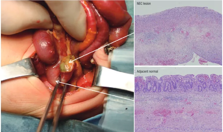
Fig. 1. NEC tissues for experiment and histological features. Arrows indicate 2 tissue sections (NEC lesion and adjacent normal regions) from the resected small bowel segment and each histological examination.
NEC = necrotizing enterocolitis.
NEC lesion
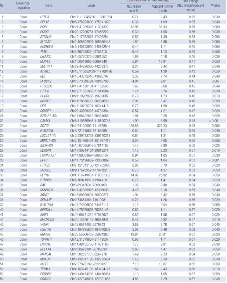
Table 1. Down-/up-regulated genes in comparison of NEC lesion and adjacent normal tissues No. Down-/up-
regulation Gene Locus
Expression level (FPKM, average) Fold change (NEC lesion/adjacent normal) P value NEC lesion (n = 5) Adjacent normal (n = 5) 1 Down HTR3A Chr11:113845796-113861034 0.71 2.43 0.29 0.030 2 Down CPLX2 Chr5:175223609-175311023 0.38 1.09 0.35 0.040 3 Down PCP4 Chr21:41239346-41301322 12.98 36.34 0.36 0.050 4 Down PCSK2 Chr20:17206751-17465222 0.39 1.08 0.36 0.030 5 Down ST8SIA6 Chr10:17362675-17496254 0.53 1.39 0.38 0.050 6 Down SLC5A7 Chr2:108602994-108630443 1.52 3.98 0.38 0.050 7 Down PCDHGA6 Chr5:140753650-140892546 0.44 1.11 0.40 0.050 8 Down TMIE Chr3:46742822-46752413 0.45 1.13 0.40 0.050 9 Down DNAJC6 Chr1:65730376-65881552 1.69 4.18 0.40 0.030 10 Down ELAVL4 Chr1:50513685-50667540 5.64 13.81 0.41 0.050 11 Down SULT4A1 Chr22:44220386-44258378 2.24 5.44 0.41 0.040 12 Down ATRNL1 Chr10:116853123-117708496 0.59 1.38 0.43 0.050 13 Down RET Chr10:43572516-43625797 3.38 7.74 0.44 0.040 14 Down SPOCK2 Chr10:73818791-73848790 4.04 9.01 0.45 0.007 15 Down PTGES3L Chr17:41120104-41132545 1.65 3.66 0.45 0.040 16 Down PTPRR Chr12:71031852-71314584 1.71 3.76 0.45 0.030 17 Down CHODL Chr21:19289656-19639687 0.79 1.73 0.46 0.010 18 Down NRXN3 Chr14:78636715-80334633 2.96 6.47 0.46 0.050 19 Down PIRT Chr17:10725791-10741418 0.73 1.58 0.46 0.050 20 Down SCUBE1 Chr22:43599228-43739394 0.57 1.21 0.47 0.020 21 Down BZRAP1-AS1 Chr17:56402810-56431088 1.07 2.25 0.48 0.020 22 Down CAMK4 Chr5:110559946-110820748 1.90 3.99 0.48 < 0.001 23 Down ACTG2 Chr2:74120092-74146780 155.56 322.23 0.48 0.050 24 Down FAM226B ChrX:72161567-72163589 0.54 1.11 0.49 0.040 25 Down LOC151174 Chr2:239133753-239140318 0.64 1.31 0.49 0.050 26 Down MBNL1-AS1 Chr3:151980404-151987415 2.53 5.05 0.50 0.050 27 Down IQCH-AS1 Chr15:67695948-67814182 1.46 2.90 0.50 0.020 28 Down CDK5R1 Chr17:30814104-30818271 1.13 2.23 0.51 0.010 29 Down FOXN3-AS1 Chr14:89883697-89886137 0.94 1.85 0.51 0.030 30 Down DPF3 Chr14:73136659-73360809 0.53 1.04 0.51 < 0.001 31 Down PTPRZ1 Chr7:121513158-121702090 2.98 5.70 0.52 0.020 32 Down SH3GL2 Chr9:17578952-17797122 0.73 1.37 0.53 0.050 33 Down SEPT6 ChrX:118749687-118827333 14.02 26.02 0.54 0.050 34 Down CTNND2 Chr5:10971951-11904110 0.76 1.41 0.54 0.020 35 Down BAI3 Chr6:69345631-70099403 1.35 2.48 0.54 0.040 36 Down RUNDC3A Chr17:42385926-42396038 4.46 8.18 0.55 0.040 37 Down ARNT2 Chr15:80696691-80890277 1.91 3.42 0.56 0.030 38 Down SEMA4F Chr2:74881354-74910981 0.71 1.26 0.56 0.020 39 Down FAM161B Chr14:74399694-74417117 1.15 2.04 0.56 0.050 40 Down RPS6KL1 Chr14:75370656-75389145 0.63 1.11 0.57 0.020 41 Down GRIP1 Chr12:66741210-67072925 0.89 1.56 0.57 0.020 42 Down MACROD2 Chr20:13976145-16033841 0.47 0.82 0.57 0.010 43 Down VAMP1 Chr12:6571403-6579843 3.88 6.76 0.57 0.040 44 Down C3orf70 Chr3:184795837-184870802 5.02 8.49 0.59 0.040 45 Down RBM38 Chr20:55966453-55984386 12.64 20.81 0.61 0.020 46 Down TSPAN11 Chr12:31079837-31149537 0.68 1.11 0.61 0.020 47 Down LRRC4C Chr11:40135750-41481186 1.73 2.81 0.62 0.030 48 Down BCL11A Chr2:60678301-60780633 2.83 4.47 0.63 0.050 49 Down MANEAL Chr1:38259773-38267278 1.49 2.33 0.64 0.050 50 Down MOXD1 Chr6:132617193-132722664 5.43 8.39 0.65 0.030 51 Down JAZF1 Chr7:27870192-28220437 7.10 10.87 0.65 0.050 52 Down TENM3 Chr4:183245136-183724177 1.61 2.43 0.66 0.010 53 Down PCDHB2 Chr5:140474236-140476964 1.10 1.65 0.67 0.050 54 Down PODXL2 Chr3:127348001-127391653 4.85 7.29 0.67 0.040
RNA-Seq and data analysis
RNA-Seq was carried out in order to identify differentially ex-pressed genes in the NEC lesion and adjacent normal tissues. The isolated mRNA from 4 μg total RNA using oligo-dT mag-netic beads was fragmented and primed at 94°C for 8 minutes, and then prepared for sequencing according to the protocol of the TruSeq stranded mRNA HT sample preparation kit (Illumi-na, Inc.). The resulting complementary DNA (cDNA) libraries were sequenced on the Illumina MiSeq system with 75 bp paired-end reads using the Illumina MiSeq sequencing kit v3 (150 cy-cles; Illumina, Inc.) following the manufacturer’s instructions. Image processing and base calling were performed using the Il-lumina Real Time Analysis Software RTA v1.9.35. FastQ sequenc-es were aligned to the human genome database (NCBI37/hg19) using TopHat v.2.0.12 with default parameters. The reads were mapped using the gene models as provided in the annotation GTF file (GRCh37.75). Gene expression values were determined using Cufflinks 2.2.1 release (http://cole-trapnell-lab.github.io/ cufflinks/), and the FPKM (fragments per kilobase of exon per million fragments mapped) values were calculated for each transcript. Cufflinks default settings were adopted, and FPKM values were computed by summing the values of different tran-scripts of the same gene. To identify differentially expressed genes, a fold change over 1.5 between any pairwise compari-sons was applied. For statistical analysis, data were examined by t-test.
Ethics statement
The present study protocol was reviewed and approved by the Institutional Review Board of Seoul National University Bundang Hospital (IRB No. B-1404-245-008). Informed consent was sub-mitted by guardians of all patients.
RESULTS
Patients
A total of 5 NEC patients, with median gestational ages of 26 weeks and 2 days, birth weight of 922 g, and birth height of 34.2 cm, were enrolled in this study. None of the patients showed other congenital anomalies in the perinatal period, and the moth-ers had no antenatal/perinatal problems except for premature delivery. The exploratory laparotomy was performed in the NEC patients at a median gestational age of 29 weeks and 1 day. The operations included a segmental resection with temporary ile-ostomy or primary anastomosis. All patients recovered from NEC and survived without complications. Following surgery, a histological examination of NEC lesion and adjacent normal tissues from the patients was performed (Fig. 1).
RNA-Seq analysis and gene expression comparison To investigate the gene expression profiles for NEC development in preterm infants, RNA-Seq analysis using the Illumina MiSeq system was performed. Mapping of sequences resulted in an average read count of 11.75 × 10⁶ (± 4.36 × 10⁶) in 10 RNA
sam-ples composed of those from 2 paired small-bowel sections (NEC lesion and adjacent normal tissues) from each of 5 NEC patients. Of the 23,972 tested genes, a total of 65 genes (57 down-regulated and 8 up-down-regulated) were observed to have significant-ly different expression levels in the comparison between NEC lesion and adjacent normal tissues, based on a significance at fold change ≥ 1.5 and P ≤ 0.05 (Table 1). As a housekeeping gene,
GAPDH was measured as 1,320.16 in NEC lesion and 1,255.20
in adjacent normal tissue (fold change = 1.05).
Among the differentially expressed genes in NEC lesions com-pared to the adjacent normal region, double PHD fingers 3 (DPF3,
P < 0.001) and calcium/calmodulin-dependent protein kinase
No. Down-/up- regulation Gene Locus
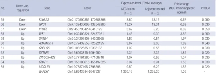
Expression level (FPKM, average) Fold change (NEC lesion/adjacent normal) P value NEC lesion (n = 5) Adjacent normal (n = 5) 55 Down KLHL23 Chr2:170590355-170608396 8.80 13.15 0.67 0.050 56 Down GPC4 ChrX:132435063-132549205 13.27 19.31 0.69 0.030 57 Down PRKCE Chr2:45879042-46415129 3.62 5.26 0.69 0.050 58 Up WT1 Chr11:32409321-32457081 1.48 0.39 3.82 0.050 59 Up SPAG4 Chr20:34203808-34208965 1.65 0.84 1.97 0.030 60 Up ADAMTS14 Chr10:72432558-72522195 3.87 2.05 1.89 0.040 61 Up GHRLOS Chr3:10322635-10335133 1.02 0.55 1.85 0.030 62 Up DSTNP2 Chr12:6993845-6994950 4.34 2.35 1.85 0.020 63 Up ZNF503-AS2 Chr10:77161285-77168740 1.07 0.68 1.57 0.030 64 Up GBAP1 Chr1:155183615-155197325 5.97 3.91 1.53 0.030 65 Up MCOLN1 Chr19:7587495-7598895 9.50 6.22 1.53 0.020 GAPDH* Chr12:6643584-6647537 1,320.16 1,255.20 1.05 -“-” in the fold change indicates down regulation.
NEC = necrotizing enterocolitis, Chr = chromosome. *GAPDH indicates a housekeeping gene.
Table 2.
Gene ontology analysis of differentially expressed genes in comparison of NEC lesion and adjacent normal tissues
Down-/up- regulation
Categor
y
Gene ontology categor
y Obser ved genes Obser ved genes number Expected genes number
Ratio of
enrichment
Significance of enrichment P value
Corrected
P value
Down
Biological process
Ner
vous system development
DPF3, LRRC4C, PCSK2, MACROD2, RET , PCP4, PCDHB2, CDK5R1, BCL11A, NRXN3, SEMA4F , SH3GL2, PTPRR, PTPRZ1, CPLX2, ARNT2, SPOCK2 17 4.71 3.61 9.3 × 10 −7 < 0.001 Down Biological process
Neuron cell-cell adhesion
CTNND2, CDK5R1, RET 3 0.03 91.44 4.1 × 10 −6 0.001 Down Biological process System development DPF3, LRRC4C, PCSK2, MACROD2, RET , CAMK4, PCP4, PCDHB2, CHODL, CDK5R1, RBM38, BCL11A, NRXN3, BAI3, SH3GL2, SEMA4F , SCUBE1, PTPRR, CPLX2, PTPRZ1, ARNT2, TMIE, SPOCK2 23 9.63 2.39 6.0 × 10 −6 0.002 Down Biological process Synaptic transmission NRXN3, SH3GL2, V AMP1, HTR3A, CPLX2, CAMK4, GRIP1, SLC5A7, PCDHB2, CTNND2 10 1.78 5.62 7.2 × 10 −6 0.003 Down Biological process
Single-multicellular organism process
DPF3, LRRC4C, PCSK2, MACROD2, RET , CAMK4, SLC5A7, PCP4, PCDHB2, CHODL, ACTG2, CDK5R1, RBM38, BCL11A, NRXN3, BAI3, SH3GL2, V AMP1, SEMA4F , SCUBE1, PTPRR, HTR3A, CPLX2, PTPRZ1, GRIP1, ARNT2, TMIE, SPOCK2, CTNND2 29 15.34 1.89 1.2 × 10 −5 0.004 Down Biological process
Multicellular organismal pro
-cess DPF3, LRRC4C, PCSK2, MACROD2, RET , CAMK4, SLC5A7, PCP4, PCDHB2, CHODL, ACTG2, CDK5R1, RBM38, BCL11A, NRXN3, BAI3, SH3GL2, V AMP1, SEMA4F , SCUBE1, PTPRR, HTR3A, CPLX2, PTPRZ1, GRIP1, ARNT2, TMIE, SPOCK2, CTNND2 29 15.43 1.88 1.4 × 10 −5 0.005 Down Biological process
Anatomical structure develop
-ment DPF3, LRRC4C, PCSK2, MACROD2, RET , CAMK4, PCP4, PCDHB2, CHODL, CDK5R1, RBM38, BCL11A, NRXN3, BAI3, SH3GL2, SEMA4F , SCUBE1, PTPRR, CPLX2, PTPRZ1, ARNT2, TMIE, SPOCK2, CTNND2 24 11.02 2.18 1.7 × 10 −5 0.006 Down Biological process
Multicellular organismal devel
-opment DPF3, LRRC4C, PCSK2, MACROD2, RET , CAMK4, PCP4, PCDHB2, CHODL, CDK5R1, RBM38, BCL11A, NRXN3, BAI3, SH3GL2, SEMA4F , SCUBE1, PTPRR, CPLX2, PTPRZ1, ARNT2, TMIE, SPOCK2, CTNND2 24 11.15 2.15 2.1 × 10 −5 0.007 Down Biological process Transmission of ner ve impulse NRXN3, SH3GL2, V AMP1, HTR3A, CPLX2, CAMK4, GRIP1, SLC5A7, PCDHB2, CTNND2 10 2.01 4.98 2.1 × 10 −5 0.007 Down Biological process
Multicellular organismal signal
-ing NRXN3, SH3GL2, V AMP1, HTR3A, CPLX2, CAMK4, GRIP1, SLC5A7, PCDHB2, CTNND2 10 2.05 4.87 2.5 × 10 −5 0.009 Down Biological process Neurotransmitter secretion NRXN3, SLC5A7, V AMP1, CPLX2 4 0.27 14.78 < 0.001 0.030 Down Biological process Cell-cell signaling NRXN3, SH3GL2, SEMA4F , V AMP1, HTR3A, CPLX2, CAMK4, GRIP1, SLC5A7, PCDHB2, CTNND2 11 3.00 3.66 < 0.001 0.030 Down Cellular component Synapse SEMA4F , V AMP1, SEPT6, HTR3A, CPLX2, GRIP1, CDK5R1, CTNND2 8 1.20 6.64 2.2 × 10 −5 0.002 Down Cellular component Synapse part GRIP1, SEMA4F , V AMP1, SEPT6, CTNND2, CDK5R1, HTR3A 7 0.91 7.68 3.0 × 10 −5 0.002 Up Biological process
Extracellular structure organi
-zation ADAMTS14, WT1 2 0.06 34.50 0.001 0.030 Up Biological process
Extracellular matrix organiza
-tion ADAMTS14, WT1 2 0.06 34.67 0.001 0.030
Gene ontology categories with corrected
P value of enrichment significance below 0.05 are shown.
NEC
=
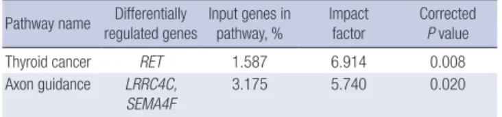
Table 3. Potential pathways affected by differentially expressed genes in comparisons of NEC lesion and adjacent normal tissues
Pathway name regulated genesDifferentially Input genes in pathway, % Impact factor Corrected P value
Thyroid cancer RET 1.587 6.914 0.008
Axon guidance LRRC4C,
SEMA4F 3.175 5.740 0.020
Corrected P value is obtained using the classical hypergeometric model (32). NEC = necrotizing enterocolitis.
IV (CAMK4, P < 0.001) showed relatively robust association
sig-nals of upregulation, whereas downregulated genes showed weak signals (Table 1). In addition, 3 genes (PCP4, PTPRR, and
WT1), which were recently reported as potential genes of NEC,
were also observed to be differentially expressed in the NEC le-sion (Table 1).
Ontology and pathway analyses of differentially expressed genes in NEC
To assess the biological functions of the differentially expressed genes in NEC lesions and adjacent normal tissues, this study per-formed a gene ontology analysis using the WEB-based GEne SeT AnaLysis Toolkit (http://bioinfo.vanderbilt.edu/webgestalt/). As a result, 16 gene ontology categories (14 in biological process-es and 2 in the cellular component) were predicted to affect NEC development in humans (Table 2), with the most significant sig-nal at nervous system development (P = 9.3 × 10⁻⁷; P
corr= 0.0003).
In additional pathway analysis using Pathway Express (http:// vortex.cs.wayne.edu/projects.htm) based on the Kyoto Ency-clopedia of Genes and Genomes (KEGG) database, genes in-volved in thyroid cancer and axon guidance showed significant associations (Table 3, Pcorr= 0.008 and 0.02, respectively).
DISCUSSION
Acquired conditions of diffuse necrotic injury to the intestinal segments are known to affect NEC development. Abnormal bac-terial colonization and formula feeding have also been impli-cated as predisposing factors for NEC in humans (23,24). In ad-dition, potential associations between NEC and environmental factors (such as microbiome, microbiome-intestinal reaction to breast milk or formula milk feeding, vaginal or cesarean section mode of delivery, and antibiotics) have been reported (10,11,23-25). Interestingly, a significant reduction of NEC in infants who were fed breast milk, compared to those who were fed formula, has been reported (26). Thus, many neonatologists have gone to great effort to manage the microbiome to prevent NEC de-velopment. Many neonatologists in Korea have changed their management protocols for preterm infants and observed a de-creased incidence of NEC during the last few years.
NEC development may be multifactorial with the interplay between intrinsic and extrinsic factors. In addition, the main
risk factor for NEC development in premature infants is thought to be intestinal immaturity (23,27), suggesting that intrinsic risk factors may be more important because premature infants have had a short exposure time to external environments. In this study, we hypothesized that global gene expression profiling may re-veal distinct genetic differences between NEC lesion and adja-cent normal region. Although candidate genes in this study did not reach great values of significance, several potential genes (such as DPF3 and CAMK4) with relatively robust association signals were identified (P < 0.001). These markers may have a role in NEC development. However, further replication and eval-uation studies are needed.
As noted, this study showed relatively robust association sig-nals at DPF3 and CAMK4. DPF3, which is known as an epigen-etic key factor for the development of heart and muscle tissue, has been reported to play a role in the neuronal differentiation process and also to take part in the disassembly of muscular fi-bers (28). In the different colon segments of Hirschsprung’s dis-ease, the gene product of DPF3 has been observed to be lowly expressed in a stenotic segment, whereas it is highly expressed in proximal anastomosis (29), suggesting that DPF3 may be dys-regulated in colonic diseases such as NEC. In the case of CAMK4, although a direct association between CAMK4 and NEC has not been reported, several connections in the literature related to necrosis can be found. In particular, CAMK4 was observed to be involved in the necrosis factor (NF)-kappaB mediated sig-naling pathway in human endothelial cells (30). These previous results and our findings suggest that dysregulated expressions of genes identified in this study may contribute to NEC devel-opment.
Recently, the first RNA-Seq for gene expression profiling in NEC was reported (31). This first RNA-Seq study used ileum tis-sues from preterm patients with other diseases for the control, and several genes associated with immune functions (in partic-ular, genes associated with Crohn’s disease) were identified as contributing factors to NEC development, together with other candidate genes. When compared to our results, PCP4 and
PT-PRR were overlapped; however, no connections in the literature
related to NEC or related cellular functions (such as necrosis) could be found. Therefore, further studies are required to eluci-date the association between these potential genes and NEC development.
So as to remove the heterogeneity of genetic background, this study excluded non-Korean parents. However, the study also has several limitations, such as insufficient sample size and lack of functional evaluation. The small sample size was due to the over-all decreased incidence of NEC. In addition, normal tissues from the small bowel segment in infants without NEC or related dis-eases would have been ideal for the comparison analysis; how-ever, it was limited to obtain these normal tissues. Although the first RNA-Seq analysis study of NEC used the ileum for the
nor-mal control (31), this study used adjacent nornor-mal tissues, and we do not rule out the possible effect of congenital diseases (such as small intestinal perforation, intestinal atresia, etc.).
In conclusion, despite study limitations, our preliminary results have identified potential involvements of certain genes (such as
DPF3 and CAMK4) in NEC development, suggesting that these
genetic factors, perhaps together with epigenetic factors such as microbiomes and breast milk, may have a role in NEC devel-opment in humans. Further validation studies are needed to de-termine clinical applications of these potential targets.
DISCLOSURE
The authors have no potential conflicts of interest to disclose.
AUTHOR CONTRIBUTION
Conceptualization: Jung K, Koh I, Shin HD. Data curation: Jung K, Park T, Nam SH, Jung SM, Sio CA, Kim SY, Jung E, Lee B, Kim HR, Jung SE, Choi CW, Kim BI, Jung E. Funding acquisition: In-vestigation: Jung K, Koh I, Shin HD. Koh I, Kim JH, Cheong HS, Shin E. Writing - original draft: Jung K, Koh I, Shin HD.
ORCID
Kyuwhan Jung http://orcid.org/0000-0001-5997-4975 InSong Koh http://orcid.org/0000-0001-6896-9748 Jeong-Hyun Kim http://orcid.org/0000-0003-4879-0075 Hyun Sub Cheong http://orcid.org/0000-0003-1749-6172 Taejin Park http://orcid.org/0000-0002-8508-2353 So Hyun Nam http://orcid.org/0000-0003-3757-4684 Soo-Min Jung http://orcid.org/0000-0002-2636-8028 Cherry Ann Sio http://orcid.org/0000-0003-0273-8239 Su Yeong Kim http://orcid.org/0000-0001-6463-0210 Euiseok Jung http://orcid.org/0000-0003-0693-5627 Byoungkook Lee http://orcid.org/0000-0001-5435-606X Hye-Rim Kim http://orcid.org/0000-0001-6445-5934 Eun Shin http://orcid.org/0000-0003-0624-8149 Sung-Eun Jung http://orcid.org/0000-0003-3155-2900 Chang Won Choi http://orcid.org/0000-0003-1911-0253 Beyong Il Kim http://orcid.org/0000-0003-0408-9279 Eunyoung Jung http://orcid.org/0000-0002-8884-1642 Hyoung Doo Shin http://orcid.org/0000-0003-1732-7838
REFERENCES
1. Bisquera JA, Cooper TR, Berseth CL. Impact of necrotizing enterocolitis on length of stay and hospital charges in very low birth weight infants. Pe-diatrics 2002; 109: 423-8.
2. Blakey JL, Lubitz L, Campbell NT, Gillam GL, Bishop RF, Barnes GL. En-teric colonization in sporadic neonatal necrotizing enterocolitis. J Pediatr
Gastroenterol Nutr 1985; 4: 591-5.
3. Hintz SR, Kendrick DE, Stoll BJ, Vohr BR, Fanaroff AA, Donovan EF, Poole WK, Blakely ML, Wright L, Higgins R; NICHD Neonatal Research Network. Neurodevelopmental and growth outcomes of extremely low birth weight infants after necrotizing enterocolitis. Pediatrics 2005; 115: 696-703.
4. Holman RC, Stoll BJ, Clarke MJ, Glass RI. The epidemiology of necrotizing enterocolitis infant mortality in the United States. Am J Public Health 1997;
87: 2026-31.
5. Lemons JA, Bauer CR, Oh W, Korones SB, Papile LA, Stoll BJ, Verter J, Tem-prosa M, Wright LL, Ehrenkranz RA, et al. Very low birth weight outcomes of the National Institute of Child health and human development neona-tal research network, January 1995 through December 1996. NICHD Neo-natal Research Network. Pediatrics 2001; 107: E1.
6. Hunter CJ, Upperman JS, Ford HR, Camerini V. Understanding the sus-ceptibility of the premature infant to necrotizing enterocolitis (NEC). Pe-diatr Res 2008; 63: 117-23.
7. Morowitz MJ, Poroyko V, Caplan M, Alverdy J, Liu DC. Redefining the role of intestinal microbes in the pathogenesis of necrotizing enterocolitis. Pe-diatrics 2010; 125: 777-85.
8. Neu J, Mihatsch W. Recent developments in necrotizing enterocolitis. JPEN J Parenter Enteral Nutr 2012; 36: 30S-35S.
9. Sharma R, Hudak ML. A clinical perspective of necrotizing enterocolitis: past, present, and future. Clin Perinatol 2013; 40: 27-51.
10. Sim K, Shaw AG, Randell P, Cox MJ, McClure ZE, Li MS, Haddad M, Lang-ford PR, Cookson WO, Moffatt MF, et al. Dysbiosis anticipating necrotiz-ing enterocolitis in very premature infants. Clin Infect Dis 2015; 60:
389-97.
11. Cortese R, Lu L, Yu Y, Ruden D, Claud EC. Epigenome-Microbiome cross-talk: a potential new paradigm influencing neonatal susceptibility to dis-ease. Epigenetics 2016; 11: 205-15.
12. Christensen RD, Lambert DK, Baer VL, Gordon PV. Necrotizing entero-colitis in term infants. Clin Perinatol 2013; 40: 69-78.
13. Stoll BJ. Epidemiology of necrotizing enterocolitis. Clin Perinatol 1994;
21: 205-18.
14. Kanto WP Jr, Wilson R, Breart GL, Zierler S, Purohit DM, Peckham GJ, El-lison RC. Perinatal events and necrotizing enterocolitis in premature in-fants. Am J Dis Child 1987; 141: 167-9.
15. Chan KY, Leung KT, Tam YH, Lam HS, Cheung HM, Ma TP, Lee KH, To KF, Li K, Ng PC. Genome-wide expression profiles of necrotizing entero-colitis versus spontaneous intestinal perforation in human intestinal tis-sues: dysregulation of functional pathways. Ann Surg 2014; 260: 1128-37.
16. Ng PC, Chan KY, Poon TC. Biomarkers for prediction and diagnosis of necrotizing enterocolitis. Clin Perinatol 2013; 40: 149-59.
17. Tian R, Liu SX, Williams C, Soltau TD, Dimmitt R, Zheng X, De Plaen IG. Characterization of a necrotizing enterocolitis model in newborn mice.
Int J Clin Exp Med 2010; 3: 293-302.
18. Hackam DG, Redelmeier DA. Translation of research evidence from ani-mals to humans. JAMA 2006; 296: 1731-2.
19. van der Worp HB, Howells DW, Sena ES, Porritt MJ, Rewell S, O’Collins V, Macleod MR. Can animal models of disease reliably inform human stud-ies? PLoS Med 2010; 7: e1000245.
20. Ioannidis JP, Allison DB, Ball CA, Coulibaly I, Cui X, Culhane AC, Falchi M, Furlanello C, Game L, Jurman G, et al. Repeatability of published mi-croarray gene expression analyses. Nat Genet 2009; 41: 149-55.
McPher-son A, Szcześniak MW, Gaffney DJ, Elo LL, Zhang X, et al. A survey of best practices for RNA-seq data analysis. Genome Biol 2016; 17: 13.
22. Wang Z, Gerstein M, Snyder M. RNA-Seq: a revolutionary tool for transcrip-tomics. Nat Rev Genet 2009; 10: 57-63.
23. Lin PW, Stoll BJ. Necrotising enterocolitis. Lancet 2006; 368: 1271-83.
24. Lucas A, Cole TJ. Breast milk and neonatal necrotising enterocolitis. Lan-cet 1990; 336: 1519-23.
25. Wang Y, Hoenig JD, Malin KJ, Qamar S, Petrof EO, Sun J, Antonopoulos DA, Chang EB, Claud EC. 16S rRNA gene-based analysis of fecal microbiota from preterm infants with and without necrotizing enterocolitis. ISME J
2009; 3: 944-54.
26. Repa A, Thanhaeuser M, Endress D, Weber M, Kreissl A, Binder C, Berger A, Haiden N. Probiotics (Lactobacillus acidophilus and Bifidobacterium infantis) prevent NEC in VLBW infants fed breast milk but not formula [corrected]. Pediatr Res 2015; 77: 381-8.
27. Claud EC. Neonatal necrotizing enterocolitis -inflammation and intesti-nal immaturity. Antiinflamm Antiallergy Agents Med Chem 2009; 8:
248-59.
28. Lange M, Kaynak B, Forster UB, Tönjes M, Fischer JJ, Grimm C, Schlesing-er J, Just S, Dunkel I, KruegSchlesing-er T, et al. Regulation of muscle development by DPF3, a novel histone acetylation and methylation reader of the BAF chromatin remodeling complex. Genes Dev 2008; 22: 2370-84.
29. Liu H, Luo Y, Li S, Wang S, Wang N, Jin X. Expression profiles of HA117 and its neighboring gene DPF3 in different colon segments of Hirschsprung’s disease. Int J Clin Exp Pathol 2014; 7: 3966-74.
30. You J, Peng W, Lin X, Huang QL, Lin JY. PLC/CAMK IV-NF-kappaB involved in the receptor for advanced glycation end products mediated signaling pathway in human endothelial cells. Mol Cell Endocrinol 2010; 320: 111-7.
31. Tremblay É, Thibault MP, Ferretti E, Babakissa C, Bertelle V, Bettolli M, Burghardt KM, Colombani JF, Grynspan D, Levy E, et al. Gene expression profiling in necrotizing enterocolitis reveals pathways common to those reported in Crohn’s disease. BMC Med Genomics 2016; 9: 6.
32. Draghici S, Khatri P, Tarca AL, Amin K, Done A, Voichita C, Georgescu C, Romero R. A systems biology approach for pathway level analysis. Genome Res 2007; 17: 1537-45.