Downloaded from http://journals.lww.com/md-journal by BhDMf5ePHKbH4TTImqenVG+tKk+L7/T70kP/uBlUid6sWuO/pDN0EALrkJkUj5dFI0pXkRU1JTM= on 09/15/2020 Downloadedfrom http://journals.lww.com/md-journalby BhDMf5ePHKbH4TTImqenVG+tKk+L7/T70kP/uBlUid6sWuO/pDN0EALrkJkUj5dFI0pXkRU1JTM=on 09/15/2020
Intramedullary nailing for subtrochanteric fracture
in autosomal dominant Type II osteopetrosis
Case report of 2 patients
Junyoung Kim, MD
a, Young Chang Park, MD
b, Hyun-Soo Moon, MD
a, Woo Sung Do, MD
a,
Kyu Hyun Yang, MD, PhD
a,∗Abstract
Rationale:Autosomal dominant type II (AD II) osteopetrosis is a rare inheritable metabolic bone disease characterized by hard but brittle bone and a narrow medullary canal. Intramedullary nailing (IMN) is a difficult but attractive option for the treatment of subtrochanteric fractures in patients with AD II osteopetrosis.
Patient concerns and diagnosis:Two patients with AD II osteopetrosis sustained subtrochanteric fractures after a fall.
Interventions:IMN was performed through the sequential use of instruments such as a 4.9-mm drill bit, small reamer, and larger reamer for over-reaming.
Outcomes:In thefirst case, IMN left some gap at the fracture site. Dynamization was performed to treat the delayed union at 6 months postoperatively. The fracture healed at 10 months after the dynamization. In the second case, IMN was successful without a gap, and the fracture healed at 8 months.
Lessons:Although IMN is difficult to perform owing to partial obliteration of the medullary canal in AD II osteopetrosis, it can be
performed with sequential widening of the medullary canal using various instruments. In addition, the fracture gap should not be left uncorrected during IMN to attain fracture union.
Abbreviations: AD= autosomal dominant, IMN = intramedullary nailing.
Keywords:intramedullary nailing, osteopetrosis, subtrochanteric fracture ;
1. Introduction
Autosomal dominant type II (AD II) osteopetrosis is a rare inheritable metabolic bone disease caused by mutation in chloride channel 7 genes and osteoclast dysfunction.[1,2]It is characterized
by hard but brittle bone, a narrow medullary canal, and a subtrochanteric fracture.[2]Owing to the biomechanical
superiori-ty of intramedullary nailing (IMN) in subtrochanteric fracture fixation, IMN is an attractive option. In ordinary IMN, the ball-tip
guide wire passes through the open medullary canal without difficulties, facilitating the reduction and reaming processes. Although IMN is the treatment of choice for subtrochanteric fractures, medullary canal deformities with obstruction and osteopetrotic bone hardness preclude ordinary IMN.[3]
Although anecdotal case reports have described the surgical management of osteopetrotic subtrochanteric femoral fracture, the treatment strategies varied among them.[4–6]In the existing case reports in the literature, IMN was rarely performed and led to unsatisfactory results due to insufficient reaming of the medullary canal. To address these issues, we report an IMN surgical technique step-by-step for patients with AD II osteope-trotic subtrochanteric fracture.
2. Case Report
2.1. Case 1
A 26-year-old man who was diagnosed as having AD II osteopetrosis at 10 years of age sustained a displaced subtrochanteric fracture without comminution after a fall. Radiography of the pelvis and bilateral femurs revealed generalized osteosclerosis with “bone-within-bone” structures and narrow medullary canals. As the intramedullary canal was traceable on plain radiography and computed tomography, we planned an IMN (Fig. 1). The patient was positioned on the fracture table under general anesthesia. The proximal fragment was derotated internally using long curved hemostatic forceps to obtain a good anteroposterior hip image.[7]A longitudinal skin
incision was made from the palpable tip of the greater trochanter and extended proximally for approximately 5 cm. The entry point was prepared just medial to the tip of the greater trochanter.
Editor: Maya Saranathan.
Written informed consent was obtained from the patients for publishing this case report, including the images.
The authors have no funding and conflicts of interest to disclose. The datasets generated during and/or analyzed during the current study are publicly available.
a
Department of Orthopedic Surgery, Gangnam Severance Hospital, Yonsei University College of Medicine, Gangnam-gu, Seoul,b
Department of Orthopedic Surgery, International St. Mary’s Hospital, Catholic Kwandong University College of Medicine, Seo-gu, Incheon, Republic of Korea.
∗
Correspondence: Kyu Hyun Yang, Department of Orthopedic Surgery, Gangnam Severance Hospital, Yonsei University College of Medicine, 211 Eonju-ro, Gangnam-gu, Seoul 06273, Republic of Korea (e-mail: [email protected]). Copyright© 2020 the Author(s). Published by Wolters Kluwer Health, Inc. This is an open access article distributed under the Creative Commons Attribution License 4.0 (CCBY), which permits unrestricted use, distribution, and reproduction in any medium, provided the original work is properly cited. How to cite this article: Kim J, Park YC, Moon H-S, Do WS, Yang KH. Intramedullary nailing for subtrochanteric fracture in autosomal dominant Type II osteopetrosis: case report of 2 patients. Medicine 2020;99:32(e21648). Received: 9 August 2019 / Received infinal form: 29 May 2020 / Accepted: 18 June 2020
http://dx.doi.org/10.1097/MD.0000000000021648
Clinical Case Report
Medicine
®
OPEN
After placement of the 3.2-mm threaded guide pin on the starting point, the entry point was double-checked in the anteroposterior and axial views (Fig. 2). This 3.2-mm guide pin could enter only a few centimeters because of the bone hardness in the trochanteric area. An 8-mm starting reamer was used to enlarge the entry site for a few centimeters along the guide pin. We then used a 4.9-mm drill bit to open the remnant medullary canal in the proximal fragment. A 4.9-mm drill bit was replaced with a 3.2-mm guide pin. The proximal fragment was reamed with a 14.5-mm tapered (trochanteric) reamer to accommodate a 13-mm nail body.
We continued to ream the distal fragment with a 4.9-mm drill bit that followed the remnant intramedullary canal. Breakage of the drill bit makes IMN impossible, so it must be advanced with special care and kept in the center of the canal. After enlarging the remnant medullary canal, a ball-tip guide wire could pass the canal. Reaming was started at 7 mm andfinished at 11.5mm in 0.5-mm increments. The reamer must advance slowly in maximum revolutions per minute with frequent saline irrigation. This procedure was repeated every 2 cm until it reached the distal
one-third of the medullary canal. Over-reaming of 1.5–2mm is necessary to advance the nail manually. Forceful impaction on the nail with a hammer may break the brittle bone; thus, it is not recommended. After nail insertion, proximal interlocking screws were inserted through the targeting guide without difficulty. However, the insertion of a distal interlocking screw was quite difficult by free hand technique owing to the hardness of the osteopetrotic bone, which caused skidding of the drill bit. The postoperative radiograms showed that we made a mistake and left a gap in the fracture site. It was caused by contact between the transitional part of the nail and the distal fragment (the arrow in Fig. 3A). The proximal part of the distal fragment was not reamed enough to accept the thickened portion (shaft-body transitional part) of the nail. As delayed union was evident at 6 months postoperatively, a dynamization procedure was per-formed (Fig. 3B, C). We exposed the proximal end of the nail, assembled a targeting guide, and removed the proximal and distal interlocking screws except for the one in the distal oval hole. The nail-targeting guide complex was pulled back until the fracture gap was closed. This movement also moved the distal locking screw to the dynamic locking mode (screw moved to the distal end of the oval hole). We then reinserted the proximal interlocking screws. Callus started to form 3 months after the dynamization. Bony union was achieved 10 months after the dynamization (16 months after the injury), and remodeling was completed 30 months after the injury (Fig. 3D, E).
2.2. Case 2
A 70-year-old woman was transferred to the emergency department because of right hip pain after a fall. A transverse subtrochanteric fracture was identified at the lower level of the lesser trochanter, without comminution, in the right femur (Fig. 4A). An incomplete subtrochanteric fracture was also identified at the same level in the left femur (Fig. 4D). The incomplete fracture started from the lateral femoral cortex and extended to the medial cortex just like an atypical femoral fracture. Prophylactic IMN was indicated on the left side and performed in the dynamic locking mode uneventfully using the technique introduced earlier. The nail was inserted in external rotation to overcome the femoral bowing.[8]The fracture achieved complete union and remodeled 8 months postoperatively (Fig. 4E). However, a short proximal fragment and associated femoral bowing precluded the IMN on the right side, and open reduction and internal
Figure 1. Typical radiography and computed tomography images of the pelvis and femur in a case of autosomal type II osteopetrosis. Generalized osteosclerosis with “bone-within-bone” structures are shown. Inlets, axial computed tomography images matched at the level of the femur.
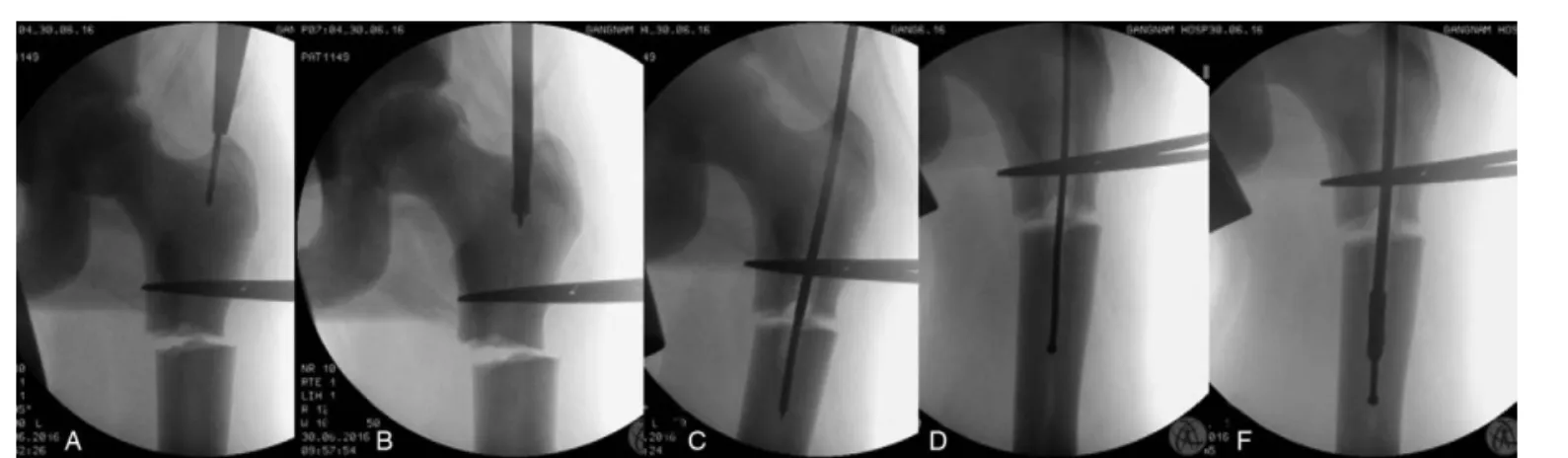
Figure 2. Sequential processes of medullary canal reaming. A. After placement of the 3.2-mm threaded guide pin on the starting point, the entry point was double-checked in the anteroposterior and axial views. B. An 8-mm starting reamer was used to enlarge the entry site for a few centimeters along the guide pin. C. A 4.9-mm-long drill bit was used to enlarge the remnant intramedullary canal. D. After enlarging the remnant medullary canal, a ball-tip guide wire could pass the canal. F. Reaming was started from 7 mm and completed at 11.5 mm in 0.5-mm increments.
Kim et al. Medicine (2020) 99:32 Medicine
Figure 4. Bilateral subtrochanteric fractures in a 70-year-old woman with autosomal dominant type II osteopetrosis. A and D. A transverse subtrochanteric fracture at the lower level of the lesser trochanter in the right femur and incomplete fracture in the left femur are shown. B, C. Open reduction and platefixation were performed on the right side. The peripheral callus is visible on plain radiography and computed tomography at 20 months postoperatively, but the fracture line remained because of delayed bone remodeling. E. The incomplete fracture line disappeared 8 months after prophylactic nailing on the left side.
Figure 3. A Subtrochanteric fracture in a 26-year-old man with autosomal dominant type II osteopetrosis. A. A subtrochanteric fracture was treated with intramedullary nailing. However, a gap in the fracture site occurred because of contact between the transitional part of the nail and the distal fragment (arrow). B. Delayed union was evident at 6 months after the operation, and dynamization (DNZ) was performed. C. Callus formation was observed at 3 months after the DNZ. D. The fracture attained union 10 months after the DNZ. F. Thefinal radiograph 24 months after the DNZ shows complete union and remodeling of the callus. POD, postoperative day.
Kim et al. Medicine (2020) 99:32 www.md-journal.com
fixation with a locking plate and screws were performed (Fig. 4B). A periosteal callus was formed at the periphery of the fracture site, but the main fracture line did not disappear even at 20 months postoperatively, probably owing to the delayed callus remodeling in osteopetrosis (Fig. 4C).
3. Discussion
Deformity of the long bone and an associated deformity of the medullary canal are usually contraindications for IMN. How-ever, IMN with or without an osteotomy is sometimes the only reliable option in cases of heritable bone disease such as osteogenesis imperfecta and X-linked hypophosphatemic rick-ets.[9,10]In AD II osteopetrosis, the long bones are usually straight
but have only traceable amounts of medullary canal. Despite the narrow medullary canal, closed IMN was possible owing to the sequential use of instruments that could keep the reamers at the center of the partially obliterated medullary canal.
Technological advances have enabled IMN in the narrow medullary canal of the femur. We used 10-mm-diameter nails in both cases to decrease the amount of reaming. However, we needed to ream the proximal fragment with a 14.5-mm trochanter reamer to accommodate a 13-mm nail body. Reaming was insufficient in the proximal part of the distal fragment to accept the thickened transitional portion of the nail in case 1. It pushed on the distal fragment, causing a gap at the subtrochanteric area. No sign of healing was observed until 6 months. To avoid this mistake, the proximal part of the femur must be reamed sufficiently with a trochanteric reamer. The fracture gap must be checked and should not be left uncorrected after nail insertion by hand. Bone union was achieved without deformity after the dynamization. We found similar mistakes in the literature, such as bursting of the fracture site due to insufficient reaming for the nail body during cephalomedullary nailing.[11]Kumbaraci et al reported bilateral subtrochanteric femoral fractures in AD II osteopetrosis, which were treated with open cephalomedullary nailing in 21-year-old female patient. They experienced bursting of the distal fragment due to the thick nail body even after open nailing. Cephalomedul-lary nails have a larger nail body (range, 15.5–16.5mm) than femoral nails, so they are more difficult to place sufficiently into the proximal femur. The bilateral subtrochanteric fractures healed with a callus on both sides but without evidence of remodeling at 12 months postoperatively.
Owing to bone hardness and reaming difficulties, many surgeons prefer open reduction and internal fixation with a plate and screws.[3,4]Although callus formation is grossly not retarded in AD II
osteopetrosis, a thick locking or non-locking plate must be kept in place for a long time owing to defects in osteoclast and bone remodeling (Fig. 4C). The risks of soft tissue irritation andfixation failure in the subtrochanteric area are higher in cases of extramedullary implants than in cases of IMN.[12,13]Thus, closed
IMN with sequential enlargement of the narrow medullary cavity by using various instruments is an attractive option for skilled surgeons in the treatment of subtrochanteric fractures in AD II osteopetrosis.
4. Conclusion
Although IMN is difficult to perform because of partial obliteration of the medullary canal in AD II osteopetrosis, it can be performed by sequential widening of the medullary canal using various instruments. In addition, the fracture gap should not be left uncorrected during IMN to attain fracture union.
Acknowledgments
We thank John Hoon Rim, M.D., and Heon Yung Gee, M.D., Ph. D., for identifying the gene mutations in these 2 patients with autosomal dominant type II osteopetrosis.
Author contributions
Junyoung Kim, Substantial contributions to the conception or design of the work; or the acquisition, analysis, or interpretation of data for the work/Final approval of the version to be published.
Young Chang Park, Drafting the work or revising it critically for important intellectual content/ Final approval of the version to be published.
Hyun-Soo Moon, Substantial contributions to the conception or design of the work; or the acquisition, analysis, or
interpretation of data for the work.
Woo Sung Do, Substantial contributions to the conception or design of the work; or the acquisition, analysis, or interpretation of data for the work.
Kyu Hyun Yang, Substantial contributions to the conception or design of the work; or the acquisition, analysis, or
interpretation of data for the work/Drafting the work or revising it critically for important intellectual content/Final approval of the version to be published.
References
[1] Cleiren E, Benichou O, Van Hul E, et al. Albers-Schonberg disease (autosomal dominant osteopetrosis, type II) results from mutations in the ClCN7 chloride channel gene. Hum Mol Genet 2001;10:2861–7. [2] Tolar J, Teitelbaum SL, Orchard PJ. Osteopetrosis. N Engl J Med
2004;351:2839–49.
[3] Aslan A, Baykal YB, Uysal E, et al. Surgical treatment of osteopetrosis-related femoral fractures: two case reports and literature review. Case Rep Orthop 2014;2014:891963. doi: 10.1155/2014/891963. [4] Amit S, Shehkar A, Vivek M, et al. Fixation of subtrochanteric fractures
in two patients with osteopetrosis using a distal femoral locking compression plate of the contralateral side. Eur J Trauma Emerg Surg 2010;36:263–9.
[5] Kumar D, Jain VK, Lal H, et al. Metachronous bilateral subtrochanteric fracture of femur in an osteopetrotic bone: a case report with technical note. J Clin Orthop Trauma 2012;3:103–6.
[6] Sen RK, Gopinathan NR, Kumar R, et al. Simple reproducible technique in treatment for osteopetrotic fractures. Musculoskelet Surg 2013;97: 117–21.
[7] Park J, Yang KH. Correction of malalignment in proximal femoral nailing-Reduction technique of displaced proximal fragment. Injury 2010;41:634–8.
[8] Park YC, Song HK, Zheng XL, et al. Intramedullary nailing for atypical femoral fracture with excessive anterolateral bowing. J Bone Joint Surg Am 2017;99:726–35.
[9] Persiani P, Martini L, Ranaldi FM, et al. Elastic intramedullary nailing of the femur fracture in patients affected by osteogenesis imperfecta type 3: Indications, limits and pitfalls. Injury 2019;50(Suppl 2):S52–6. [10] Song HR, Soma Raju VV, Kumar S, et al. Deformity correction by
externalfixation and/or intramedullary nailing in hypophosphatemic rickets. Acta Orthop 2006;77:307–14.
[11] Kumbaraci M, Karapinar L, Incesu M, et al. Treatment of bilateral simultaneous subtrochanteric femur fractures with proximal femoral nail antirotation (PFNA) in a patient with osteopetrosis: case report and review of the literature. J Orthop Sci 2013;18:486–9.
[12] Chhabra A, Westerlund LE, Kline AJ, et al. Management of proximal femoral shaft fractures in osteopetrosis: a case series using internal fixation. Orthopedics 2005;28:587–92.
[13] Sonohata M, Okubo T, Ono H, et al. Bipolar hip arthroplasty for subtrochanteric femoral nonunion in an adult with autosomal dominant osteopetrosis type II. J Orthop Sci 2011;16:652–5.
Kim et al. Medicine (2020) 99:32 Medicine