저작자표시-비영리-변경금지 2.0 대한민국 이용자는 아래의 조건을 따르는 경우에 한하여 자유롭게 l 이 저작물을 복제, 배포, 전송, 전시, 공연 및 방송할 수 있습니다. 다음과 같은 조건을 따라야 합니다: l 귀하는, 이 저작물의 재이용이나 배포의 경우, 이 저작물에 적용된 이용허락조건 을 명확하게 나타내어야 합니다. l 저작권자로부터 별도의 허가를 받으면 이러한 조건들은 적용되지 않습니다. 저작권법에 따른 이용자의 권리는 위의 내용에 의하여 영향을 받지 않습니다. 이것은 이용허락규약(Legal Code)을 이해하기 쉽게 요약한 것입니다. Disclaimer 저작자표시. 귀하는 원저작자를 표시하여야 합니다. 비영리. 귀하는 이 저작물을 영리 목적으로 이용할 수 없습니다. 변경금지. 귀하는 이 저작물을 개작, 변형 또는 가공할 수 없습니다.
ERK Regulates NeuroD1-Mediated
Neurite Outgrowth via Proteasomal
Degradation
by
Tae-Young Lee
Major in Neuroscience
Department of Biomedical Science
The Graduate School, Ajou University
ERK Regulates NeuroD1-Mediated
Neurite Outgrowth via Proteasomal
Degradation
by Tae-Young Lee
Supervised by
Haeyoung Suh-Kim, Ph.D.
A Dissertation Submitted to The Graduate School of
Ajou University in Partial Fulfillment of the Requirements
for The Degree of Ph.D. in Neuroscience
August, 2020
Major in Neuroscience
Department of Biomdical Science
The Graduate School, Ajou University
This certifies that the dissertation of
Tae-Young Lee is approved
SUPERVISORY COMMITTEE
Sun Ah Park
Sung-Rae Cho
Sung-Soo Kim
Byung Gon Kim
Haeyoung Suh-Kim
The Graduate School, Ajou University
July, 17th, 2020
Acknowledgement
We would like to thank Dr. Jacqueline E. Lee (Dep. Molecular, Cellular & Developmental Biology, University of Colorado, Boulder, USA) and Dr. Kang Ho Chul (Dep. Physiol. Ajou University, Suwon, Korea) for providing Myc-tagged NeuroD1 and HA-tagged Ubiquitin constructs, respectively.
i Abstract
Neurogenic differentiation 1 (NeuroD1) is a class B basic helix-loop-helix (bHLH) transcription factor and regulates differentiation and survival of neuronal and endocrine cells by means of several protein kinases, including extracellular signal-regulated kinase (ERK). However, the effect of phosphorylation on the functions of NeuroD1 by ERK has sparked controversy based on context-dependent differences across diverse species and cell types. Here, we evidenced that ERK-dependent phosphorylation controlled the stability of NeuroD1 and consequently, regulated proneural activity in neuronal cells. A null mutation at the ERK-dependent phosphorylation site, S274A, increased the half-life of NeuroD1 by blocking its ubiquitin-dependent proteasomal degradation. The S274A mutation did not interfere with either the nuclear translocation of NeuroD1 or its heterodimerization with E47, its ubiquitous partner and class A bHLH transcription factor. However, the S274A mutant increased transactivation of the E-box-mediated gene and neurite outgrowth in F11 neuroblastoma cells, compared to the wild-type NeuroD1. Transcriptome and Gene Ontology enrichment analyses indicated that genes involved in axonogenesis and dendrite development were downregulated in NeuroD1 knockout (KO) mice. Overexpression of the S274A mutant salvaged neurite outgrowth in NeuroD1-deficient mice, whereas neurite outgrowth was minimal with S274D, a phosphomimicking mutant. Our data indicated that a longer protein half-life enhanced the overall activity of NeuroD1 in stimulating downstream genes and neuronal differentiation. We propose that blocking ubiquitin-dependent proteasomal degradation may serve as a strategy to promote neuronal activity by stimulating the expression of neuron-specific genes in differentiating neurons.
ii
Keywords: bHLH transcription factor, NeuroD1, Phosphorylation, Ubiquitination, Neurite outgrowth
iii
TABLE OF CONTENS
ACKNOWLEDGEMENT
ABSTRACT ... i
TABLE OF CONTENS ... iii
LIST OF FIGURES ... v
LIST OF TABLES ... vii
I. INTRODUCTION... 1
II. MATERIALS AND METHODS ... 5
1. Animals ... 5
2. Cell culture and Transfection ... 5
3. Preparation of Lentiviral Vectors ... 6
4. Subcellular Fractionation and Western Blotting Analysis ... 6
5. Cycloheximide (CHX) Chase Assay ... 7
6. Ubiquitination Assay... 8
7. Luciferase Assay ... 8
8. Neurite Outgrowth Analysis ... 9
9. RNA Sequencing Analysis... 11
10. Quantitative RT-PCR ... 12
iv
III. RESULTS ... 13
1. Deletion of NeuroD1 reduced the neurite developmental genes ... 13
2. Deletion of NeuroD1 Reduces Neurite Outgrowth ... 24
3. ERK Regulates NeuroD1 Protein Stability ... 26
4. NeuroD1 protein stability regulates via the ubiquitin-proteasome system (UPS) ... ... 29
5. NeuroD1 Nuclear Localization is Reduced in the S274D Mutant ... 31
6. WT and mutant NeuroD1 proteins interaction with E47 protein ... 34
7. S274A Mutant NeuroD1 increase the transcriptional activity ... 36
8. S274 Phosphorylation Attenuates Transactivation Potential of NeuroD1 ... 41
9. WT and S274A Rescue Neurite Outgrowth in NeuroD1 KO Neurons... 44
IV. DISCUSSIONS ... 49
REFERENCES ... 58
v
LIST OF FIGURES
Fig. 1. The flowchart of NeuroD1 knockout mouse RNA-seq ... 15
Fig. 2. Volcano plot analysis of differentially expressed genes (DEG) ... 16
Fig. 3. RNA-sequencing functional analysis and validation in NeuroD1-/- mice ... 18
Fig. 4. Gene network analysis about NeuroD1 and axon development-related genes using GeneMANIA web software ... 19
Fig. 5. qRT-PCR validation of mRNA expression level about axon development-related genes ... 21
Fig. 6. Protein to protein interaction analysis about NeuroD1 and axon development-related genes using STRING web software... 23
Fig. 7. Neurite outgrowth assay using microfluidics chamber in NeuroD1 KO mouse cortical Neurons ... 25
Fig. 8. Erk signaling inhibitor, PD98059, increases NeuroD1 protein stability ... 27
Fig. 9. Erk-mediated phosphorylation S274 site mutation blocks the NeuroD1 protein degradation ... 28
Fig. 10. Extracellular signal-regulated kinase (ERK) pathway regulates the NeuroD1 protein stability via ubiquitination ... 30
Fig. 11. NeuroD1 Nuclear Localization is Reduced in the S274D Mutant ... 32
Fig. 12. NeuroD1 proteins subcellular localization in 293T cells ... 33
Fig. 13. NeuroD1 protein heterodimer formation with E47 ... 35 Fig. 14. Schematic diagram of the WT and S274 mutants in the lentivirus transfer vector ...
vi
... 38 Fig. 15. Transactivation activity of NeuroD1 wild type (WT) and mutants in F11 cells ... ... 39 Fig. 16. Wild-type and mutant NeuroD1 proteins Rat Insulin Promoter Element 3 (RIPE3) luciferase assay in various cell lines ... 40 Fig. 17. Neurite-promoting activity of NeuroD1 wild type (WT) and mutants in F11 cells .. ... 42 Fig. 18. qRT-PCR for Gbx2 mRNA expression in transiently overexpressed NeuroD1 F11 cells ... 43 Fig. 19. Schematic representation of the experimental process for transducing primary cortical neurons from the brain of NeuroD1 knockout (KO) embryos... 46 Fig. 20. Rescue experiments to supplement neurogenic differentiation 1 (NeuroD1) deficiency ... 47 Fig. 21. Schematic model for physiological roles of ERK-dependent phosphorylation of NeuroD1. ... 48 Fig. 22. Schematic model for future hypothesis of physiological roles of ERK-dependent phosphorylation of NeuroD1. ... 53
vii
LIST OF TABLES
Table 1. NeuroD1 LacZ(KO) mouse Differentially Expressed Genes (DEGs) ... 17
Table 2. Neurite outgrowth relates genes ... 20
Table 3. qRT-PCR Primer List ... 22
1
I. INTRODUCTION
Neurogenic differentiation factor 1 (NeuroD1) is a tissue-specific (class B) member of the basic helix-loop-helix (bHLH) protein family and plays a critical role in the commitment of neuronal precursors to neuronal differentiation (Dennis et al., 2019). NeuroD1 was first identified as a neurogenic transcription factor that could induce cell fate change after it was observed that presumptive epidermal cells could develop into fully differentiated neurons in Xenopus embryos (Lee et al., 1995). In mice, NeuroD1 is highly expressed in several regions of the developing nervous system, as well as in adult neurons (Lee et al., 1995; Liu et al., 2000; Kim et al., 2001). The targeted mutation of NeuroD1 induces a dramatic loss of neurons, due to extensive cell death, in the cerebellum, hippocampal dentate gyrus, and the inner ear (Miyata et al., 1999; Schwab et al., 2000; Gao et al., 2009). Knockdown of NeuroD1 in primary cerebellar granule neurons dramatically reduces dendritic growth and arborization without altering axonal growth. These findings indicate that NeuroD1 is essential for the survival and maturation of adult-born neurons, particularly with regard to dendrite morphogenesis.
NeuroD1 (also known as NeuroD and BETA2) is expressed in developing enteroendocrine cells of the pancreas and intestine and plays a critical role in the differentiation and survival of these cells (Naya et al., 1997; Huang et al., 2000; Schonhoff et al., 2004). NeuroD1 forms heterodimers with class A bHLH partners, such as E47 and induces cell-type-specific gene expression in mature neuroendocrine cells. NeuroD1 also induces insulin and secretin gene expression in pancreatic β cells and intestinal
2
enteroendocrine cells, respectively (Naya et al., 1995; Mutoh et al., 1997; Huang et al., 2000). Therefore, the global deletion of NeuroD1 causes neonatal diabetes, whereas the overexpression of NeuroD1 together with pancreatic and duodenal homeobox 1 (PDX1) and MAF BZIP Transcription Factor A (MafA) converts hepatocytes or intestinal cells into insulin-secreting cells (Song et al., 2007; Ham et al., 2013; Luo et al., 2014; Lee et al., 2017). Extracellular signal-regulated kinases 1/2 (ERK1/2) appear to integrate long- and short-term nutrient sensing information in the nucleus of β cells to maintain insulin homeostasis. The modulation of NeuroD1 by ERK1/2 has been studied extensively in insulinoma cell lines, such as ßTC, INS-1, and MIN6 cells. Glucose induces ERK-dependent phosphorylation of four Ser residues, namely S162, S259, S266, and S274, in NeuroD1 in a Ca2+-dependent manner. Among Ser to Ala mutations at these sites (S162A, S259A, S266A, and S274A), S274A exerts the most prominent effect (Khoo et al., 2003). Indeed, S274 is involved in the nuclear translocation of NeuroD1 in response to stimulating glucose levels (Petersen et al., 2002). The abovementioned mutations decrease promoter activity of the insulin gene when tested as chimeric proteins with GAL4 DNA-binding domain. In contrast to what was observed in insulinoma cell lines, S162A, S259A, S266A, and S274A mutants of Xenopus NeuroD1 (xNeuroD1) are significantly more capable of forming ectopic neurons (Dufton et al., 2005). In addition, S266A and S274A resulted in accumulation of xNeuroD protein in the injected embryos. Despite the contradicting findings of the interactions between ERK1/2 and NeuroD1, the mechanism of context-dependent NeuroD1 activity has remained inconclusive.
multi-3
ubiquitin chain onto a target substrate (Hershko and Ciechanover, 1998). Ubiquitin is first activated in an ATP-consuming reaction by an E1 ubiquitin-activating enzyme, to which it becomes attached by a high-energy thioester bond. Subsequently, the activated ubiquitin is transferred to the active site Cys of a second protein, an E2 ubiquitin-conjugating enzyme. With the aid of a third enzyme, called E3 or ubiquitin-protein ligase, E2 catalyzes the transfer of polyubiquitin onto the protein that is destined for degradation (Ravid and Hochstrasser, 2008). This multi-ubiquitin chain promotes the protein’s unfolding and degradation by the 26S proteasome. How E3 ubiquitin-ligases recognize specific substrates, it is important for understanding the regulation of the ubiquitin pathway. One of famous factor, which is regulated by ubiquitination, is c-myc. c-Myc is a basic-helix–loop–helix/leucine zipper (bHLH/Zip) transcription factor that can form heterodimers with a variety of DNA binding proteins – including Max, Mad, and Mnt – to bind consensus DNA sequences located in 50 regulatory regions within promoters (Levens, 2003). Recent work has established Fbw7/Ago as the primary mechanism for phosphorylation-dependent degradation of c-Myc in Drosophila and human cells via the GSK3b pathway (Moberg et al., 2004; Welcker et al., 2004; Yada et al., 2004). Furthermore, the polyubiquitination of many proteins targeted for destruction by the ubiquitin proteasome system (UPS) is regulated by phosphorylation. This is certainly true of members of the bHLH family, such as MyoD, Myf5, and E12(Kho et al., 1997; Song et al., 1998; Nie et al., 2003; Doucet et al., 2005). Neurogenin1, upstream factor of NeuroD1, identified a similar potential CKII site in NGN, namely Thr118. The T118A mutant of NGN had a similar half-life to the WT protein on its own (Vosper et al., 2007). NeuroD1 is similar to c-myc protein, NeuroD1 also famous bHLH transcription family
4
protein, and has many phosphorylation sites. In those studies, suggested the possibility of NeuroD1 protein stability regulated through the ubiquitination.
In this study, we investigated the molecular mechanisms underlying the context-dependent activity of NeuroD1 in neuronal cells with respect to its ubiquitin-context-dependent proteolysis, nuclear localization, and heterodimer formation with E47, and the transactivation of target genes. We propose that the ERK-dependent phosphorylation of NeuroD1 may be a key mechanism for maintaining neural progenitor pools during early brain development and for switching the neurogenic-to-gliogenic competence in neural progenitor cells during late brain development. Additionally, we suggest that blocking ubiquitin-dependent proteasomal degradation, thereby stimulating the expression of neuron-specific genes during neuronal differentiation, may be used to promote neuronal activity.
5
II. MATERIAS & METHODS
1. Animals
All experimental procedures using animals were approved by the Institutional Animal Care and Use Committee (IACUC) of Ajou University School of Medicine, South Korea (ethics number: 2015-0043). NeuroD1+/LacZ mice were interbred to obtain NeuroD1+/+ and NeuroD1LacZ/LacZ littermates (Naya et al., 1997). Genotypes were determined by PCR-based analysis of genomic tail DNA using primers NeuroD1-F (5′-CTT GAA GCC ATG AAT GCA-3′) and NeuroD1-R (5′-TGA CAG AGC CCA GAT GTA-3′) for the wild type (WT) allele and B2.lacZ (5′-ATC GAT CTC GCC ATA CAG-3′) for the mutant. The mice were housed in groups of 3–4 per cage, with ad libitum access to food and water, and maintained in a 12:12 h light-dark cycle. All mice were anesthetized with 2,2,2-Tribromoethanol (Sigma-Aldrich, T48402) (250 mg/kg) and euthanized by CO2 asphyxiation.
2. Cell Culture and Transfection
HEK 293T and F11 cells were maintained in Dulbecco's modified Eagle’s medium (DMEM) supplemented with 10% fetal bovine serum (FBS), 100 units/mL penicillin, and 100 μg/mL streptomycin and incubated in a humidified 5% CO2 incubator at 37℃. Cell lines with a maximum of ten passages were used. Cells were transiently transfected using polyethyleneimine (PEI; Polysciences, 23966) as a DNA carrier. To measure the half-life of NeuroD1, 100 μg/mL cycloheximide (Sigma-Aldrich, C4859) was added to the culture at
6
the 0, 30, 60 and 120 min before harvest. Furthermore, 20μM PD 98059 (Sigma-Aldrich, P215) or 10μM N-acetyl-Leu-Leu-norleucinal (ALLN) (Sigma-Aldrich, A6185) was added to the cell culture 30 min or 2 h prior to harvesting to inhibit the ERK or proteasome pathway, respectively.
3. Preparation of Lentiviral Vectors
Lentiviral transfer vectors, encoding the Flag-tagged WT and mutant NeuroD1 together with green fluorescent protein (GFP), were generated by replacing the hepatocyte growth factor (HGF) cDNA in pLenti-GIII-CMV-GFP-2A-Puro (Abm, LV180162) with the corresponding cDNA from the abovementioned proteins. Using PEI, HEK 293T cells (2 × 10⁶ cells in a 150-mm2 dish) were transfected with 7.5 μg of each transfer plasmid; 5 μg of each packaging plasmid, pPACKH1-GAG and pPACKH1-REV; and 2.5 μg of pVSV-G (Systembio, LV500A-1). Thereafter, the conditioned medium was harvested twice, after 40 and 60 h, placed in a tabletop centrifuge with 10% sucrose-containing buffer, and centrifuged at 14,000 g overnight, following the method described previously (Jiang et al., 2015).
4. Subcellular Fractionation and Western Blotting Analysis
HEK 293T cells cells (1 × 10⁶ cells in a 100-mm2 dish) were transfected with NeuroD1 expression vectors and cultured to confluence. The cell lysate-enriched cytosolic fraction was prepared by scraping the cells into RIPA Lysis and Extraction Buffer (Thermo Scientific, 89901) containing Protease Inhibitor Cocktail (Sigma-Aldrich, P2714). Subcellular fractionation was performed using the Subcellular Protein Fractionation Kit
7
(Thermo Scientific, 78840), according to the manufacturer's instructions. Protein concentration was determined using the PierceTM bicinchoninic acid (BCA) Protein Assay Kit (Thermo Fisher Scientific, 23225). Proteins were separated by sodium dodecyl sulfate polyacrylamide gel electrophoresis (SDS-PAGE) using 8% or 10% gel and transferred onto AmershamTM HybondTM 0.2-μm polyvinylidene difluoride (PVDF) membranes (GE Healthcare, 1060021). The primary antibodies used were Flag epitope (Sigma-Aldrich, F1804), glyceraldehyde-3-phosphate dehydrogenase (GAPDH; Santa Cruz Biotechnology, sc-32233), Lamin B (Santa Cruz Biotechnology, sc-6217), E12/E47 (Santa Cruz Biotechnology, sc-365261), c-Myc (Thermo Fisher Scientific, 13-2500), hemagglutinin A (HA; Thermo Fisher Scientific, 71-5500), Histone H3 (Abcam, ab1791), and actin (EMD Millipore, MAB1501). Bound primary antibodies were detected using horseradish peroxidase-conjugated anti-mouse or -rabbit IgG (Sigma-Aldrich, A9044, or Thermo Fisher Scientific, G-21234, respectively) and SuperSignalTM West Pico PLUS Chemiluminescent Substrate (Thermo Fisher Scientific, 34580).
5. Cycloheximide (CHX) Chase Assay
HEK 293T cells were seeded in Dulbecco's modified Eagle’s medium (DMEM) supplemented with 10% fetal bovine serum (FBS), 100 units/mL penicillin, and 100 μg/mL streptomycin at a density of 105 cells per well of a 6-well plate. One day later, PEI was used to transiently transfect cells with 3 μg of each plasmid encoding the WT and mutant NeuroD1 strains. To block novel protein synthesis, CHX was added to the culture, 48 h after transfection, to obtain a final concentration of 100 μg/mL. Additionally, 30 min prior to
8
CHX addition, a mitogen-activated protein kinase (MAPK) kinase (MEK) inhibitor, PD 98059, was added, to yield a final concentration of 0.1 mg/mL, to inhibit the ERK pathway. Thereafter, cells were harvested at 0, 30, 60, and 120 min following CHX addition, lysed in RIPA Lysis and Extraction Buffer containing the Protease Inhibitor Cocktail, and subjected to western blot analysis.
6. Ubiquitination Assay
HEK 293T cells (105 cells per well of a 6-well plate) were transfected with 8 μg of each plasmid encoding the Myc-tagged WT (c-Myc-tag method was previously described (Dufton et al., 2005) or NeuroD1 mutants and 2 μg of pCHA-ubiquitin vector per 106 cells. Forty-eight hours after transfection, ALLN, a proteasome inhibitor, was added to the culture to obtain a final concentration of 20 μM. Two hours later, cell lysates were prepared in phosphate-buffered saline (PBS) with 1% TritonTM X-100 (Sigma-Aldrich, T8787) and Protease Inhibitor Cocktail. A total of 500 μg of protein was incubated with 2 μg of anti-Myc antibody to precipitate NeuroD1 proteins. In order to precipitate the immune-complex, it was added to 20 μL of Protein A Sepharose CL-4B slurry (GE Healthcare, 17-0780-01) and incubated at 4°C for 1 h. Precipitates were separated on SDS-polyacrylamide gel and immunoblotted with anti-HA antibody to detect ubiquitin conjugates. To validate the input of NeuroD1 proteins, 10 μg of each lysate was subjected to western blot analysis using anti-Myc antibodies.
9
One day prior to transfection, F11, P19, INS-1, and HeLa cells were seeded at a density of 1 × 10⁵ cells per well of a 6-well plate. Transfection was carried out with 1 μg of each reporter plasmid of pGL3-NeuroD1, containing the 2.2 kb-long flanking sequences of the translation initiation site of NeuroD1 (Huang et al., 2000), and pGL3-RIPE3, containing three copies of the rat insulin promoter E-box sequence (Hwung et al., 1990). The expression vectors encoding WT and S274 mutants (0.5 μg of each) were co-transfected with 0.2 μg of pCR3.1-E47 (Huang et al., 2000) using 2 μg/mL PEI. Forty hours after transfection, cell lysates were harvested in 1× Passive Lysis BufferTM (Promega, E1941) and luciferase activity was determined using the Dual-Luciferase Reporter Assay SystemTM (Promega, E1960). During transfection, the total amount of DNA was kept constant by adding pcDNA3 (Invitrogen), and 0.1 μg of thymidine kinase promoter-Renilla luciferase reporter plasmid (Promega) was used as an internal control to normalize transfection efficiency. Normalized luciferase activity was presented as a fold ratio with respect to the basal activity of the reporter gene in the absence of expression vectors.
8. Neurite Outgrowth Analysis
Cultures were prepared from mouse primary cortical neurons at embryonic day 16.5 (E16.5). Briefly, the embryonic brains of NeuroD1+/+ and NeuroD1-/- littermates were isolated and the surrounding meninges were removed. The cortices were minced and dissociated by incubation in Accumax solutionTM (Sigma-Aldrich, A7089) at 37°C for 15 min. Dissociated cells were washed and plated on poly-D-lysine-coated coverslips, placed in the wells of a 12-well plate, at a seeding density of 1 × 10⁵ cells per well. The cells were
10
subsequently incubated in minimum essential medium alpha (MEMα), supplemented with GlutaMAXTM, 10% (v/v) FBS, 0.6% glucose, and 1% penicillin-streptomycin, at 37°C in a humidified atmosphere of 5% CO2 and 95% ambient air for 2 h. All culture reagents, except Accumax solutionTM, were obtained from Thermo Fisher Scientific. Thereafter, the medium was replaced with serum-free B27/neurobasal medium, and half of the volume was replaced with fresh medium of the same composition every other day. Lentiviral vectors coexpressing WT or S274 mutants and green fluorescent protein (GFP) were transduced into the primary cortical neurons in microfluidic chambers (Xona, XC450) or Φ 18 mm cover glasses. Following 12 h of transduction, the medium was replaced with fresh medium. Cell cultures were maintained for 4–8 d by replacing the medium with fresh medium every other day, after which cells were fixed with 4 % paraformaldehyde. Immunocytochemistry was performed using anti-microtubule-associated protein 2 (MAP2) (1:500 dilution; Millipore, AB2290,) and anti-TurboGFP (1:500 dilution; Invitrogen, PA5-22688) antibodies. After washing cells with PBS containing 0.1% Triton X-100 for 1 h at 25°C, either Alexa Fluor 488-conjugated goat anti-rabbit (1:500 dilution; Thermo Fisher Scientific, A-11034) or Alexa Fluor 568-conjugated goat anti-mouse (1:500 dilution; Thermo Fisher Scientific, A-11004) antibody was added to the sample. Additionally, nuclei were stained with Hoechst 33258 (1:10000 dilution; Thermo Fisher Scientific, H3569). The coverslips were mounted with Fluoromount-G® Mounting Medium (SouthernBiotech, 0100-01), and images were acquired using the confocal microscope (ZEISS, LSM710).
To assess neurite outgrowth, F11 cells were plated in growth medium (DMEM with 10% FBS, 100 units/mL penicillin, and 100 μg/mL streptomycin) and transiently
co-11
transfected with lentiviral plasmids encoding the WT or S274 mutants, together with GFP, using PEI. Twenty-four hours later, transfected cells were re-plated into 6-well plates in a differentiation medium (DMEM with 0.5% (v/v) FBS, 100 units/mL penicillin, and 100 μg/mL streptomycin). Neurite outgrowth was monitored daily for up to 4 d (Cho et al., 2001). Fluorescent live cell images were captured without immunostaining and analyzed using the NeuronJ plugin (version 1.4.3; Biomedical Imaging Group, Rotterdam, Netherlands) for ImageJ software (version 1.52; NIH, Bethesda, MD, USA). Data are presented in terms of the 25th, 50th, and 75th percentiles and standard error (S.E.) of more than 200 cells.
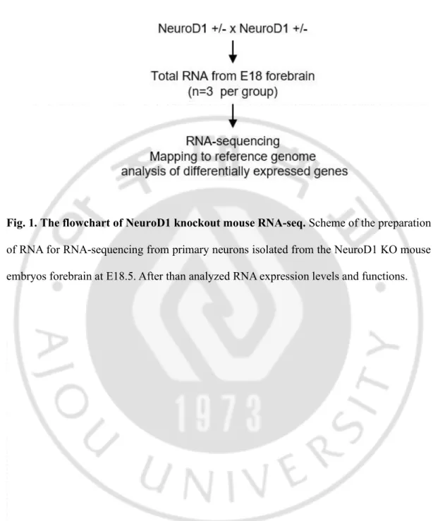
9. RNA Sequencing Analysis
Three biological replicates of NeuroD1+/+ and NeuroD1-/- RNA sets were derived from mouse forebrains, including the neocortex and hippocampus, at E18. To determine differentially expressed genes among NeuroD1+/+ and NeuroD1-/- samples, preprocessed RNA-seq reads from the respective samples were aligned to the reference genome and quantified for each gene expression. To determine the potential roles of these differentially expressed genes, data from the statistical evaluation of labeled gene sets were represented in the form of volcano plots, overlapped with functional categories (including biological processes of Gene Ontology (GO) and the KEGG pathway), and profiled using g:Profiler ver. 0.6.7 (Reimand et al., 2016). The association between labeled genes and reference genes were analyzed using GeneMANIA software (Montojo et al., 2010).
12 10. Quantitative RT-PCR
Total RNA of cultured cells and homogenized mouse forebrain was extracted using
Hybrid-RTM RNA extraction kit (GeneAll, 305-101) and reverse-transcribed with
SuperScriptTM III First-Strand Synthesis System (Invitrogen, 18080051). The transcripts
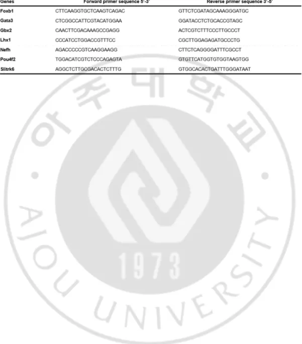
were quantified by qPCR using PowerSYBR® Green PCR MasterMix (ABI, 4367659) on the StepOnePlus Real-Time PCR System (ABI). Mouse GAPDH primers were used for the normalization of RNA expression. The sequences of all primers used in this study are provided in Table 3.
11. Statistical Analysis
All data are presented as mean ± standard error (S.E.). All statistical analyses were performed using SigmaPlotTM v14 software (Systat Software, Inc., San Jose, CA, USA). One-way ANOVA and Holm-Sidak or Dunn’s tests were used, when deemed appropriate, to compare differences between the various groups. When necessary, the t-test and Mann-Whitney U test were also performed. To assess the normality of data, the Shapiro–Wilk test (p < 0.05) was used.
13 III. RESULTS
1. Deletion of NeuroD1 reduced the neurite developmental genes
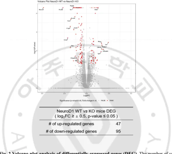
To investigate the role of NeuroD1 in differentiated neurons, we assessed the RNA profiles at gestational age 18d (E18), the latest period to consistently obtain the NeuroD1 knockout (KO) brains. RNA was extracted from the forebrains, including regions of the neocortex and hippocampus, of E18 NeuroD1 KO and WT littermates (Fig. 1). Differentially expressed genes between KO and WT mice are represented as red dots and labelled in the volcano plot. A total of 47 genes were upregulated and 95 genes were
downregulated (Log2 Fold Change (FC) cutoff value ≥ 0.5 and P-value ≤ 0.05) (Fig. 2). A
detailed gene list is provided in Table 1.
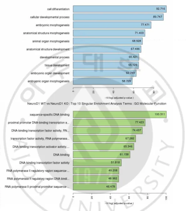
Gene Ontology (GO) enrichment analysis revealed that downregulated genes were enriched in biological processes and molecular functions for cellular development and promoter binding/transcription, respectively (Fig. 3). Since NeuroD1 is exclusively expressed in the nervous tissue during embryonic development, the data suggests that
NeuroD1 deletion impaired neuronal transcription activity and neuronal
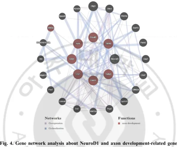
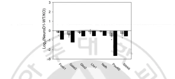
development/differentiation. We further analyzed the relationship among the 95 downregulated genes using GeneMANIA web software (Montojo et al., 2010) to delineate NeuroD1 functions specific for axon and telencephalic development. Seven genes, known to be co-expressed and co-localized with NeuroD1 (Fig. 4), were detected, namely Gata3, Gbx2, Foxb1, Lhx1, Nefh, Pou4f2, and Slitrk6. A detailed gene information is provided in
14
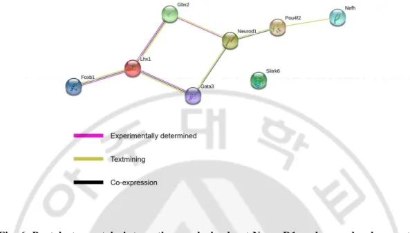
Table 2. The downregulation of these genes, triggered by NeuroD1 deletion, was further validated by quantitative reverse transcription polymerase chain reaction (qRT-PCR) analysis of RNA isolated from the embryonic forebrain using (Fig. 5). Protein network analysis using STRING database revealed that six of the abovementioned proteins, excluding Slitrk6, interacted with NeuroD1 (Szklarczyk et al., 2019) (Fig. 6). Gata3, Gbx2, Foxb1, Lhx1, and Pou4f2 are transcription factors that regulate neuronal differentiation in a diverse region of the brain during development and promote neurite outgrowth. Nefh and Slitrk6 are structural protein that regulates axonal elongation and stabilization (see the Discussion). Our finding of potential targets that regulate neurite outgrowth suggests that NeuroD1 may be a crucial factor during brain development, particularly for neuritogenesis.
15
Fig. 1. The flowchart of NeuroD1 knockout mouse RNA-seq. Scheme of the preparation
of RNA for RNA-sequencing from primary neurons isolated from the NeuroD1 KO mouse embryos forebrain at E18.5. After than analyzed RNA expression levels and functions.
16
Fig. 2 Volcano plot analysis of differentially expressed genes (DEG). The number of up
and downregulated genes are shown in the Table (Log2FC ≥ 0.5; p-value ≤ 0.05; for 3 animals per group).
18
Fig. 3. RNA-sequencing functional analysis and validation in NeuroD1-/- mice. TOP 10
19
Fig. 4. Gene network analysis about NeuroD1 and axon development-related genes using GeneMANIA web software. The network of downregulated genes, identified by
GeneMANIA software, evidencing seven key genes that are related to the function of NeuroD1 regarding axon development. Blue lines connect genes that are co-localized, and purple lines indicate co-expression. Outside circle genes present connect each gene about NeuroD1 and axon development-related genes.
21
Fig. 5. qRT-PCR validation of mRNA expression level about axon development-related genes. In qRT-PCR data also seven genes mRNA showed similar patterns like RNA-seq
data, mRNAs expression reduced in NeuroD1 knockout mouse compared to wild-type mouse mRNA.
22
23
Fig. 6. Protein to protein interaction analysis about NeuroD1 and axon development-related genes using STRING web software. NeuroD1 and Gbx2 proteins are
experimentally determined, and Pout4f2 and Gata3 co-expression. Other proteins are also linked with each other. Only the Slitrk6 protein is unknown protein to protein interaction.
24
2. Deletion of NeuroD1 Reduces Neurite Outgrowth
The neurite outgrowth in KO and WT primarily cultured neurons was assessed to investigate the role of NeuroD1 in differentiated neurons. An equal number of primary cortical neurons prepared from the forebrains of KO and WT embryos at E16.5 was plated in the cell body compartment of microfluidic chambers. At the 8th day in vitro (DIV), cells were fixed and stained for MAP2, a dendritic marker. Neurites extended through the 450 μm-wide microgrooves to reach the neurite compartment (Fig 7A). Although NeuroD1-KO cortical neurons extended neurites, neurite length in the neurite compartment was significantly shorter than that of WT cortical neurons (p < 0.005). The number of cells in the cell body compartment was similar between KO and WT cultures, suggesting that NeuroD1 does not affect the survival of differentiated neurons during short-term culture (Fig. 7B and C). These results indicate that NeuroD1 promotes neuron-specific functions, such as neurite outgrowth; thus, neurite outgrowth is delayed in the absence of NeuroD1.
25
Fig. 7. Neurite outgrowth assay using microfluidics chamber in NeuroD1 KO mouse cortical Neurons. (A) Representative structure of microfluidic chambers with primary
cortical neurons, plated in the cell body compartment (left). Neurite lengths in the neurite compartment (right) separated by microgrooves (450 μm long, 10 μm wide) were measured at the 8th day in vitro (DIV). (B and C) Cortical neurons, at 8 DIV, were immunostained by
microtubule-associated protein 2 (MAP2) (green) for dendrites and Hoechst (blue) for nuclei. Data are represented as means ± S.E. from 27 WT and 21 KO neurons. (*** p < 0.001, compared to the value of WT).
26 3. ERK Regulates NeuroD1 Protein Stability
The effects of ERK on the functioning of NeuroD1 are distinct for insulinoma cell lines and Xenopus embryos in a context-dependent manner (Petersen et al., 2002; Dufton et al., 2005). Non-phosphorylated forms of S266A and S274A resulted in the accumulation of xNeuroD protein in the injected Xenopus embryos. Before investigating whether ERK modifies the ability of NeuroD1 to promote neurite outgrowth, we first determined the effect of ERK-dependent phosphorylation on NeuroD1 protein stability using CHX chase assays. The addition of PD98059 to transfected cells, to suppress ERK-dependent phosphorylation, increased NeuroD1 protein stability (Fig. 8). Among three potential serine residues (i.e., S259, S266, and S274), the Ser to Ala mutation at S274 (S274A) profoundly reduced the protein decay rate following CHX treatment (Fig. 9). Mutations at the other two sites, S259A or S266A, did not reduce the decay rate. These results suggest that S274 is the predominant site for ERK-dependent phosphorylation. Interestingly, the addition of ALLN, a proteasome inhibitor, dramatically increased the stability of the WT and all the mutant proteins, suggesting that NeuroD1 is a potential substrate of ubiquitination.
27
Fig. 8. Erk signaling inhibitor, PD98059, increases NeuroD1 protein stability. (A)
Western blot analysis of NeuroD1 protein in the absence and presence of PD 98059, a mitogen-activated protein kinase (MEK) inhibitor. Cycloheximide (CHX) was added 30 min prior to the addition of PD 98059. (B) The NeuroD1 protein intensity shown in (a) was presented with respect to the value at t = 0 (n = 3).
28
Fig. 9. Erk-mediated phosphorylation S274 site mutation blocks the NeuroD1 protein degradation. (A) Western blot analysis of Myc-tagged mutants of NeuroD1 protein at ERK
phosphorylation sites with and without N-acetyl-Leu-Leu-norleucinal (ALLN), a proteasome inhibitor. CHX was added 30 min prior to adding ALLN. (B and C) NeuroD1 protein intensity shown in (a) was presented with respect to the value at t = 0 (n = 3). Note that the S274A mutant exhibited the highest half-life and ALLN increased the half-lives of the wild type (WT) as well as all mutants.
A
29
4. NeuroD1 protein stability regulates via the ubiquitin-proteasome system (UPS)
S724A NeuroD1 protein doesn't eliminate even without proteasome inhibitor, ALLN. So, we hypothesized NeuroD1 protein degradation by the ubiquitin-proteasome system (UPS), and this activity is turn on by ERK-dependent phosphorylation at S274 site on NeuroD1 protein. Next, we performed ubiquitination assay with a co-transfected HA-tagged ubiquitin construct to confirm the hypothesis. Multiple ubiquitin chains were identified in NeuroD1-containing precipitates, which were partially diminished by PD98059 (Fig. 10A). Importantly, the polyubiquitin chain entirely disappeared in the S274A mutant, whereas some chains remained in S259A or S266A mutants (Fig. 10B). These results indicated that NeuroD1 is a target of ubiquitin-proteasome-dependent degradation induced by ERK-dependent phosphorylation at S274.
30
Fig. 10. Extracellular signal-regulated kinase (ERK) pathway regulates the NeuroD1 protein stability via ubiquitination. (A) Western blot analysis showing the ubiquitination
of NeuroD1 WT in the presence of ALLN. PD 98059 partially attenuated ubiquitination. (B) Myc-tagged mutants of S259A, S266A, S274A, or Triple-A were analyzed for ubiquitination. All figures are representative of 3 independent experiments. Note that S274A abolished ubiquitination, whereas S266A showed a lower level of ubiquitination compared to the WT.
A
31
5. NeuroD1 Nuclear Localization is Reduced in the S274D Mutant
A previous study showed that S274 mutation affects nuclear localization in insulinoma cell lines (Petersen et al., 2002). We tested whether the effects of ERK-dependent phosphorylation are reproduced equally in cells other than those of insulinoma cell lines. We transfected HEK 293T cells with expression vectors for WT and mutants, and the cell lysate was fractionated to isolate three compartments: cytosolic, nuclear, and chromatin-bound extracts. Subcellular fractionation methods were validated by the presence of GAPDH (cytosol), Lamin B (nucleus), and Histone H3 (chromatin-bound) (Fig. 11A). The WT and S274A mutant were expressed in both the cytosol and nucleus, which were expressed at higher levels in the nucleus. This tendency was more pronounced with regards to the S274A mutant (Fig. 11B). In contrast, S274D is mostly expressed in the cytosol but barely present in the nuclear fraction. Immunostaining with an anti-Flag antibody revealed strong signals for WT and S274A in the nucleus, but weak signals for S274D (Fig. 12). These results suggest that the nuclear translocation of NeuroD1 is retarded following ERK-phosphorylation.
32
Fig. 11. NeuroD1 protein fractionation in 293T cells. (A) Flag-tagged neurogenic
differentiation 1 (NeuroD1) wild type (WT) and mutants were expressed in HEK 293T cells and subcellular proteins were fractionated. Glyceraldehyde 3-phosphate dehydrogenase (GAPDH), LaminB, or Histone H3 were used as internal controls for the cytoplasmic extract (CE), nuclear extract (NE), and chromatin bound extract (ChE). (B) Data from (a) are represented as means ± S.E. from 3 independent experiments. (# p < 0.05, compared to the value of the WT CE; * p < 0.05; ** p < 0.01, compared to the value of WT NE).
A
33
Fig. 12. NeuroD1 proteins subcellular localization in 293T cells. Immunocytochemistry
showing Flag-tagged WT, S274A, and S274D in HEK 293T cells. Flag (red); Turbo green fluorescent protein (GFP) (green); Hoechst (blue) for nuclei. The fluorescence intensity of Flag-tagged NeuroD1/GFP was quantified using the ImageJ software program. Data from (a) are represented as means ± S.E. with respect to the value of WT (* p < 0.05).
WT S274A S274D Flag GFP Hoechst merge Scale bar 20μm * B A Flag GFP Hoechst merge Flag GFP Hoechst merge
34
6. WT and mutant NeuroD1 proteins interaction with E47 protein.
To determine whether S274 mutations alter the ability of NeuroD1 proteins to heterodimerize with class A protein families, such as E47, coimmunoprecipitation assays were performed. HEK 293T cells were co-transfected with expression vectors for the WT and mutant NeuroD1, together with E47. Since both NeuroD1 and E47 independently contain nuclear localization signals and form heterodimers in the nucleus, we used cytosolic fractions for co-immunoprecipitation. The immunoprecipitates containing Flag-tagged WT, S274A, and S274D were immunoblotted to detect the presence of E47. The immunoprecipitate content of E47 was proportional to the amount of total immune-complex in the cytosol. This suggests that S274 mutations do not significantly alter the heterodimer-binding activity of NeuroD1 and E47 in the cytosol (Fig. 13). By comparison, the NeuroD1 content in E47-containing immunoprecipitates appeared slightly higher in case of the WT than that in case of the mutants. It is not practically feasible to delineate the underlying mechanisms for this at present, as several factors may influence the coimmunoprecipitation efficiency, including relative protein amounts, E47 homodimer formation, and steric hindrance caused by other protein interactions. Importantly, S274 phosphorylation, as demonstrated by S274D, does not significantly alter the ability of NeuroD1 to form heterodimers with E47.
35
Fig. 13. NeuroD1 protein heterodimer formation with E47. Co-immunoprecipitation
assay was used to assess heterodimer formation of NeuroD1 and E47. Note that the WT, S274A, and S274D retain similar binding activity to E47 protein. Actin was used as a loading control.
36
7. S274A Mutant NeuroD1 increase the transcriptional activity
We tested whether ERK-induced proteolysis correlated with the ability of NeuroD1 to transactivate downstream genes in neuronal cells. Previous studies evidenced ERK transactivated the C-terminus of NeuroD1 (155-355 aa) that was conjugated to the GAL4 DNA-binding domain in insulinoma cell lines, such as MIN6, bTC3, or INS-1 (Petersen et al., 2002; Khoo et al., 2003). Unlike previous studies, we used full length (1-355 aa) NeuroD1 proteins in the neuronal cell line, F11. For this, we generated pLenti (lentivirus transfer) vectors for concomitant expression of the WT or S274 mutants together with GFP protein. Concomitant expression of GFP and Flag-tagged NeuroD1 proteins was validated by western blot analysis (Fig. 14). Since class B bHLH transcription factors, such as NeuroD1, form heterodimers with their class A partners, such as E47 (Glick et al., 2000), transfection was performed with and without E47. A reporter gene containing rat insulin promoter element 3 (RIPE3), an E-box containing enhancer that is known to be activated by NeuroD1, was used (Naya et al., 1995; Huang et al., 2000). In contrast to that of insulinoma cell lines, the S274A mutant activated RIPE3, containing the reporter gene, by 5-fold, which further increased to 11-fold when transfection was performed with E47. In contrast, S274D, a non-phosphorylated mutant, only activated RIPE3 by 2-fold, even when transfected with E47. To further verify the NeuroD’s functions, a reporter gene containing the 2.2-kb promoter region of NeuroD1 (Huang et al., 2000) was used as it was evidenced to be autoregulated by NeuroD1 through E-box sequences (Fang et al., 2010). The 2.2 kb fragment of NeuroD1 promoter was also activated by S274A by 40- and 18-fold with and without E47, respectively (Fig. 15). NeuroD1-mediated gene activation is cell-type specific. S274A
37
enhanced the expression of both reporter genes in pluripotent P19 cells, or INS-1 insulinoma cells, and in F11 cells but not in HeLa cells (Fig. 16). Importantly, the full length NeuroD1 can transactivate the E-box containing promoter in both F11 neuronal and INS-1 insulinoma cells. These results clearly indicate that ERK-dependent phosphorylation at S274 exerts negative effects on the NeuroD1 protein.
38
Fig. 14. Schematic diagram of the WT and S274 mutants in the lentivirus transfer vector. (A) WT and Mutants NeuroD1 lentiviral constructs Flag-tagged and co-expressed
green fluorescent protein (GFP). (B) Validation of both green fluorescent protein (GFP) and Flag-tagged NeuroD1 proteins by western blot analysis.
B A
39
Fig. 15. Transactivation activity of NeuroD1 wild type (WT) and mutants in F11 cells.
(A and B) Schematic representation of the luciferase reporter gene of rat insulin promoter element 3 (RIPE3). Schematic representation of the 2.2 kb promoter region of NeuroD1 containing at least two E-box sequences. F11 cells were transfected with expression vectors for WT and mutants with and without E47. Luciferase activity was presented as the relative ratio to the value of mock transfection. Note that the stimulatory effect of full length S274A was the highest in the presence of E47, whereas the effect of the S274D mutant was minimal. Data are represented as means ± S.E. from at least 3 independent experiments (** p < 0.01; *** p < 0.001, compared to the value of the WT without E47; # p < 0.05; ## p < 0.01; ### p < 0.001, compared to the value of the WT with E47).
40
Fig. 16. Wild-type and mutant NeuroD1 proteins Rat Insulin Promoter Element 3 (RIPE3) luciferase assay in various cell lines. S274A mutant NeuroD1 significantly
increase transcriptional activity, S274D mutant NeuroD1 was the opposite. ** p < 0.01, *** p < 0.001, compare to the value of wild-type NeuroD1. Data are means ± SEM; n=3 in HeLa and P19, n=11 in INS-1, n=number of independent cell culture preparations.
41
8. S274 Phosphorylation Attenuates Transactivation Potential of NeuroD1
We used F11 cells to investigate whether increased NeuroD1 activity promotes neurite outgrowth in differentiated neurons. F11 cells provide an in vitro system for assessing the proneural functions of NeuroD1 and Neurogenin 1 (Ngn1), which can be observed through neurite outgrowth (Cho et al., 2001). F11 cells were transfected with pLenti-plasmids encoding NeuroD1 proteins together with GFP, and neurite outgrowth was assessed in GFP-expressing cells. In the presence of low serum levels, neurite outgrowth occurred slowly in F11 cells with the GFP control vector and were only evident on day 4 (Fig. 17A). Overexpression of WT and S274A mutant accelerated neurite outgrowth significantly from day 2, and neurite length was maximal at day 4. Moreover, the stimulatory effect was higher with S274A than the WT. By comparison, overexpression of S274D caused a delay in neurite outgrowth on days 2 and 3, but reached a level similar to that of the WT on day 4 (Fig. 17B).
Previously we identified seven genes, which are related to axon development in knockout NeuroD1 mouse brain (Fig. 4). We thought that these genes also could affect neurite outgrowth in F11 cells by NeuroD1 or not. To determine whether S274A NeuroD1 increases, neurite outgrowth related gene mRNAs qPT-PCR was performed. In S274A mutant NeuroD1 overexpressed F11 cells, it showed Gbx2 mRNA level higher than others on differentiation day 3 (Fig. 18). The other six genes were no significant differences (data not shown). These results suggest that transactivation activity of NeuroD1, demonstrated by S274A and S274D, correlates with the transactivation function of NeuroD1 in mature neurons.
42
Fig. 17. Neurite-promoting activity of NeuroD1 wild type (WT) and mutants in F11 cells. (A) F11 cells were transfected with lentiviral transfer vectors encoding GFP with WT
or S274 mutants. Following transfection, fluorescence images were obtained at the indicated time post-transfection. (B) Neurite length was quantified by the NeuronJ plugin of the ImageJ software program. Data are represented as means ± S.E. from at least 50 transfected cells per group. (* p < 0.05; ** p < 0.01; *** p < 0.001, between groups).
43
Fig. 18. qRT-PCR for Gbx2 mRNA expression in transiently overexpressed NeuroD1 F11 cells. S274A NeuroD1 significantly increases the Gbx2 mRNA level after differentiate
day 3 in F11 cells. * p < 0.05 compare to the value of wild-type NeuroD1. Data are means ± SEM; n=3, n=number of independent cell culture preparations.
44
9. WT and S274A Rescue Neurite Outgrowth in NeuroD1 KO Neurons
To determine whether the stimulatory effect of NeuroD1 observed in F11 neuronal cells is also conserved in primary neurons, rescue experiments were performed with the WT and mutants from primary cortical neurons isolated from NeuroD1 KO embryos. On day 4, total and longest neurite lengths were reduced in KO neurons compared to those of the WT, indicating that our approach of using a 4-day culture was as valid as the 8-day long microfluidic chamber assay in terms of dissociated cultures. Several types of lentiviral vectors, encoding GFP in conjunction with WT or mutant NeuroD1 proteins, were transduced to KO neurons to assess the effect of ERK-dependent phosphorylation (Fig 19). Expression of the WT rescued neurite outgrowth in KO neurons, which was similar to the case for WT neurons (p < 0.01). Importantly, the S274A mutant also significantly enhanced total and longest neurite lengths similar to that of WT neurons (p < 0.05). By comparison, S274D expression could increase neurite length to a lesser degree; thus, the difference between S274D and the GFP control was statistically insignificant (p = 0.295). The number of neurites was around 4 per cell in all experimental groups (Fig. 20). The results suggest that ERK-dependent phosphorylation of S274 may attenuate the function of NeuroD1 in neurite extension without affecting the formation of new neurites.
This study shows that NeuorD1 deficiency mouse has abnormal neurite length compare to wild type, and it can be overcome rescue by NeuroD1. Axon development related genes, such as GATA3, Gbx2, Foxb1, Pou4f2, Lhx1, Nefh, and Slitrk6, mRNA level are reduced in NeuroD1 knockout mouse forebrain. And we also showed that phosphorylation on NeuroD1 protein controls stability dynamics via the ERK signaling pathway. NeuroD1
45
protein stabilized in the presence of PD98059, which is ERK signaling inhibitor, and this data indicated that NeuroD1 protein stability regulates through the ERK signaling pathway. This ERK signaling pathway promotes phosphorylation on NeuroD1 S259, S266, and S274 amino acid site, protein stability effect showed only at S274 site. When we inhibited this phosphorylation site of the NeuroD1, such as S274A mutant form, protein doesn’t go to degradation even without PD98059. After that, this stabilized NeuroD1 (S274A) more exist in nuclear localization than wild-type, enhanced the transcriptional activity. Furthermore, this phenomenon appears phenotype of neurite outgrowth in neuroblastoma cells, F11, and primary cortical neurons (Fig. 21)
46
Fig. 19. Schematic representation of the experimental process for transducing primary cortical neurons from the brain of NeuroD1 knockout (KO) embryos. One day after
plating, cortical neurons were transduced with lentiviral vectors encoding green fluorescent protein (GFP) together with wild type (WT) or S274 mutants. Cells were fixed at the 5th day
47
Fig. 20. Rescue experiments to supplement neurogenic differentiation 1 (NeuroD1) deficiency. (A) Cells were immunostained with anti-GFP antibody (green) to detect
lentivirus transduced neurons and Hoechst (blue) for nuclei. (B-D) Neurite length was quantified from images shown in (a) using the NeuronJ plugin of the ImageJ software program. The number of neurites, total neurite length, and longest neurite length per cell were counted and presented. Data are represented as means ± S.E.; at least 50 neurons were counted per group. (* p < 0.05; ** p < 0.01, compared to the value of KO neurons expressing GFP as a control).
48
Fig. 21. Schematic model for physiological roles of ERK-dependent phosphorylation of NeuroD1. Erk signaling activation promotes NeuroD1 phosphorylation, specific S274 site,
this brings out proteasomal degradation. On the other hand, when inhibits the Erk signaling, such as PD98059, NeuroD1 goes to nuclear localization with an E47 partner protein. This NeuroD1 can enhance the transcription of downstream target genes, these genes contribute to facilitate neurite outgrowth.
49
IV. DISCUSSION
NeuroD1 is a tissue-specific transcription factor and its functions are regulated by several kinases that transmit the growth- and activity-related extracellular signals to the nucleus. Several studies conducted over the past decade have evidenced that transcriptional activity of NeuroD1 appears to be oppositely regulated by ERK in neurons and pancreatic β cells. In insulinoma cell lines, ERK, triggered by glucose stimulation, appeared to activate the transactivation potential of NeuroD1, when functioning as a hybrid protein with the GAL4 DNA-binding domain. In contrast, a phosphorylation-null mutation at the ERK phosphorylation site increased proneural functions of NeuroD1 in early embryogenesis. The present study has clarified a long-standing enigma regarding the impact of ERK phosphorylation on functions of NeuroD1.
Like other members of the class B bHLH transcription factor family (Lingbeck et al., 2005; Hindley et al., 2012), the NeuroD1 protein undergoes rapid decay via ubiquitin-dependent degradation (Fig. 10). The degradation rate declines when the ERK pathway is blocked with a MEK inhibitor. Ala substitution at the ERK phosphorylation site (S274A) also lowers the protein decay rate and increases the amount of protein in the nuclear compartment (Fig. 11 and 12). Unlike previous studies using a hybrid of NeuroD1 (156-355) with Gal4DNA-binding domains, the current study evidenced that full-length NeuroD1 proteins are capable of upregulating the expression of downstream reporter genes and that the stimulatory effect is higher with the S274A mutation compared to the WT (Fig. 15). Similar results were obtained with full-length NeuroD1 in P19, an embryonic carcinoma cell
50
line, and INS-1, a rat insulinoma cell line (Fig. 16), suggesting that the ERK pathway negatively regulates the functions of NeuroD1. We summarized of the effect S274A mutation in diverse cell types in Table 4. Likewise, neurite outgrowth is accelerated with the S274A mutation than that of the WT in F11 neuronal cells (Fig. 17). The S274A mutation does not alter the innate activity of the NeuroD1 protein with regards to nuclear localization or heterodimer formation with E47 (Fig. 13). Thus, the increased activity of S274A can be explained by its high content, due to reduced decay rates or increased protein stability.
Table 4. Summary of the effect of S274A mutation in diverse cell types.
In contrast, S274D, a phosphomimetic mutant of ERK, demonstrated poor activity in transactivating reporter genes or inducing neurite outgrowth compared to that of the WT protein. Although the cytosolic content of S274D was relatively high compared to that of the WT or S274A, the nuclear localization of S274D was significantly attenuated (Fig. 11 and 12). Thus, the poor activity of S274D in transactivation or neurite promotion is due to
51
the low content of functional complexes containing the S274D protein in the nuclear compartment, and consequently low occupancy of the promoter. E47 contains nuclear localization sequences at 165–169 aa and 539–554 aa; thus, it is able to direct nuclear localization of a partner protein in heterodimer complexes containing a tissue-restricted class B bHLH protein partner, such as MyoD (Lingbeck et al., 2005). Since S274D in the cytosolic compartment can interact and form a heterodimer with E47 (Fig. 13), the low content of S274D in the nuclear compartment implies that the S274D-containing complex is prohibited from the nuclear compartment, which cannot be negated by the nuclear localization sequences of E47. Further studies are necessary to reveal whether and, if so, how ERK-dependent phosphorylation induces tethering of the heterodimer complex to cytosolic adaptor(s).
Although this study has revealed the molecular mechanism of Erk-dependent NeuroD1 regulation via observing neurite outgrowth in differentiated neuronal cells, this finding can be expanded to elaborate cell fate determination of the uncommitted stem cells. During the development of forebrain, ERK signals exert opposite effects on neuronal and astroglial differentiation. ERK suppresses neuronal differentiation by inhibiting the expression of Neurogenins 2 (Neurog2), a proneural transcription factor, in dorsal progenitor cells during cell cycle progression. At the commitment stages of progenitor cells, transcription of Neurog2 and NeuroD1 (the target gene of Neurog2) increases as ERK signals decline. As a result, the cells in the dorsal forebrain exit the cell cycle and terminally differentiate into glutamatergic neurons (Li et al., 2014). Indeed, NeuroD1 is known to activate a cyclin-dependent kinase inhibitor, p21 and thereby induce cell cycle exit (Mutoh
52
et al., 1998). Thus, the negative regulation of Neurog2 at the transcriptional level and NeuroD1 at the post-translational level by ERK shown in the present study may demonstrate potential mechanisms for prohibiting premature cell cycle exit while maintaining cortical progenitor pools in the mid-embryonic stages. After entering late embryonic stages, the dorsal progenitor cells acquire astroglial competence and generate astrocytes at the expense of neurons. Suppression of RAS-ERK pathway by deletion of Mek1/2 (also known as MAPK/ERK kinase 1/2) or deletion of ERK1/2 induces depletion of astrocytes in postnatal brain (Imamura et al., 2010; Li et al., 2012), suggesting that ERK plays a role in the astroglial lineage progression of dorsal progenitor cells. Our finding of the negative regulation of NeuroD1 protein by ERK may provide a clue to elucidate the previously unexplored mechanisms for driving the genetic switch of dorsal progenitor cells from neurogenic-to-gliogenic competence after Neurog2 expression declines in the late stage of development (Fig. 22) (Kriegstein and Alvarez-Buylla, 2009; Tabata, 2015; Beattie and Hippenmeyer, 2017). Impairments in lineage progression caused by malfunction of RAS-ERK signaling lead to alterations in the cortical cytoarchitecture, which is thought to represent the major underlying cause of several neurological disorders, including microcephaly or megalocephaly, and more subtle neurodevelopmental diseases, including schizophrenia, autism, epilepsy, and RASopathies (Desikan and Barkovich, 2016; Silbereis et al., 2016; Kang and Lee, 2019).
53
Fig. 22. Schematic model for future hypothesis of physiological roles of ERK-dependent phosphorylation of NeuroD1. NeuroD1 proteins heterodimerize with E47, translocate to
the nuclear compartment, and transactivate downstream target genes. Consequently, NeuroD1 induces cell cycle exit, neuronal differentiation, and neurite outgrowth. ERK-dependent phosphorylation at S274 facilitates ubiquitination and proteasomal degradation of NeuroD1. By negatively regulating NeuroD1, Erk maintains the progenitor pools at mid-stage brain development and promotes glial cell differentiation of progenitor cells at the expense of neurogenesis at late-stage brain development.
Transcriptome analysis and GO enrichment analysis indicated that the genes involved in axonogenesis and dendrite development were downregulated in NeuroD1 KO mice (Fig. 3 and 4). In the primary neurons obtained from NeuroD1 KO embryos, neurite
54
outgrowth is attenuated, suggesting that NeuroD1 plays a role in proneural activity regarding the development of neuronal processes. F11 cells are a hybridoma cell line between mouse neuroblastoma and rat dorsal root ganglion (pseudo-unipolar) cells and extend bipolar neurites in the presence of dibutyryl-cAMP. Without it, the F11 cells barely extend neurites up to day 3 (Fig. 17), suggesting that innate neurogenic factors are limited in F11 cells. Overexpression of WT, S274D, and S274A NeuroD1 can promote neurite outgrowth in F11 cells with the highest magnitude for S274A and the lowest for S274D (Fig. 17). The data are consistent with the proteins’ half-lives (Fig. 9) and transactivation potential of downstream reporter genes (Fig. 15).
By comparison, the effect of S274A is not prominent compared to the WT in NeuroD1 -/- primary neurons (Fig. 20). It can be partly due to that axonal and dendritic morphogenesis in cortical neurons is regulated by pleiotropic factors and the loss of NeuroD1 can be compensated by other bHLH proteins, such as NeuroD2, MATH2, MATH3, and Atoh7 as shown in NeuroD1 knockout mice (Miyata et al., 1999; Cho and Tsai, 2006; Mao et al., 2013). Thus, NeuroD1 -/- cortical neurons can extend neurites, although to a lesser degree, compared to WT neurons (Fig. 7 and 20). Therefore, the influence of S274A may be minimal in the rescue experiments using NeuroD1 -/- cortical neurons compared to the one in F11 cells. Importantly, S274D is less effective to rescue the NeuroD1’s functions (Fig. 20), supporting our hypothesis that phosphorylation of S274 is important to determine the neuritogenic function of NeuroD1.
activity-55
induced protein kinase, CaMKII, at S336 and the S336A mutation destroyed the ability of NeuroD1 to stimulate dendritic growth in Bcl2-overexpressed cerebellar granule neurons (Gaudilliere et al., 2004; Lee et al., 2019). However, similar to that of insulinoma cells (Khoo et al., 2003), the transactivation activity of NeuroD1 is not altered by S336 mutations when tested with chimeric proteins fused to the Gal4 DNA-binding domain (Gaudilliere et al., 2004). Further studies using the full length NeuroD1 protein may be helpful in revealing how NeuroD1 phosphorylation, triggered by CaMKII, upregulates dendritic growth in mature neurons.
The RNA sequencing analysis of 95 downregulated genes revealed the functions of NeuroD1 with respect to the GO terms of promoter binding, RNA polymerase II, and cellular developmental processes (Fig. 3). Subsequent analysis using GeneMANIA revealed seven core genes related to the GO term of neurite outgrowth (Fig. 4). Gata3, Gbx2, Foxb1, Lhx1, and Pou4f2 are transcription factors that regulate neuronal differentiation in a diverse region of the brain during development and promote neurite outgrowth (Alvarez-Bolado et al., 2000; Wang et al., 2002; Chen et al., 2010; Appler et al., 2013; Lui et al., 2017). Nefh (Neurofilament, Heavy polypeptide) is a cytoskeletal element of axons and Slitrk6 (SLIT and NTRK like family member 6) is an integral membrane protein, both of which regulates axonal elongation and stabilization (Katayama et al., 2009; Lee and Shea, 2014). Interestingly, all these genes contain potential E-box sequences within 1 kb upstream of the transcription start site (data not shown). These 7 genes were not included in the list of previously identified target genes of NeuroD1 (Seo et al., 2007). A possible explanation is
56
that two different approaches were used. Previous studies adopted a gain-of-function approach by overexpressing NeuroD1 in uncommitted cells such as early Xenopus embryos or pluripotent P19 cells, whereas we adopted a loss-of-function approach with mid-stage embryonic development when neurogenesis occurs most actively. Since the NeuroD1’s neurogenic function is prominent during mid-stage embryos, it would be interesting to determine whether the newly identified genes are physiologically relevant target candidates
of NeuroD1 in the mammalian brain.Indeed, overexpression of S274A NeuroD1 induced
the expression of Gbx2 in F11 cells on day 3, when neurite outgrowth was most prominently enhanced by S274A (Fig. 18). The expression of the other six genes was not directly affected by the overexpression of NeuroD1 proteins in F11 cells (data not shown). Gbx2 is a homeobox gene and plays a key role in patterning both vestibular and auditory components of the inner ear (Lin et al., 2005) and NeuroD1 is expressed for the proper development of sensory neurons in the inner ear (Liu et al., 2000; Chatterjee et al., 2010). Further study may reveal whether and how NeuroD1 and Gbx2 interact with each other and regulate inner ear development.
Recently, NeuroD1 has emerged as an important genetic therapeutic strategy to treat the diseased brain where adult neurogenesis or synaptic integration of newborn neurons are hampered. In Alzheimer’s disease model (5xFAD), overexpression of NeuroD1 using a retroviral vector can induce reprogramming of reactive astroglial cells into functional neurons (Guo et al., 2014). Similarly, overexpression of NeuroD1 also can rescue spatial memory in aged AD mice (APPxPS1) by accelerating neuronal maturation of hippocampal
57
progenitor cells and promote functional integration of newborn neurons (Richetin et al., 2015; Richetin et al., 2017). S274A has a longer protein half-life than WT NeuroD1, making it a better choice for overexpressing NeuroD1 to alleviate neuronal loss in a diseased brain. By blocking the ubiquitin-dependent degradation of endogenous NeuroD1 protein may provide an alternative therapeutic strategy that promotes neuronal activity by enhancing the expression of NeuroD1’s target genes.
58 References
1. Alvarez-Bolado G, Zhou X, Voss AK, Thomas T, Gruss P: Winged helix transcription factor Foxb1 is essential for access of mammillothalamic axons to the thalamus.
Development 127: 1029-1038, 2000
2. Appler JM, Lu CC, Druckenbrod NR, Yu WM, Koundakjian EJ, Goodrich LV: Gata3 is a critical regulator of cochlear wiring. J Neurosci 33: 3679-3691, 2013
3. Beattie R, Hippenmeyer S: Mechanisms of radial glia progenitor cell lineage progression. FEBS Lett 591: 3993-4008, 2017
4. Chatterjee S, Kraus P, Lufkin T: A symphony of inner ear developmental control genes. BMC Genet 11: 68, 2010
5. Chen L, Chatterjee M, Li JY: The mouse homeobox gene Gbx2 is required for the development of cholinergic interneurons in the striatum. J Neurosci 30: 14824-14834, 2010
6. Cho JH, Kwon IS, Kim S, Ghil SH, Tsai MJ, Kim YS, Lee YD, Suh-Kim H: Overexpression of BETA2/NeuroD induces neurite outgrowth in F11 neuroblastoma cells. J Neurochem 77: 103-109, 2001
7. Cho JH, Tsai MJ: Preferential posterior cerebellum defect in BETA2/NeuroD1 knockout mice is the result of differential expression of BETA2/NeuroD1 along anterior-posterior axis. Dev Biol 290: 125-138, 2006
8. Dennis DJ, Han S, Schuurmans C: bHLH transcription factors in neural development, disease, and reprogramming. Brain Res 1705: 48-65, 2019
9. Desikan RS, Barkovich AJ: Malformations of cortical development. Ann Neurol 80: 797-810, 2016
10. Doucet C, Gutierrez GJ, Lindon C, Lorca T, Lledo G, Pinset C, Coux O: Multiple phosphorylation events control mitotic degradation of the muscle transcription factor Myf5. BMC Biochem 6: 27, 2005
11. Dufton C, Marcora E, Chae JH, McCullough J, Eby J, Hausburg M, Stein GH, Khoo S, Cobb MH, Lee JE: Context-dependent regulation of NeuroD activity and protein accumulation. Mol Cell Neurosci 28: 727-736, 2005