Pro187Ser Polymorphism of NAD(P)H:quinone oxidoreductase 1 and Prognosis of Non-small
Cell Lung Cancer after Radiation Therapy
Si Yeol Song1, Heon Joo Park3, Yun-Chul Hong4,Jong-Eun Lee6, Sang Min Yoon1, Seong Soo Shin1, Seung Do Ahn1, Jin Hee Kim4, Jung Shin Lee2, Charn Il Park5, and Eun Kyung Choi1
Departments of 1Radiation Oncology and 2Medical Oncology, Asan Medical Center, College of Medicine, University of Ulsan, Seoul, Korea
3Department of Microbiology, College of Medicine, Inha University, Incheon, Korea
Departments of 4Preventive Medicine and 5Radiation Oncology, College of Medicine, Seoul National University, Seoul, Korea
6DNA Link Inc., Seoul, Korea
*Correspondence: Eun Kyung Choi, e-mail: [email protected]
1. Introduction
NAD(P)H:quinone oxidoreductase 1 (NQO1) has been known to function on reduction of oxidative status as a cytosolic flavoenzyme that catalyzes the electron reduction of substrates [1]. It was reported to play a role in the prognosis of lung cancer patients treated with chemotherapy. Single nucleotide polymorphisms (SNPs) make up about ninety percent of human DNA polymorphisms, and they are a major focus of study about the individual differences for the risk of cancer and for anti-cancer treatment. A point mutation in exon 6 of the NQO1 gene is a C-to-T base pair substitution at position 609 of the NQO1 cDNA, and this codes for a proline-to-serine change at position 187 in the amino acid sequence of the protein. We hypothesized that
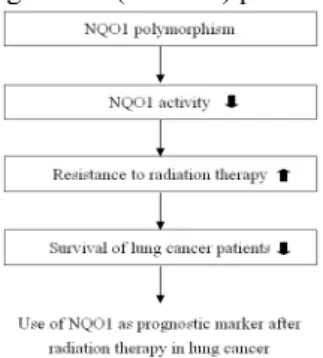
NQO1 polymorphism could have an adverse influence
on the survival of NSCLC patients treated with radiation therapy and/or surgery (Figure 1), and so we tried to discover whether the NQO1 polymorphism could be a predictive or prognostic marker for determining treatment outcome of radiotherapy in non-small cell lung cancer (NSCLC) patients.
Figure 1. Hypothesis: NQO 1 polymorphism & survival 2. Materials and Methods
2.1 Study populations and Radiation therapy
Patients who had been treated with radiation therapy from 2000 to 2003 were recruited at the Asan Medical Center. NSCLC patients (stage I-III) who were histologically proven as squamous cell carcinoma and adenocarcinoma were recruited, and they had a good
performance (ECOG 2 or below). Patients with the history of another neoplasm or radiation therapy were excluded. Radiation therapy was performed with 15-MV X-rays from a linear accelerator. 1.8Gy per fraction was delivered once a day to the dose of 50.4Gy over 5 weeks postoperatively and 70.2Gy over 8 weeks for curative aim.
2.2 Sampling and Genotyping assay
Sampled blood was bottled in tubes that contain 1.5ml of anticoagulant acid citrate dextrose solution (ACD) A. DNA was extracted from the lymphocytes contained within whole blood sample with the use of a QIAamp DNA Blood Mini Kit (Qiagen, Valencia, CA, USA). DNA was amplified by polymerase chain reaction (PCR), and PCR products were then denatured. The primer set for the NQO1 Pro187Ser polymorphism was 5΄-TCC TCA GAG TGG CAT TCT GC-3΄ [2], and the denatured PCR products was annealed, reacted with buffer mixture, and then tagged with fluorescent dye. The genotyping procedure was performed using a ABI PRISM SNaPshotTM Multiplex Kit with the aid of an ABI 3700 automatic sequencer (Applied Biosystmes, CA, USA).
2.3 Immunohistochemical staining (IHC staining)
Formalin-fixed paraffin-embedded tissue sections were deparaffinised in xylene and rehydrated through graded ethanol washes to diluted water. Primary and secondary antibody incubations, and streptavidin complex (DAKO LSAB kit, UK) were amplified. Immunocomplex visualization was performed using diaminobenzidine (Figure 2). Immunohistochemical staining results were scored semiquantitatively as four grades. The score was reported according to the staining intensity and distribution: 0 (no staining), 1+ (weak positive), 2+ (moderate positive), 3+ (strong positive).
A B
Transactions of the Korean Nuclear Society Autumn Meeting Busan, Korea, October 27-28, 2005
Figure 2. IHC staining (A: NQO1(+), B: NQO1(-))
2.4 Statistical analysis
The survival time was counted from the start of the curative treatment, operation or radiation therapy. Kaplan-Meier survival curves and the log-rank test were used to analyze the survival.
3. Results and Discussion
3.1 NQO1 polymorphism and patients’ characteristics
The NQO1 polymorphism was divided into three groups according to presence of point mutation: 1) the wild type, Pro/Pro, 2) the mutated heterozygote, Pro/Ser, and 3) the mutated homozygote, Ser/Ser, and the numbers of the patients having the different polymorphisms were 56, 106 and 37, respectively. No significant relationship was found between NQO1 polymorphism and the baseline characteristics of the recruited patients
3.2 Univariate and multivariate analysis for the survival of NSCLC patients
The effect on the progression-free survival (PFS) and overall survival (OS) were significantly different according to the cancer stage, histology and the treatment protocol (progression-free survival; P < .001,
P = 0.004, and P < .001, overall survival; P = 0.010, P
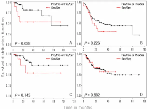
= 0.051, and P < .001), but there was no difference according to gender, age, ECOG and weight loss (progression-free survival; P = 0.932, P = 0.842, P = 0.238, and P = 0.740, overall survival; P = 0.823, P = 0.564, P = 0.752, and P = 0.812). The effect of NQO1 polymorphism was significantly different for the progression-free survival (P = 0.033), but not for the overall survival (P = 0.452). Survival rate was shown on figure 3, and multivariate analysis was listed on table 1.
Figure 3. PFS and OS according to the NQO1 genotype
A. PFS at stage I and II B. PFS at stage III C. OS at stage I and II D. OS at stage III
Table 1. Multivariate analysis
3.3 Immunohistochemical analysis
IHC staining was performed only in 38 patients who had adequate formalin-fixed tissue among 199 patients. Fourteen patients have shown the mutated homozygote, no activity of NQO1, and the other 24 patients have shown the wild type NQO1 in genotyping assay using blood-sampling. The comparison of NQO1 polymorphism between genotyping assay from blood and IHC staining (IHC_NQO1) from tissue specimen were performed. The result showed that NQO1 expression and activity, whether the mutated homozygote or the wild type, were not identical (correlation coefficient=0.029, P=0.879). However this NQO1 result, p53 from tissue specimen (IHC_p53) had an analogue in the distribution between two studies, although it didn’t reach the statistical significance (correlation coefficient=0.291, P=0.076). Progression-free survival according to IHC_NQO1 and IHC_p53 were not different (P=0.45 and 0.53), and overall-survival were also not different between groups (P=0.66 and 0.64).
3.4 NQO1 polymorphism and Radiation therapy
We tried to reveal the difference of survival for patients after radiation therapy according to the NQO1 genotypes. This study showed that NQO1 polymorphism contributed to the decreased progression-free survival of patients with NSCLC. As a result, the NQO1 polymorphism resulted in reduced survival and reduced radiosensitivity, and this result was consistent with Fleming’s report [3] about the chemosensitivity. This report is the first to suggest the possibility that NQO1 polymorphism may be used as a prognostic marker for NSCLC patients who are undergoing radiotherapy. On the basis of the expected outcome of patients, both the predictive and prognostic factors may be important in the choice of treatment protocol.
3.5 Genotyping versus Immunohistochemical staining
For the analysis of the NQO1 polymorphism in NSCLC patients, genotyping assay from sampled blood was mainly used and immunohistochemical staining was additionally performed only in some patients. The results showed that the NQO1 expression and its activity were not identical in same patients between two methods. We could not clearly interpret this result, and no study or data comparing the genotyping assay with immunohistochemical staining for polymorphism has been reported. Further investigation and more specimens should be provided, and then we might revise the result of immunohistochemical staining.
NAD(P)H:quinone oxidoreductase1 pro187ser
polymorphism could be used as a prognostic marker for those non-small cell lung cancer patients who were treated with radiotherapy, although the further study about the immunohistochemical analysis should be provided.
Acknowledgements
This study was supported by a grant from the National R&D Program for Cancer Control (Grant No. 0320333-2), Ministry of Health and Welfare, Korea and also supported by 2003 Basic Atomic Energy Research Institute (BAERI).
References
[1] Ross D, Kepa JK, Winski SL, et al. NAD(P)H: quinone oxidoreductase1 (NQO1): chemoprotection, bioactivation, gene regulation and genetic polymorphisms. Chem Biol Interact 129(2000) 77-97. [2] Traver RD, Siegel D, Beall HD, et al. Characterization of a polymorphism NAD(P)H: quinone oxidoreductase (DT-diaphorase). Brit J Cancer 75(1997) 69-75.
[3] Fleming RA, Drees J, Loggie BW, et al. Clinical significance of a NAD(P)H: quinone oxidoreductase 1 polymorphism in patients with disseminated peritoneal cancer receiving intraperitoneal hyperthermic chemotherapy with mitomycin C. Pharmacogenetics 12(2002) 31-7