서 론
뇌심부자극술(deep brain stimulation, DBS)은 본태성 진 전(essential tremor) 또는 파킨슨병(Parkinson disease)과 관 련된 진동을 극적으로 완화할 수 있는 외과적 수술로 다양 한 신경 상태를 가진 사람들에게 이점을 제공하는 뇌심부
자극술이 임상에서 적용되고 있다. 이러한 뇌심부자극술은
전극(electrode)이 시상밑핵(subthalamic nucleus)이나 담창 구(globus pallidus)의 내부 분절을 자극하면 파킨슨병 환자
의 느린 움직임, 근육의 떨림, 경직 및 보행 장애를 크게 줄
일 수 있다. 뇌심부자극술의 전극의 정확한 위치 삽입에 대
한 연구는 임상 치료법을 넘어서는 중요성을 가지고 있다 (Cuny et al. 2002; Papavassilou et al. 2008). 뇌심부자극술의 행동 기제에 대한 조사는 뇌 회로의 기능적 해부학 및 그 회로와 행동의 활동 사이의 관계와 같은 근본적인 문제를 명확히 할 잠재력을 가지고 있다(Perlmutter et al. 2006).
Jong Seok Lee
1and Dae Cheol Kweon
2,*
1Department of Radiological Technology, Wonkwang Health University,
Iksan 54538, Republic of Korea
2Department of Radiological Science, College of Bioecological Health, Shinhan University,
Uijeongbu 11644, Republic of Korea
Abstract - The purpose of this study was to evaluate the accuracy of the electrode images of the subthalamic nucleus by using preoperative MR imaging and postoperative CT imaging in patients with deep brain stimulation. After electrode implantation, CT images of the deep brain stimulation patients were analyzed and evaluated before surgery. Preoperative MR imaging of deep brain stimulation was performed on AC-PC(anterior commissure-posterior commissure) line as T2-weighted images. CT scans after deep brain stimulation was scanned on the AC-PC line. The images were fused by using image merge channels function of MR images and CT images using ImageJ (NIH, Bethesda, MD, USA), and the accuracy of the presence of electrodes in the hypothalamic nucleus was confirmed. Post-operative CT images of deep brain stimulation were acquired and the images were subjected to the color synthesis function of the ImageJ program. MR and CT images were inserted into various red, green, blue, and gray functions in order to confirm the precise location of the electrodes. In the program, both the electrodes and thalamus nucleus were revealed in the synthesis of red(CT image) and green(MR image). Fusion of MR and CT images is a safe and practical application and accuracy in identifying the location of electrodes in deep brain stimulation patients. Such as, CT and MRI fusion images can be applied in clinical practice.
Key words : CT, Deep brain stimulation, Fusion, MR imaging
─ 133 ─
* Corresponding author: Dae Cheol Kweon, Tel. +82-31-870-3411, Fax. +82-31-870-3419, E-mail. [email protected]
뇌심부자극술은 작은 구조인 시상밑핵에 전극를 삽입하 여 치료에 응용되기 때문에 정확한 위치에 전극을 안착하 는 게 매우 중요하다(Hamid et al. 2005). 현재 임상에서는 뇌심부자극술의 전극을 삽입하기 위해 컴퓨터 프로그래밍 치료계획의 일환으로 미세전극 모니터링에 의한 전극 위치 를 확인하고 전극 자극을 통한 환자의 증상 및 변화를 관찰 하고 있다. 또한 뇌심부자극술에서 수술 전의 MR 영상(Cui et al. 2016; Salvin et al., 2016)과 수술 후의 CT(computed tomography) 영상은 합병증을 평가하고 정확한 전극 위치 를 평가하는 데 중요한 요소이다(Saleh et al. 2016). 뇌심부자극술에서 CT 영상은 높은 기하학적 정확도와 안 전문제를 제기하지 않고 쉽게 영상을 획득하는 장점을 가지 고 있지만 뇌의 심부 구조를 식별하는 데 필요한 연부조직의 대조도가 부족하고 금속 전극의 인공물로 인한 영상의 식별 이 어려운 점이 있다(Sumanaweera et al. 1994; Pinsker et al. 2008). 뇌심부자극술의 수술 전의 MRI와 수술 후의 CT 영상
의 융합은 신경외과 의사가 사용하는 일반적인 방법이다(Shin et al. 2007; Lee et al. 2010). 이러한 뇌심부자극술 환자에게 다양한 컴퓨터 프로그램을 이용하여 전극의 위치의 좌표를 확 인하기 위해 수술 전의 MR 영상과 수술 후의 CT 영상을 합성 하여 평가하였다(Engelhardt et al. 2018). 이 연구는 뇌심부자극술 환자를 대상으로 하여 수술 전 MR 영상과 수술 후 획득한 CT 영상을 프로그램을 이용해 합성하여 시상밑핵 전극 영상의 정확성을 평가하는 데 목적 이 있다.
대상 및 방법
1. 영상 획득 연구는 뇌심부심자극 수술 전 환자에게 MR 스캔하여 영 상을 획득하고, 수술 후에 CT를 스캔하여 영상을 DICOM(a) (b)
Fig. 1. MR pre-operative scout image of anterior commissure and posterior commissure line(a) and MR T2 axial image of anterior
commis-sure(arrow) and posterior commissure(arrowhead)(b).
(a) (b)
(c) (d)
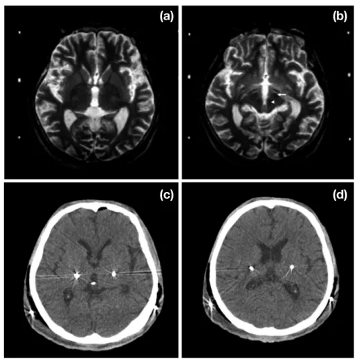
Fig. 3. Deep brain stimulation surgery of pre-operative of axial T2-weighted MR image(a) and axial T2-weighthed image of subthalamic
nucleus(arrow) and red nucleus(arrowhead) and post-operative axial CT image with air bilateral subthalamic nucleus and deep brain
stimulation(c, d).
Fig. 4. Pre-operative MR and postoperative CT merge showing tips of ImageJ used of electrodes fused to bilateral trajectories of planned subthalamic nucleus and deep brain stimulation. ImageJ program used of color merge function display of CT and MR image.
(digital imaging and communications in medicine) 파일 및 raw 데이터로 획득한다. 뇌심부자극술 환자에게 전극을 주 입한 수술 후 CT 영상과 수술 전의 MR 영상을 융합한 영상 을 분석하고 평가하였다. 파킨슨병으로 진단하여 뇌심부자극술 환자를 대상으 로 CT 및 MR을 스캔하기 위해 정보 제공 동의 및 연구윤 리 지침에 따라 작성하였다. 뇌심부자극술 환자의 수술 전
에 3 T MRI(Philips Intera, Eindhoven, the Netherlands)으 로 스캔하였고, 수술 후에는 MDCT(Sensation 64, Siemens, Erlangen, Germany)을 이용하여 스캔하였다.
뇌심부자극술의 수술 전의 MR 영상의 스캔은 기준선 인 AC-PC(anterior commissure-posterior commissure) line
Fig. 5. CT and MR image fusion of red(MR) and green(CT)(a), green(CT) and blue(MR)(b), red(CT) and gray(MR)(c), red(MR) and
gray(CT)(d), red(MR) and blue(CT)(e), and red(CT) and green(MR)(f).
(a) (b)
(c) (d)
득은 경구개(hard palate)에서 12°에 위치하는 AC-PC line 을 기준으로 하여(Kim et al. 2009) 촬영 조건은120kV, dose modulation(100~250mA), slice thickness differed between 0.6 and 1.25mm로 스캔하였다(Fig. 2).
2. 영상 합성 및 분석
뇌심부자극술의 수술 전의 MR 영상과 수술 후의 CT 영
상으로 전극의 위치 및 시상밑핵을 비교하기 위해 ImageJ (NIH, Bethesda, MD, USA) 프로그램을 이용하여 MR 영상 과 CT 영상을 image color의 merge channels 기능을 이용하 여 영상을 융합하여 영상에서 전극의 위치가 시상밑핵에 정 확하게 존재하는지를 융합 영상으로 확인하였다.
결 과
연구결과는 뇌심부자극술 시술 환자에 대한 영상 정보를 제공하고 전극 삽입에 대한 정확한 정보를 제공하여 임상 에서 치료에 적용할 수 있다. 이에 뇌심부자극술의 수술전 의 MR 영상에서 시상밑핵과 적핵(red nucleus)을 확인하였 다(Fig. 3). 뇌심부자극술의 수술 후의 CT 영상을 획득하여 영상을 ImageJ 프로그램에서 제공하는 color의 합성 기능을 이용하여 영상을 합성하였다(Fig. 3). CT 및 MRI 스캔의 융 합 영상 평가는 뇌심부자극술의 전극의 위치를 안전하고 실 제적으로 정확하게 제공하였다.ImageJ 프로그램의 color의 merge channels 합성 기능을 이용하여 영상의 전극의 위치의 정확한 묘출을 확인하기 위 해 다양한 red, green, blue, gray 기능에 뇌심부자극술의 수
술 전 MR 영상과 수술 후의 CT 영상을 삽입하여 융합 영상
을 묘출하였다(Fig. 4).
ImageJ 프로그램에서 묘출된 영상에서 red(MR 영상)와 green(CT 영상)의 합성은 Fig. 5a이고, green(CT 영상) 과 blue(MR 영상)은 Fig. 5b, red(CT 영상)와 gray(MR 영상)
고 찰
뇌심부자극술에서 전극의 위치를 시각화하여 정확한 목 표를 확보할 수 있게 하여 수정할 필요성이 없어 임상에서 는 정확한 정보의 제공이 필수적이다(Kremer et al. 2018). 이러한 전극의 정확한 정보의 제공은 향후 파킨슨병의 치료 계획에 유용하게 적용할 수 있어 뇌심부자극술에서 전극의 시상밑핵에 위치하는 명확한 정보의 제공이 필수적이다. 뇌심부자극술의 전극의 위치를 확인할 수 있는 비침 습적인 방법은 초음파(Walter et al. 2011), 수술 중 엑스 선(Verhagen et al. 2012), 수술 중의 3D 엑스선(Weise et al. 2012), Medtronic O-arm(Caire et al. 2010; Smith et al. 2011), 수술 중의 MRI(Huston et al. 2011; Cui et al. 2016) 또는 CT(Fiegele et al. 2008; Shahlaie et al. 2011) 검사가 임 상에서 현재의 유용한 정보를 제공할 수 있으며 정확한 전 극의 식별을 가능하게 한다. 이러한 검사 방법 중에서 뇌심 부자극술에서 현재 stereotactic frame을 삽입한 상태의 수술 후 CT가 frame이 없는 post-operative CT보다 우수하다는 증거는 없다고 보고하였다(Saleh et al. 2016). 이러한 뇌심 부자극술 환자의 수술 후의 CT 영상은 전극의 위치를 확인 하는 중요한 방법으로 임상에서 적용되고 있다. 또한 뇌심 부자극술의 전극에 대한 발열현상에 대한 고주파 출력에 대 한 제한기준의 보고가 있어 이러한 수술법은 치료계획에 주 의가 요구된다(Yeon et al. 2017). MR 및 CT 영상의 융합은 뇌심부자극술에 따른 전극 의 위치를 정확하게 알 수 있고 수술 전 및 수술 후의 전 극 위치를 직접적으로 비교할 수 있게 한다(Barnaure et al. 2015). CT와 MR 영상의 융합에서 CT는 안전성과 공간분해 능을 제공하고 MR 영상은 조직의 우수한 대조도의 이점이 있으나, 이러한 CT 및 MR의 두 가지 영상의 융합은 오차의 요인을 수반하고 있다(Ferroli et al. 2004; Pinsker et al. 2008). 기존의 뇌심부자극술의 CT와 MR 영상의 융합 연구
는 위치의 정확성에 맞춘 연구를 집중적으로 연구하여 보고 하였다(Ferroli et al. 2004; Thani et al. 2011). 이러한 뇌심부 자극술의 전극의 정확성은 아티팩트와 여러 알고리즘에 관
련된 요인에 따라 달라진다. 이번 연구에서는 뇌심부자극술
의 CT와 MR 영상에서 시상밑핵과 전극의 묘출 여부를 확
인하기 위해 ImageJ 프로그램으로 MR 영상과 CT 영상을 image color의 merge channels 기능을 이용하여 영상을 융합 하여 영상에서 정확한 전극이 시상밑핵에 존재 여부의 결과 는 red(CT 영상)와 green(MR 영상)의 합성에서 Fig. 5f 같 이 전극과 시상밑핵이 정확한 위치의 정보가 모두 묘출되었 다. 이러한 융합적인 영상은 전극의 궤적을 직접 비교할 수 있으므로 전극의 거리를 측정할 필요가 없는 장점이 있다. 또한, 수술 후 MRI와 관련된 위험을 피하는 것 외에도 MRI 와 비교하여 CT를 빠른 시간에 스캔하여 영상을 획득하여 환자에게 편안함과 함께 잠재적인 움직임의 아티팩트를 줄 이는 데 효과적으로 적용할 수 있다. 검사실 내에 설치된 워크스테이션 및 3D 영상 프로그램 을 이용하는 방법을 임상에서 적용하여 응용하고 있지만 이 러한 ImageJ의 영상 융합기능을 이용하여 뇌심부자극술에 서의 전극의 위치를 식별하고 확인하는 데 본 연구의 결과 를 응용할 수 있다. 이러한 영상의 융합에 대한 기존 연구에 서는 CT와 MR 영상의 융합의 차이는 통계적으로 유의하지 않다고 보고하였다(Yoshida et al. 2008). 향후 수술 후의 전 극의 위치에 대한 정보의 경과를 확인하는 데에도 적용할 수 있다(Lee et al. 2010). 연구결과의 활용은 뇌질환 환자에서 뇌심부자극술을 시 행한 CT 영상과 MR 영상의 정보를 임상에서 적용할 수 있 고 진단 및 치료에 응용할 수 있는 연구결과가 기대된다. 연구의 제한점은 첫째, 다양한 및 3D 영상 기법 및 MPR (multiplanar reformation)를 적용하여 정확한 전극의 위치를 확인하는 추가적인 연구가 필요하다. 둘째는 CT와 MR 영 상에서 x축, y축, z축의 차이를 측정하여 통계적으로 유의성 을 검정할 필요가 있다. 셋째 MR 영상의 획득을 다양한 펄 스 시퀀스를 이용하여 영상을 획득하여 CT와 MR 영상의 융합 영상에 대한 정량화의 연구가 필요하고, 마지막으로 다양한 환자를 대상으로 임상에서 획득한 영상으로 추가적 인 연구의 필요성이 있다.
결 론
프로그램을 이용한 MR 및 CT 영상의 융합은 뇌심부자극 술 환자에서 전극의 위치를 확인하는 데 안전하고 실용적으 로 응용할 수 있는 기술이며 정확도를 제공한다. 이러한 CT 와 MR의 융합 정보의 영상을 임상에서 적용할 수 있다.사 사
본 연구는 2019학년도 원광보건대학교 연구비 지원에 의 하여 연구되었음.참 고 문 헌
Barnaure I, Pollak P, Momjian S, Horvath J, Lovblad KO, Boëx C, Remuinan J, Burkhard P and Vargas MI. 2015. Evaluation of electrode position in deep brain stimulation by image fusion(MRI and CT). Neuroradiology 57(9):903-908.
Caire F, Gantois C, Torny F, Ranoux D. Maubon A and Moreau JJ. 2010. Intraoperative use of the Medtronic O-arm for deep brain stimulation procedures. Stereotact. Funct. Neu-rosurg. 88(2):109-114.
Cui Z, Pan L, Song H, Xu X, Xu B, Yu X and Ling Z. 2016. Intraoperative MRI for optimizing electrode placement for deep brain stimulation of the subthalamic nucleus in Par-kinson disease. J. Neurosurg. 124:62-69.
Cuny E, Guehl D, Burbaud P, Gross C, Dousset V and Rougier A. 2002. Lack of agreement between direct magnetic res-onance imaging and statistical determination of a subtha-lamic target: the role of electrophysiological guidance. J. Neurosurg. 97(3):591-597.
Engelhardt J, Guehl D, Damon-Perrière N, Branchard O, Bur-baud P and Cuny E. 2018. Localization of deep brain stim-ulation electrode by image registration is software depen-dent: a comparative study between four widely used soft-ware programs. Stereotact. Funct. Neurosurg. 96(6):364-369.
Ferroli P, Franzini A, Marras C, Maccagnano E, D’Incerti L and Broggi G. 2004. A simple method to assess accuracy of deep brain stimulation electrode placement: pre-operative stereotactic CT+postoperative MR image fusion. Stereo-tact. Funct. Neurosurg. 82(1):14-19.
Fiegele T, Feuchtner G, Sohm F, Bauer R, Anto JV, Gotwald T, Twerdy K and Einser W. 2008. Accuracy of stereotactic electrode placement in deep brain stimulation by intraoper-ative computed tomography. Parkinsonism Relat. Disord.
14(8):595-599.
Hamid H, Mitchell R, Mocroft P, Milner J and Pall H. 2005. Tar-geting the subthalamic nucleus for deep brain stimulation: technical approach and fusion of pre- and postoperative MR images to define accuracy of lead placement. J. Neurol. Neu-rosurg. Psychiatry 76(3):409-414.
Huston OO, Watson RE, Bernstein MA, McGee KP, Stead SM, Gorman DA, Lee KH and Huston J. 2011. Intraoperative
CT in subthalamic nucleus stimulation in Parkinson disease. Clin. Neurol. Neurosurg. 112(1):47-53.
Papavassiliou E, Rau G, Heath S, Abosch A, Barbaro NM, Larson PS, Lamborn K and Starr PA. 2004. Thalamic deep brain stimulation for essential tremor: relation of lead loca-tion to outcome. Neurosurgery 54(5):1120-1130.
Perlmutter JS and Mink JW. 2006. Deep brain stimulation. Annu. Rev. Neurosci. 29:229-257.
Pinsker MO, Herzog J, Falk D, Volkmann J, Deuschl G and Mehdorn M. 2008. Accuracy and distortion of deep brain stimulation electrodes on postoperative MRI and CT. Zen-tralbl. Neurochir. 69(3):144-147.
Saleh C, Dooms G, Berthold C and Hertel F. 2016. Post-opera-tive imaging in deep brain stimulation: A controversial issue. Neuroradiol. J. 29(4):244-249.
Shahlaie K, Larson PS and Starr PA. 2011. Intraoperative computed tomography for deep brain stimulation surgery: Technique and accuracy assessment. Neurosurgery 68:S114- S124.
Shin M, Lefaucheur JP, Penholate MF, Brugires P, Gurruchaga JM and Nguyen JP. 2007. Subthalamic nucleus stimulation in Parkinson’s disease: postoperative CT-MRI fusion imag-es confirm accuracy of electrode placement using intraop-erative multiunit recording. Neurophysiol. Clin. 37(6):457-466.
Slavin KV, Thulborn KR, Wess C and Nersesyan H. 2006.
Di-Verhagen Metman L, Pilitsis JG, Stebbins GT, Bot M and Bakay RA. 2012. Intraoperative x-ray to measure distance between DBS leads: a reliability study. Mov. Disord. 27(8):1056-1059.
Walter U, Kirsch M, Wittstock M, Müller JU, Benecke R and Wolters A. 2011. Transcranial sonographic localization of deep brain stimulation electrodes is safe, reliable and pre-dicts clinical outcome. Ultrasound Med. Biol. 37(9):1382-1391.
Weise L, Eibach S, Seifert V and Setzer M. 2012. Intraopera-tive 3D fluoroscopy in stereotactic surgery. Acta. Neurochi.
154(5):815-821.
Yeon KJ, Chang YA, Lee SK and Lee TS. 2017. Evaluation of clinical usefulness of radio-frequency power limitation in brain MRI of patients with deep brain stimulation. J. Radi-at. Indt. 11(3):139-144.
Yoshida F, Miyagi Y, Morioka T, Hashiguchi K, Murakami N, Matsumoto K, Nagata S and Sasaki T. 2008. Assessment of contact location in subthalamic stimulation for Parkinson’s disease by coregistration of computed tomography images. Stereotact. Funct. Neurosurg. 86(3):162-166.
Received: 24 May 2019 Revised: 12 June 2019 Revision accepted: 21 June 2019