저작자표시-비영리-변경금지 2.0 대한민국 이용자는 아래의 조건을 따르는 경우에 한하여 자유롭게 l 이 저작물을 복제, 배포, 전송, 전시, 공연 및 방송할 수 있습니다. 다음과 같은 조건을 따라야 합니다: l 귀하는, 이 저작물의 재이용이나 배포의 경우, 이 저작물에 적용된 이용허락조건 을 명확하게 나타내어야 합니다. l 저작권자로부터 별도의 허가를 받으면 이러한 조건들은 적용되지 않습니다. 저작권법에 따른 이용자의 권리는 위의 내용에 의하여 영향을 받지 않습니다. 이것은 이용허락규약(Legal Code)을 이해하기 쉽게 요약한 것입니다. Disclaimer 저작자표시. 귀하는 원저작자를 표시하여야 합니다. 비영리. 귀하는 이 저작물을 영리 목적으로 이용할 수 없습니다. 변경금지. 귀하는 이 저작물을 개작, 변형 또는 가공할 수 없습니다.
A Doctoral Dissertation
Study on Ca
2+
signaling-mediated
regulation of somatic K
+
outward currents
contributing to neuronal excitability in
dissociated hippocampal neurons of rats
Yoon-sil Yang
Department of Medicine
Graduate School
Jeju National University
초대 배양한 흰쥐 해마 신경세포에서 신경세포
흥분성에 관여하는 세포체 칼륨 통로 조절
과정의 칼슘 매개 기전에 대한 연구
지도교수: 정 성 철
양 윤 실
이 논문을 의학 박사학위 논문으로 제출함
2015년 2월
양윤실의 의학 박사학위 논문을 인준함
심사위원장
위 원
위 원
위 원
위 원
제주대학교 대학원
2015년 2월
Study on Ca
2+signaling-mediated regulation of somatic
K
+outward currents contributing to neuronal
excitability in dissociated hippocampal neurons of rats
Yoon-Sil Yang
(Supervised by professor Sung-Cherl Jung)
A thesis submitted in partial fulfillment of the requirement for the
degree of doctor of philosophy in medicine
February, 2015
This thesis has been examined and approved.
Date
Department of Medicine
Graduate School
Jeju National University
1
ABSTRACT
In hippocampal neurons, intrinsic excitability (IE) modulated by dynamic synaptic activity is important to determining neuronal functions such as information processing and
cognition. Voltage-dependent K+ channels (KV channels) showing outward K
+
currents participate in both IE and synaptic excitability by regulating membrane conductance and
potential.Therefore, it is necessary to identify the mechanism regulating expression and
kinetics of KV channels. KV channels consist of two types of currents. One is rapidly
inactivated or transient current (A-type current, IA), which is known to contribute to
learning and memory mechanisms by regulating both somatic and dendritic excitability.
The other is slowly inactivated or delayed rectifier current (IDR), which participates in
neuronal firing.
Recently, the downregulation of somatic IA channels is systemically targeted by Ca
2+
influx via synaptic NMDA receptors (NMDARs). However, spatial restriction between synapses and soma in hippocampal neurons strongly suggests that there may be possible
cellular links responsible for synaptic Ca2+ influx for somatic IA downregulation.
Therefore, in the first theme of the current paper, testing was performed to determine
whether two major receptors of endoplasmic reticulum (ER), ryanodine and IP3 receptors
(RyRs and IP3Rs, respectively), participate in Ca
2+
-mediated IA downregulation in
dissociated hippocampal neurons. Downregulation of IA channels was induced by high
Ca2+ (3.6 mM, for 24 hrs to culture media) or glycine (200 μM), which induces chemical
long-term potentiation (LTP), and electrophysiological measurement of the peak of IA was
performed using a whole-cell patch. In results, high Ca2+ effect in reducing IA peak was
clearly abolished by antagonists of NMDARs or voltage-dependent Ca2+ channels
2
synaptic activities involving NMDARs and VDCCs. In this cellular processing, blocking
RyRs (by Ryanodine, 10 μM) completely abolished IA downregulation, while blocking
IP3Rs (by 2APB, 100 μM) did not show any effects. In addition, treatment with Ryanodine
also resulted in reduced Ca2+-increased activity of protein kinase A (PKA, cyclic
AMP-dependent protein kinase), indicating that subsequential signaling cascades including ER
and PKA play roles in regulation of somatic IA. I suggest here that contribution of RyRs to
Ca2+-induced Ca2+ release (CICR) is required for regulation of neuronal excitability via IA
channel trafficking.
IDR channels act to maintain homeostasis by regulating membrane excitabilities in
pathological conditions such as epileptic seizure. However, it is not clear how their kinases or phosphatases are activated during pathological condition. To address this issue,
I attempted to confirm mechanisms for regulation of IDR for prevention of overexcitability
under high Ca2+ condition (3.6 mM, for 24 hrs) in dissociated hippocampal neurons. In
results, it was proved that IDR was enhanced by high Ca2+ treatment without change of
kinetic properties. This increment of IDR was clearly inhibited by nimodipine (VDCCs
antagonist, 10 μM) but not APV (NMDARs antagonist, 100 μM), suggesting that the
regulation of IDR is targeted by Ca
2+
influx through VDCCs with the independence on the
downregulation of IA. It is also confirmed that small conductance Ca
2+
-activated K+
channels (SK channels) are not involved in IDR upregulation. In addition, results from use
of PP2 (1 μM) to block Src family tyrosine kinases (SFKs) showed that the enhancement
of IDR is dependent on SFKs activation. These results indicate that SFKs activated by Ca
2+
influx via VDCCs participate in IDR upregulation to protect neurons from overexcitability
conditions.
Consequently, the regulation of K+ outward currents in local somatic area of neurons is
quietly correlated in synaptic activities which occur in distant. However, hippocampal neurons are not questionable to actively reflect the dynamic but systemic signaling
3
cascades linking synaptic and somatic processings. These successive cellular processings provide a possibility that somatic membrane excitability is directly regulated by synaptic
plasticities such as LTP via regulation of K+ channels as well as Ca2+ signaling,
determining neuronal excitabilities under various physiological and pathological conditions.
4
CONTENTS
ABSTRACT ………
1
CONTENTS ………...
4
LIST OF FIGURES ………...
5
LIST OF TABLES ………...
8
CONTENTS OF PART Ⅰ ………...
9
CONTENTS OF PART Ⅱ ………...
11
PART Ⅰ ………
13
PART Ⅱ ………
50
ABSTRACT IN KOREAN ………
76
5
LIST OF FIGURES
Figure 1.
Effects of the high Ca
2+treatment on cell viability in cultured
hippocampal neurons ...
24
Figure 2.
The peaks of somatic I
Awere decreased by the enhancement
of synaptic Ca
2+influx through NMDARs and VDCCs ...
25
Figure 3.
Activation and inactivation properties of I
Achannels are not
changed by high Ca
2+treatment ...
26
Figure 4.
CICR showed the signaling specificity dependent on RyRs but
not IP
3Rs in somatic I
Adownregulation ...
29
Figure 5.
Downregulation of somatic I
Ainduced by chemical LTP
resulted from RyRs activation ...
30
Figure 6.
RyRs induce faster enhancement of [Ca
2+]
ithan IP
3Rs in
hippocampal neurons ...
33
Figure 7.
Downregulation of I
Aby opening RyRs did not require
6
Figure 8.
RyRs-mediated I
Adownregulation required the activation of
PKA ...
38
Figure 9.
Active PKA was reduced by blocking RyRs in cultured
hippocampal neurons ...
39
Figure 10.
A simplified model of Ca
2+-mediated downregulation of
somatic I
Achannels induced by synaptic activation ...
41
Figure 11.
I
DRare increased by high Ca
2+application in dissociated
hippocampal neurons ...
56
Figure 12.
Upregulating I
DRby high Ca
2+does not involve significant
alteration of activation properties in hippocampal neurons ...
57
Figure 13.
I
DRupregulation to prevent neuronal overexcitation requires
VDCCs ...
60
Figure 14.
The blockade of I
DRupregulation by nimodipine does not
exhibit any changes of I
DRactivation property ...
61
7
Figure 16.
No effects of nimodipine alone on the density of I
DRchannels
were observed in hippocampal neurons ...
65
Figure 17.
Activation of RyRs and PKA did not affect the upregulation of
I
DRchannels under the high Ca
2+condition ...
66
Figure 18.
SK channels were not involved in the upregulation of
sustained currents under high Ca
2+condition ...
68
Figure 19.
SFKs played important role in upregulation of I
DRchannels under high Ca
2+condition in dissociated
hippocampal neurons ...
69
8
LIST OF TABLES
Table 1.
The changes of A-type currents induced by cLTP ...
31
9
CONTENTS OF PART Ⅰ
1. INTRODUCTION ...
14
2. MATERIALS AND METHODS ...
17
2.1. Materials
2.2. Experimental animals
2.3. Hippocampal primary cultures
2.4. Measurement of neuronal viability
2.5. Electrophysiology
2.6. Intracellular calcium measurements
2.7. PKA activity
2.8 Statistical analysis
3. RESULTS ...
22
3.1. Ca
2+influx through NMDARs and VDCCs can modulate somatic
excitability by downregulating I
Achannels
10
3.2. Ryanodine receptors of intracellular Ca
2+store, but not IP
3receptors, are crucial for I
Adownregulation
3.3. RyRs of ER more rapidly release Ca
2+than IP
3Rs in dissociated
hippocampal neurons
3.4. Activation of RyRs independently downregulates I
Awithout
synaptic activity
3.5. RyRs-mediated signaling absolutely requires PKA phosphorylation
for I
Adownregulation
4. DISCUSSION ...
42
11
CONTENTS OF PART Ⅱ
1. INTRODUCTION ...
51
2. MATERIALS AND METHODS ...
53
2.1. Materials
2.2. Hippocampal primary cultures
2.3. Electrophysiology
2.4. Statistical analysis
3. RESULTS ...
55
3.1. Enhancement of I
DRunder the high Ca
2+condition did not include
any changes of kinetic properties in hippocampal neurons derived from
DIV 6-9
3.2. Upregulation of I
DRunder high Ca
2+condition is mediated with
12
3.3. Nimodipine, alone, does not affect the density of I
DRin dissociated
hippocampal neurons
3.4. Upregulation of I
DRchannels is independent of Ca
2+signaling-mediated regulation of I
Achannels
3.5. No involvement of small conductance Ca
2+-activated K
+channels in
increased sustained outward current under high Ca
2+condition
3.6. High Ca
2+increases the density of I
DRthrough activation of SFKs in
the dissociated hippocampal neurons
4. DISCUSSION ...
70
13
Part Ⅰ
Ryanodine receptors of Ca
2+store are required
for downregulation of somatic A-type K
+channels
induced by the activation of synaptic NMDARs
to regulate excitability in neurons
14
1. Introduction
Hippocampus is extremely important to memory processes by consolidating information. Cellular mechanisms to explain the learning and memory functions are dominantly dependent on synaptic plasticity such as long-term potentiation (LTP) or depression (LTD) (Hebb, 1949; Maren and Baudry, 1995; Yang et al., 2014). These physiological synaptic adaptations to given issues are quietly considerable as a potent model to trigger the initial formation of memory at the cellular level but not enough to support neuronal mechanisms for storage of long-lasting information in the central nervous system (CNS). Many reports have demonstrated that the alteration of neuronal intrinsic excitability (IE), which is an ability to make action potentials (APs) with given inputs, may possibly play a role in cellular information storage (Aizenman and Linden, 2000; Daoudal and Debanne, 2003; Oh et al., 2003; Zhang and Linden, 2003; Xu et al., 2005; Kim and Linden, 2007). Therefore, verification of mechanical or functional links between synaptic and somatic alterations seems to be important for completion of a memory mechanism but difficult because of local restriction of synaptic plasticity (Frick et al., 2004). In this study, I attempted to determine
whether and how somatic excitability could be altered by synaptic activities through Ca2+
signaling cascades.
Potassium outward currents through various voltage-dependent or independent K+
channels are crucial factors in regulation of neuronal excitability and in determining the level
and pattern of neuronal responsiveness in hippocampus. Total outward K+ currents are
generally classified according to a transient or rapidly inactivating current, known as A-type
(IA) current, and a sustained or slow/non-inactivating current (delayed rectifier K
+
currents,
IDR) (Hoffman et al., 1997).
In the first section, I investigated the mechanism to explain Ca2+-mediated somatic IA
15
important for activity-dependent regulation of glutamatergic transmissions in dendritic processings and synaptic plasticity and their internalization from active spines, which are dependent on N-methyl-D-aspartate receptors (also known as the NMDA receptors or
NMDARs) activation (Kim et al., 2007; Hammond et al., 2008). It has been shown that Ca2+
-signaling dependent channel trafficking in both dendrites and spines is rapid but locally
restricted, because Ca2+ influx through NMDARs in active areas is not sufficient to activate
total kinases and auxiliary proteins associated with channel trafficking in a whole neuron.
However, the internalization of IA channels from active spines during glycine-induced
chemical LTP was also accompanied by the reduction of somatic IA, which was
electrophysiologically observed (Kim et al., 2007). In addition, synaptic LTP of CA1 neurons
induced by the paired pulse stimulation resulted in the reduction of somatic IA, which was
lasting at least during LTP (Jung and Hoffman, 2009). It is possible that the activation of synaptic NMDARs may contribute to the regulation of somatic excitability via leading the
internalization of somatic IA channels because either chemical or electrical LTP in
hippocampus are mediated with the activation of synaptic NMDARs. However it is still not
clear how glutamatergic activation in local synapses affects the trafficking of somatic IA
channels.
Synaptic and extrasynaptic NMDARs in mechanisms of synaptic plasticities observed in
mammalian CNS, show distinctive and opposite functions in the modulation of Ca2+
signaling and glutamatergic responses. Despite some arguments, synaptic and extrasynaptic NMDARs are commonly thought to participate in LTP and LTD, respectively (Lu et al.,
2001). In CA1 neurons, Ca2+ influx through synaptic NMDARs contributes to upregulation
of α-amino-3-hydroxy-5-methyl-4-isoxazolepropionic acid receptor (also known as AMPA
receptors or AMPARs), resulting in LTP. In particular, the amount of Ca2+ entering synaptic
sites determines the potentiation level and duration of LTP, indicating the potential role of
16
and inositol-tri-phophate receptors (IP3Rs) in intracellular Ca
2+
store are also involved in NMDA-dependent synaptic plasticities of both LTP and LTD (Schiegg et al., 1995; Reyes
and Stanton, 1996; Adasme et al., 2011). Furthermore, opening Ca2+ stores may be especially
considerable as a dominant factor in triggering widespread cellular events following LTP
induction because synaptic NMDAR activation increases cytosolic Ca2+ levels by opening
RyRs and IP3Rs (Emptage et al., 1999; Nishiyama et al., 2000; Taufiq et al., 2005). These
results indicate that Ca2+ influx occurring locally in synapses may open Ca2+ stores to extend
their cellular functions. Therefore, revealing the alteration of outward flow via somatic K+
channels by synaptic modulations is critically important for understanding how voltage-dependent channels decide neuronal functions in synaptic plasticity for learning and memory mechanism.
17
2. Materials and Methods
2.1. Materials
Minimal essential medium (MEM), Neurobasal medium, fetal bovine serum (FBS), penicillin/streptomycin, B-27 serum-free supplement, and L-glutamine were purchased from Gibco (Gland Island, NY, USA). Nimodipine, 2-Aminoethoxydiphenylborane (2APB), Ryanodine, Tetrodotoxin (TTX), and H89 were purchased from Tocris (Ellisville, MO, USA). Fura-2 acetoxymethyl ester (Fura-2 AM) was purchased from Invitrogen (Carlsbad, CA, USA). All other reagents were purchased from Sigma-Aldrich (St Louis, MO, USA), unless indicated otherwise.
2.2. Experimental animals
Sprague-Dawley (SD) rats were used in this experiment were bred in the animal facility of the Medical School of Jeju National University. The environment of the breeding room was automatically controlled at a temperature of 23 ± 3℃ and 50 ± 10 % humidity. The lighting system was set to repetitively turn on and off for 12 hours a day. Food and water were given ad libitum. Two females and a male SD rats were mated in a cage for breeding.
All experiments and procedures with animals were performed with permission from the Animal Care and Use Committee of Jeju National University.
2.3. Hippocampal primary cultures
18
embryos removed from deeply anesthetized pregnant rats were transferred to an ice-cold
normal tyrode solution containing the following (in mM): 140 NaCl, 5.4 KCl, 2.3 MgCl2, 10
HEPES, 5 glucose, pH 7.4 adjusted with NaOH. Hippocampi isolated from embryonic rat brains were transferred to ice-cold MEM containing Earle’s salts and glutamine with 10% FBS, 0.45% glucose, 1 mM sodium pyruvate, 25 μM glutamate, and antibiotics, and then
triturated. For electrophysiological studies and intracellular Ca2+ recordings, the cells were
counted and seeded on glass coverslips (Fisher Scientific) coated with poly-L-lysine at a
density of 9 X 10⁴ cells/ml and maintained at 37°C in 95% air and 5% CO2. After 7
hours, the whole plating medium was changed to Neurobasal medium containing B-27, and half of the medium was changed twice per week. To examine neuronal viability, dissociated hippocampal neurons were seeded on poly-L-lysine coated 24 well plates at a density of 1.8
X 105 cells/ml and 1 μM arabinofuranosyl cytidine (Ara-C) was added to prevent glial
proliferation at DIV 4 and 8.
2.4. Measurement of neuronal viability
To assess effects of high Ca2+ on cell viability, DIV 9 cultured hippocampal neurons 3.6
mM CaCl2 was treated for 24 hrs, followed by addition of 0.4 mg/ml MTT
(3-(4,5-methylthiazol-2-yl)-2,5-diphenyl-tetrazolium bromide). One hour later, medium was gently removed and DMSO was added to each well to dissolve formazan crystals. The neuronal viability was obtained by reading absorbance at 550 nm using a microplate reader (Model 550, Bio-rad, USA).
19
Primary dissociated culture neurons of DIV 6-8 were used for patch-clamp recordings of
transient A-type K+ channels. Coverslips containing these young hippocampal neurons were
transferred to a recording chamber with a continuous flow of recording solution containing
the following (in mM) : 145 NaCl, 5 KCl, 2 CaCl2, 1.3 MgCl2, 10 HEPES, 10 glucose, pH
7.4 with NaOH, and bubbled with 95% O2 and 5% CO2. TTX (0.5 μM) was added to the
recording solution to block the voltage-dependent Na+ channels. The patch pipettes (4-6 MΩ)
were filled with an internal solution containing the following (in mM) : 20 KCl, 125 K+
-gluconate, 4 NaCl, 10 HEPES, 0.5 EGTA, 4 ATP, 0.3 tris-GTP, and 10 phosphocreatin, and pH 7.2 was adjusted with KOH. During recordings, the series resistance varied between 8~30 MΩ, and recordings where the series resistance varied by more than 10% were rejected.
Transient and sustained K+ currents were digitally separated using a prepulse protocol after
subtracting leak currents. Peak currents were measured at +60 mV after 200 ms prepulse to either -120 mV or -20 mV. All electrophysiological data were recorded using an Axopatch 200B amplifier (Axon Instruments, Foster City, CA, USA), and command pulse generation, data acquisition and analysis were performed using Digidata 1322A convertor (Axon Instruments), pClamp 8 (Axon Instruments), and IGOR Pro (Wavemetrics, Lake Oswego, OR) software.
2.6. Intracellular calcium measurements
DIV 9 dissociated culture neurons were loaded with a fluorescent Ca2+ indicator Fura-2AM (5 μM) and 0.1% pluronic F127at 37°C for 45min in a dark incubator with or without ryanodine or 2APB. The loaded neurons on coverslips were transferred to a recording chamber with a continuous flow of recording solution and imaged using an inverted Olympus IX71 microscope (Olympus, Japan). Fluorescence excitation was measured using 340 and 380 nm filter sets and controlled with a high-speed filter switching device (Sutter
20
Instruments, Lambda DG-4). Digitized fluorescence image were acquired at 6s intervals using a cooled-charged device (CCP) camera (Roper Scientific, USA). To confirm changes
of Ca2+ levels after enhancing synaptic activity, 20 mM KCl and 100 μM Glycine were added
to the recording solution. Ratio images were obtained by acquiring pairs of images at alternate excitation wavelengths (340/380 nm) and analyzed using Metafluor software (Universal Imaging, Sunnyvale, CA, USA).
2.7. PKA activity
DIV 7 hippocampal neurons treated with antagonist of RyRs or IP3Rs for 24 hrs were
lysed with modified radio-immunoprecipitation assay (RIPA) buffer containing protease inhibitors and kept on ice for 15 min. The lysates were centrifuged at 13000 rpm for 15 min at 4°C and supernatants were collected from the lysates. Protein concentrations were determined using bio-rad protein assay (Bio-Rad, Hercules, CA, USA). PKA activity was measured using a non-radioactive PKA Kinase activity assay kit (Enzo life science, Plymouth Meeting, PA). Kinase assay dilution buffer (50 µl) was added to the PKA substrate pre-coated microplate wells for 10 min and removed. The kinase reaction was then initiated by addition of 0.05 μg proteins and ATP for 30 min at 30°C. After emptying the contents of each well, the microplate was incubated with a phosphosubstrate–specific antibody for 30 min at room temperature, followed by washing four times with wash buffer. The peroxidase-conjugated secondary antibody was added, followed by incubation for 30 min at room temperature. The color was developed with tetramethylbenzidine (TMB) substrate and stopped with stop solution. The intensity of the color was measured at 450 nm.
21
Data analysis was performed and statistical significance was determined using Excel (Microsoft, USA) software and data were expressed as mean value ± standard error of mean (SEM). The Student’s t-test was used, and significance was indicated for p values less than 0.05 or 0.01.
22
3. Results
3.1. Ca2+ influx through NMDARs and VDCCs can modulate somatic excitability by downregulating IA channels
In previous study, synaptic activation induced the downregulation of somatic IA channels
(Jung and Hoffman, 2009). However, it is not clear how synaptic excitation affects somatic
IA channels. I tested this issue using DIV6-9 dissociated hippocampal neurons. The neurons
are exposed to high Ca2+ (3.6 mM CaCl2) for 24 hours to induce synaptic excitability (Jung
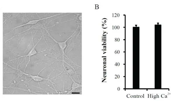
et al., 2008). Figure 1 shows DIV9 dissociated hippocampal neurons with glia and their viability (A and B). The neuronal viability was determined by measuring mitochondria activity in Ara-C treated neurons. No difference of neuronal viability was observed between
control and high Ca2+ application groups (Control= 100 ± 3.40 %, n= 48; High Ca2+= 103.71
± 3.16 %, n= 45, p= 0.43 compared with control), suggesting that high Ca2+ treatment did not
induce neuronal cell death, at least in this study.
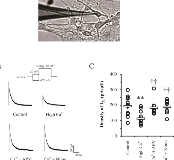
The synaptic activation by high Ca2+ treatment clearly reduced the amplitude of the
somatic IA peak (Figure 2). The high Ca
2+
group showed a significant decrease of
approximately 40 % IA density compared with that of the control group (Control= 193.28 ±
15.08 pA/pF, n = 14; High Ca2+= 113.58 ± 12.03 pA/pF, n = 12, p<0.01). To confirm whether
the downregulation of IA channels was dependent on synaptic activity, antagonists of sources
of synaptic Ca2+ influx such as NMDARs and VDCCs were applied. In neurons treated with
NMDAR antagonist APV under the condition of high Ca2+ for 24 hrs, the decrease of IA peak
was completely abolished (APV = 183.72 ± 20.23 pA/pF, n= 7, p= 0.727 and 0.008
compared with control and high Ca2+, respectively). Furthermore, the downregulation of IA
channels also appeared to be dependent on VDCCs in presynapse, as VDCCs antagonist
23
pA/pF, n= 8, p= 0.87 compared with control). These results indicate that the activation and enhancement of synaptic transmission can actively regulate intrinsic excitability by
decreasing density of IA channels in soma via Ca
2+
signaling pathways.
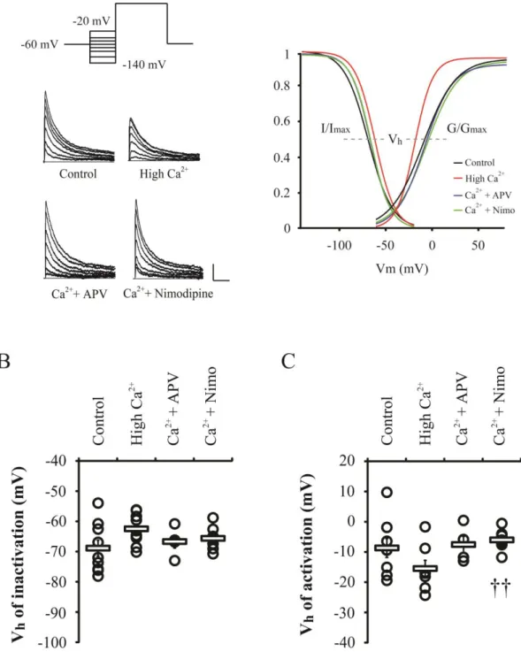
Figure 3 shows the activation and inactivation properties of IA channels in high Ca
2+
treated neurons with APV or nimodipine. It was observed that a slight depolarized-shift of IA
inactivation curve was induced by high Ca2+ and voltage half (Vh) of IA inactivation was
increased, but it was not significant (Control= -68.87 ± 2.61 mV; High Ca2+= -62.48 ± 1.62
mV, p= 0.07). Activation curve of IA was shifted to the left by high Ca
2+
and Vh of activation
was slightly decreased (Control= -8.68 ± 3.14 mV; High Ca2+= -15.51 ± 2.73 mV, p= 0.16).
Both NMDARs and VDCCs antagonists did not change activation and inactivation
properties of IA channels (Vh of inactivation: APV= -66.70 ± 2.15 mV; Nimodipine= -65.63
± 1.23, Vh of activation: APV= -7.54 ± 2.67 mV; Nimodipine= -6.01 ± 1.07). This finding
suggests that the downregulation of IA channels by high Ca2+ application may result from the
24
Figure 1. Effects of the high Ca2+ treatment on cell viability in cultured hippocampal neurons. A. DIV 9 cultured hippocampal neurons. The morphology was observed by using
Metamorph software. Scale bar; 100 μm. B. Neuronal viability was not affected by high Ca2+
application. The high Ca2+ (3.6 mM CaCl2) was applied to Ara-C exposed hippocampal
neurons for 24hrs and neuronal viability was evaluated by using MTT assay. Error bars represent SEM.
25
Figure 2. The peaks of somatic IA were decreased by the enhancement of synaptic Ca 2+
influx through NMDARs and VDCCs. A. Whole-cell patch clamp in DIV 7 primary
hippocampal neurons. The image was obtained by using RS image (Roper Scientific). B.
Example traces of IA recorded in dissociated neurons (DIV 6-9) after high Ca
2+
(3.6 mM) treatment with APV (100 μM) or nimodipine (10 μM) in culture media for 24 hrs. The APV and nimodipine can block NMDARs and VDCCs, respectively. Scale bars; 500 pA, 100 ms.
C. Individual (circle) and averaged (square) transient current densities. Error bars represent
26
Figure 3. Activation and inactivation properties of IA channels are not changed by high
Ca2+ treatment. The high Ca2+ (3.6 mM) was treated with APV (100 μM) or nimodipine (10 μM) and then currents kinetics were electrophyically recorded. The inactivation properties were measured at 60 mV after 200 ms prepulse (-140 to -20 mV with 20 or 40 mV steps) and
27
activation properties at -60 to 80 mV with 20 or 40 mV steps (prepulse: -140 mV for 200 ms).
A. Examples of inactivation traces and boltzmann fitted gating kinetics of IA channels. Scale
bars 200 pA, 50ms. B and C. The voltage half (Vh) values of inactivation and activation
properties, indicated with thick gray dotted lines in A. Circles and squares are individuals
and averaged Vh value, respectively. Error bars represent SEM. p <0.01 compared with high
28
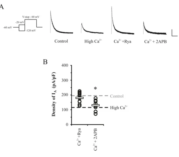
3.2. Ryanodine receptors of intracellular Ca2+ store, but not IP3 receptors, are crucial
for IA downregulation.
In this experiment, a role of ER Ca2+ store to find a Ca2+ mediator between synapse and
soma of neurons was tested by applying 10 μM Ryanodine or 100 μM 2APB under the high
Ca2+ condition for 24 hrs. Ryanodine and 2APB were used as antagonists of RyRs and IP3Rs,
respectively, which are major Ca2+ outflux channels in ER. As shown in Figure 4, the effect
of high Ca2+ in reducing IA peaks was completely blocked by Ryanodine application while
2APB did not affect the downregulation of IA (Ryanodine = 180.63 ± 9.7 pA/pF, p= 0.53;
2APB= 130.34 ± 12.27 pA/pF, p= 0.01). This suggests a possibility that Ca2+ influx during
synaptic enhancement primarily targets the RyRs to downregulate somatic IA channels.
Similarly, Ryanodine also prevented the reduction of IA peaks when chemical LTPs (cLTPs)
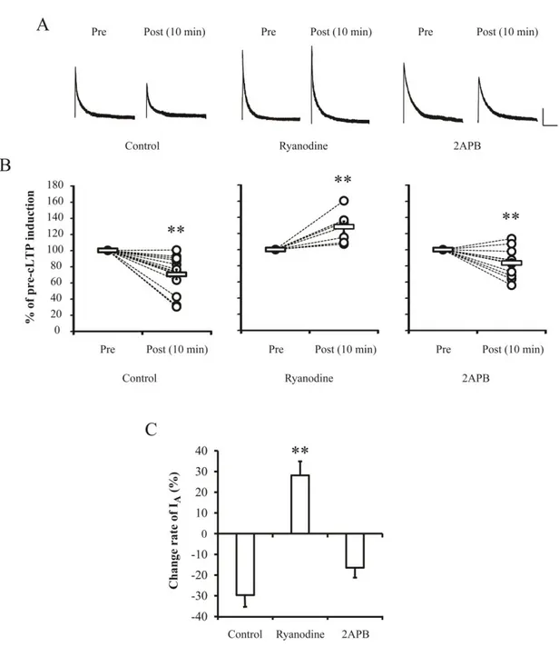
were induced by 200 μM glycine (Figure 5, before= 100 ± 0 %, 10 min after cLTP= 128.16 ±
6.71 %, p= 0.008 compared with before cLTPs) which significantly reduced IA peak
amplitude by approximately 30 % in the control (before= 100 ± 0 %; 10 min after cLTP= 70.35 ± 6.07 %, p= 0.001) or 2APB group (before= 100 ± 0 %; 10 min after cLTP= 83.53
± 4.74 %, p= 0.007). Table 1 shows the densities of IA before and 10 min after cLTP
induction. Interestingly, before cLTP induction, density of IA in Ryanodine-treated neurons
was significantly higher than that of the control group (Control= 1.66 ± 0.13 pA/pF; Ryanodine= 2.28 ± 0.28 pA/pF, p= 0.04). This indicates the existence of RyRs' roles to
participate in trafficking of somatic IA channels under the normal condition. These results
indicate that Ca2+ release from ER via RyRs induced by activation of postsynaptic NMDARs
may regulate somatic excitability by acting as cellular linkers between synaptic and somatic areas.
29
Figure 4. CICR showed the signaling specificity dependent on RyRs but not IP3Rs in
somatic IA downregulation. Ca 2+
-induced downregulation of IA was abolished by Ryanodine
(10 μM) to block RyRs of ER, while an IP3R blocker (2APB, 100 μM) did not show any
effects. A. Example traces of IA after high Ca
2+
treatment with Ryanodine or 2APB to culture
media for 24 hrs. Scale bars 500 pA, 100 ms. B. Averaged (square bars) densities of IA with
30
Figure 5. Downregulation of somatic IA induced by chemical LTP resulted from RyRs
activation. Ryanodine completely blocked the downregulation of IA shown in case of
chemical LTPs (cLTPs) which were induced by adding 200 μM glycine to recording solution.
A. Example traces of IA before and after 10 min of cLTP induction. Scale bars 500 pA, 100
ms. B. Normalized changes of IA peaks induced by cLTP. Open circles and square bars
indicate individuals and averaged values, respectively. C. Change rate of IA peak after cLTPs
32
3.3. RyRs of ER more rapidly release Ca2+ than IP3Rs in dissociated hippocampal
neurons.
Intracellular Ca2+ levels were measured to evaluate how RyRs of ER, not IP3Rs, are
involved in IA downregulation. To observe changes of somatic free Ca
2+
level after synaptic enhancement, 20 mM KCl and 100 μM glycine were applied to cultured neurons loaded with
fluorescent Ca2+ indicator Fura-2AM (Figure 6). Intracellular Ca2+ levels in
Ryanodine-treated neurons were increased slowly, compared with control and 2APB Ryanodine-treated neurons (Figure 6.D, Control= 196.86 ± 4.26 sec, n= 42; Ryanodine= 216.63 ± 4.45 sec, n= 38; 2APB= 201.6 ± 4.89, n= 40; Ryanodine with 2APB= 214.7 ± 3.9, n= 46). However, no
significant changes of Ca2+ wave peak and the level of late phase (5 min after synaptic
activation) were observed among each group (Peak of F1/F0: Control= 2.17 ± 0.06; Ryanodine= 2.11 ± 0.08; 2APB= 2.24 ± 0.08, 5 min after synaptic activation; Control= 1.71 ± 0.04, Ryanodine= 1.72 ± 0.06, 2APB= 1.76 ± 0.06). These results suggest that the
increasing rate of intracellular Ca2+ levels is more important than the total amount for
34
Figure 6. RyRs induce faster enhancement of [Ca2+]i than IP3Rs in hippocampal
neurons. Intracellular Ca2+ levels were more rapidly increased through RyRs of ER,
compared with IP3Rs. Ca
2+
levels were recorded in cultured hippocampal neurons by loading
fluorescent Ca2+ indicator Fura-2AM (5 μM). High K+ (20 mM) and glycine (100 μM) were
treated to induce neurotransmitter release and NMDARs activation in synaptic sites. A.
Cultured hippocampal neurons loaded with fura-2AM before and after high K+ and glycine
treatment. Ryanodine and 2APB were added to culture media for 45 min before recording. Scale bars 50 μm. B. Averaged F1/F0 of 340/380 ratio. C and D. Averaged peak of F1/F0 and time to peak. Error bars represent SEM. p < 0.01
35
3.4. Activation of RyRs independently downregulates IA without synaptic activity
It is clear that RyRs of Ca2+ stores are targeted by the activation of synaptic NMDA
receptors to regulate somatic excitability from the above. Those results suggest one more
question; ‘Can active RyRs independently induce IA downregulation?’. To answer this
question, RyRs agonists, Caffeine (5 mM) and 4CmC (50 μM), were hired. Caffeine was added to internal solution to prevent effects of adenosine receptors in plasma membrane and
4CmC was applied extracellularly to culture media for 24 hrs. The density of somatic IA was
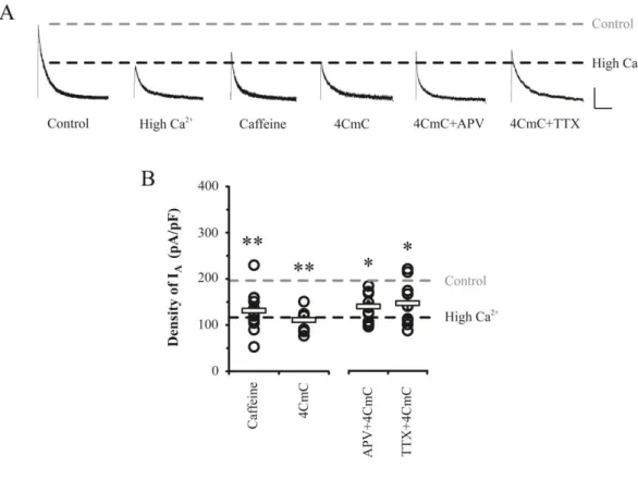
significantly reduced by either caffeine or 4CmC without the enhancement of synaptic transmission (Figure 7, caffeine= 131.17 ± 13.1 pA/pF, n= 11, p= 0.008; 4CmC= 110.88 ± 9.85 pA/pF, n= 8, p= 0.001). In addition, 4CmC effect was also observed in cases in which 100 μM APV or 0.5 μM TTX was added to abolish synaptic transmission(APV= 139.35 ± 8.93 pA/pF, n=11, TTX= 146.43 ± 13.96 pA/pF, n=10, P<0.05). These results indicate that
36
Figure 7. Downregulation of IA by opening RyRs did not require synaptic activity.
Caffeine or 4CmC treatment to open Ca2+ store via activating RyRs alone induced the
downregulation of IA without synaptic activities, as reduced IA channel densities were not
restored by APV (100 μM) or Na+
channel blocker TTX (0.5 μM). A. Example traces of IA
currents after caffeine (5 mM) or 4CmC (50 μM) treatments with/without APV and TTX to culture media for 24 hrs in pyramidal neurons. Scale bars 500 pA, 100 ms. B. Averaged
density of IA (square bars) with individual values (open circles). Error bars represent SEM. p
37
3.5. RyRs-mediated signaling absolutely requires PKA phosphorylation for IA
downregulation.
It is well known that the downregulation of IA channels is mediated by PKA activity
(Hoffman and Johnston, 1998; Schrader et al., 2002). Therefore, in the present study, PKA
effects were also tested by observation of IA changes after treatment with PKA antagonist
and a solid phase enzyme-linked immune-absorbent assay (ELISA). As shown in Figure 8,
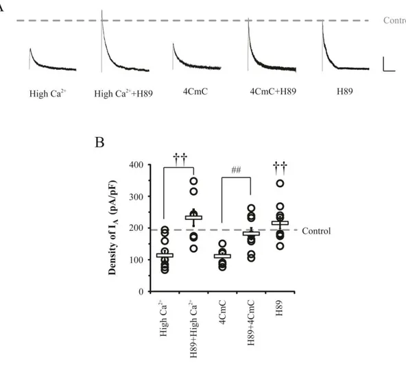
effects of high Ca2+ and 4CmC on density of somatic IA were significantly abolished by
adding PKA antagonist H89 (10 μM). H89 restored somatic IA peaks up to control levels
under conditions of high Ca2+ (232.32 ± 27.49 pA/pF, p<0.001 compared with high Ca2+) as
well as 4CmC application (182.13 ± 19 pA/pF p<0.01), while it did not affect IA densities in
normal condition (215.60 ± 19.24 pA/pF, p= 0.39). In addition, the effects of RyRs and IP3Rs
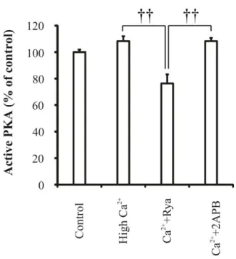
on PKA activity were observed by using ELISA under high Ca2+ condition (Figure 9). In
neurons treated with high Ca2+, PKA activity was slightly but not significantly increased
(Control= 100 ± 1.89 %, high Ca2+= 108.28 ± 3.71, P= 0.18). However, Ryanodine-treated
neurons showed a significant reduction of PKA activity under high Ca2+ condition
(Ryanodine= 76.38 ± 6.8 %, p= 0.05 and 0.03 compared with control and high Ca2+,
respectively), while 2APB did not affect the degree of active PKA (2APB= 108.28 ± 2.4 %).
These results indicate that RyRs-mediated IA downregulation absolutely involves PKA
38
Figure 8. RyRs-mediated IA downregulation required the activation of PKA. The
activation of PKA was crucial for neuronal excitability via IA downregulation in both
synaptic dependent and independent conditions. A. Example traces of IA after H89 (10 μM,
PKA inhibitors) treatment to culture media for 24hrs in various conditions. Scale bars 500
pA, 100 ms. B. Averaged densities of IA (square bars) with individual values (open circles).
39
Figure 9. Active PKA was reduced by blocking RyRs in cultured hippocampal neurons.
Normalized levels of PKA activities in each group were measured by using Elisa Assay. Error bars represent SEM. p < 0.01
41
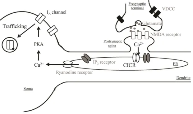
Figure 10. A simplified model of Ca2+-mediated downregulation of somatic IA channels
induced by synaptic activation. The enhancement of Ca2+ influx through synaptic NMDARs acts as a modulator to activate CICR via opening RyRs of ER. Opened RyRs more
rapidly releases Ca2+ from ER than IP3Rs and then Ca
2+
phosphorylates PKA .
Subsequentially, the activation of PKA induces the downregulation of IA channels in soma.
Consequentially, synaptic activation possibly regulates intrinsic excitability by internalizing
42
4. Discussion
A number of studies have investigated regulatory mechanisms of IA channels modulating
intrinsic excitability and synaptic plasticity since 1961 (Hagiwara, 1961; Kang et al., 2014).
Downregulation of IA channels is mediated by intracellular Ca
2+
signaling cascades activated
by Ca2+ influx through NMDARs of active postsynaptic sites. This IA downregulation is
absolutely required for the formation of synaptic plasticity induced by synaptic specific protocols and enhancement of intrinsic excitability (Ramakers and Storm, 2002; Watanabe et al., 2002; Jung et al., 2008; Jung and Hoffman, 2009). However, it is not clear how synaptic
activity influence somatic IA channels. In this study, it was confirmed that synaptic activity
participates in somatic excitability by regulating IA channels via activating RyRs of Ca
2+
store and PKA signaling in hippocampal neurons. IA channel density of plasma membrane
under the condition of high Ca2+ was significantly reduced and these changes were
dramatically blocked by postsynaptic NMDARs antagonist (Figure 2). This finding indicates
that postsynaptic Ca2+ influx through NMDARs is required for downregulation of IA
channels. VDCCs participating in neurotransmitter release in presynaptic terminals
(Tomizawa et al., 2002; Uriu et al., 2010) also seem to be crucial regulating IA channels, as
shown in experiments using VDCCs antagonist, nimodipine. This suggests that IA channels
are internalized from somatic membrane by Ca2+ influx through postsynaptic NMDARs
activated by glutamate released by presynaptic VDCCs activation.
How does synaptic activity influence somatic excitability by downregulating IA channels?
For this issue, ER Ca2+ store was focused as a Ca2+ mediator to make the correlation between
synapse and soma of neurons. ER Ca2+ stores play important roles in dynamic neuronal
functions such as neuronal excitability, neurotransmitter release, and synaptic plasticity by
43
2005). ER has two major Ca2+ release channels, one is a Ca2+-gated Ca2+ channels known as
RyRs that mainly plays important roles in CICR, and the other is InsP3-gated Ca
2+
channels
referred to as IP3Rs. Previous studies have showed that RyRs correlated with both synaptic
(LTP and LTD) and somatic modification in normal and pathologic condition such as Alzheimer’s disease (Kumar and Foster, 2005; Thibault et al., 2007; Grigoryan et al., 2012).
In the present study, RyRs participated in downregulation of IA channels by releasing Ca
2+
more quickly than IP3 receptors (Figure 4-6). Miyazaki and Ross (2013) recently reported
that RyRs mediate Ca2+ “sparks” by spontaneous events without IP3Rs activation and IP3Rs
induce Ca2+ “puff” by a synaptic tetanus in hippocampal neurons. They also demonstrated
that rise time of Ca2+ via RyRs was faster than that of IP3Rs, consistent with results observed
in the current study. It is considered that rapid Ca2+ increase in soma through RyRs occurs
because opened RyRs by binding Ca2+ activate surrounding RyRs in ER. However, it is still
not clear how rapid efflux of Ca2+ through RyRs modulates somatic excitability by regulation
of IA channels. RyRs agonists such as caffeine and 4CmC significantly reduced the density
of somatic IA without synaptic enhancement (Figure 7), indicating that the activation of RyRs
is necessary and sufficient for modulation of neuronal excitability via downregulation of IA
channels. In addition, blocking the RyRs enhanced the density of somatic IA before induction
of synaptic plasticity while inactivated IP3Rs did not change it (Table 1). The result shows
that RyRs also participate in turn-over of IA channels in normal condition as well as synaptic
plasticity.
In previous studies, the trafficking of IA channels was dependent on PKA activation
induced by Ca2+ signaling (Hoffman and Johnston, 1998; Schrader et al., 2002). To test
whether increased Ca2+ levels by RyRs activation regulate the neuronal excitability by
phosphorylation of PKA, H89, selective inhibitor of PKA, was treated with high Ca2+ or
4CmC. RyRs-mediated IA downregulation was confirmed to result from PKA activation,
44
Table 2 shows whole-cell parameters of neurons recorded in the present study. The values of whole cell capacitance (WC) under various conditions were similar to those of control, but differences were observed in 2APB-, caffeine- and H89-treated neurons. To compensate for
difference of WC, density of IA channels was analyzed in all recordings. Resting membrane
potentials (RMPs) were also not affected under various conditions except in the case of RyRs activation. It is considerable that elevated RMPs in caffeine- and 4CmC-treated neurons may
be due to over-efflux of Ca2+ from ER.
In conclusion, it is possible that the synaptic enhancement via activation of NMDARs and
VDCCs directly induces the downregulation of IA channels by releasing sufficient Ca
2+
through RyRs of ER (Figure 10). Here, I show that the RyRs act as a linker between
synapses and soma and then participate in PKA signaling to regulate the trafficking of IA
45
5. Reference
Adasme T, Haeger P, Paula-Lima AC, Espinoza I, Casas-Alarcon MM, Carrasco MA, Hidalgo C (2011) Involvement of ryanodine receptors in neurotrophin-induced hippocampal synaptic plasticity and spatial memory formation. Proc. Natl. Acad. Sci. U S A 108:3029-3034.
Aizenman CD, Linden DJ (2000) Rapid, synaptically driven increases in the intrinsic excitability of cerebellar deep nuclear neurons. Nat .Neurosci. 3:109-111.
Daoudal G, Debanne D (2003) Long-term plasticity of intrinsic excitability: learning rules and mechanisms. Learn. Mem. 10:456-465.
Emptage N, Bliss TV, Fine A (1999) Single synaptic events evoke NMDA receptor-mediated release of calcium from internal stores in hippocampal dendritic spines. Neuron 22:115-124.
Frick A, Magee J, Johnston D (2004) LTP is accompanied by an enhanced local excitability of pyramidal neuron dendrites. Nat. Neurosci. 7:126-135.
Grigoryan G, Korkotian E, Segal M (2012) Selective facilitation of LTP in the ventral hippocampus by calcium stores. Hippocampus 22:1635-1644.
46
Hammond RS, Lin L, Sidorov MS, Wikenheiser AM, Hoffman DA (2008) Protein kinase a mediates activity-dependent Kv4.2 channel trafficking. J. Neurosci. 28:7513-7519.
Hebb DO (1949) The organization of behavior. New York:Wiley.
Hoffman DA, Johnston D (1998) Downregulation of transient K+ channels in dendrites of
hippocampal CA1 pyramidal neurons by activation of PKA and PKC. J. Neurosci. 18:3521-3528.
Hoffman DA, Magee JC, Colbert CM, Johnston D (1997) K+ channel regulation of signal
propagation in dendrites of hippocampal pyramidal neurons. Nature 387:869-875.
Jung SC, Hoffman DA (2009) Biphasic somatic A-type K channel downregulation mediates intrinsic plasticity in hippocampal CA1 pyramidal neurons. PLoS One 4:e6549.
Jung SC, Kim J, Hoffman DA (2008) Rapid, bidirectional remodeling of synaptic NMDA
receptor subunit composition by A-type K+ channel activity in hippocampal CA1
pyramidal neurons. Neuron 60:657-671.
Kang MS, Yang YS, Kim SH, Park JM, Eun SY, Jung SC (2014) The Downregulation of
Somatic A-Type K+ Channels Requires the Activation of Synaptic NMDA Receptors
in Young Hippocampal Neurons of Rats. Korean J. Physiol. Pharmacol. 18:135-141.
Kim J, Jung SC, Clemens AM, Petralia RS, Hoffman DA (2007) Regulation of dendritic
excitability by activity-dependent trafficking of the A-type K+ channel subunit Kv4.2
47
Kim SJ, Linden DJ (2007) Ubiquitous plasticity and memory storage. Neuron 56:582-592.
Kumar A, Foster TC (2005) Intracellular calcium stores contribute to increased susceptibility to LTD induction during aging. Brain Res 1031:125-128.
Lu W, Man H, Ju W, Trimble WS, MacDonald JF, Wang YT (2001) Activation of synaptic NMDA receptors induces membrane insertion of new AMPA receptors and LTP in cultured hippocampal neurons. Neuron 29:243-254.
Maren S, Baudry M (1995) Properties and mechanisms of long-term synaptic plasticity in the mammalian brain: relationships to learning and memory. Neurobiol. Learn. Mem. 63:1-18.
Miyazaki K, Ross WN (2013) Ca2+ sparks and puffs are generated and interact in rat
hippocampal CA1 pyramidal neuron dendrites. J. Neurosci. 33:17777-17788.
Nishiyama M, Hong K, Mikoshiba K, Poo MM, Kato K (2000) Calcium stores regulate the polarity and input specificity of synaptic modification. Nature 408:584-588.
Oh MM, Kuo AG, Wu WW, Sametsky EA, Disterhoft JF (2003) Watermaze learning enhances excitability of CA1 pyramidal neurons. J. Neurophysiol. 90:2171-2179.
Ramakers GM, Storm JF (2002) A postsynaptic transient K+ current modulated by
arachidonic acid regulates synaptic integration and threshold for LTP induction in hippocampal pyramidal cells. Proc. Natl. Acad. Sci. U S A 99:10144-10149.
48
Reyes M, Stanton PK (1996) Induction of hippocampal long-term depression requires
release of Ca2+ from separate presynaptic and postsynaptic intracellular stores. J.
Neurosci. 16:5951-5960.
Schiegg A, Gerstner W, Ritz R, van Hemmen JL (1995) Intracellular Ca2+ stores can account
for the time course of LTP induction: a model of Ca2+ dynamics in dendritic spines. J.
Neurophysiol. 74:1046-1055.
Schrader LA, Anderson AE, Mayne A, Pfaffinger PJ, Sweatt JD (2002) PKA modulation of Kv4.2-encoded A-type potassium channels requires formation of a supramolecular complex. J. Neurosci. 22:10123-10133.
Sharma G, Vijayaraghavan S (2003) Modulation of presynaptic store calcium induces release of glutamate and postsynaptic firing. Neuron 38:929-939.
Taufiq AM, Fujii S, Yamazaki Y, Sasaki H, Kaneko K, Li J, Kato H, Mikoshiba K (2005)
Involvement of IP3 receptors in LTP and LTD induction in guinea pig hippocampal
CA1 neurons. Learn. Mem. 12:594-600.
Thibault O, Gant JC, Landfield PW (2007) Expansion of the calcium hypothesis of brain aging and Alzheimer's disease: minding the store. Aging Cell 6:307-317.
Tomizawa K, Ohta J, Matsushita M, Moriwaki A, Li ST, Takei K, Matsui H (2002) Cdk5/p35 regulates neurotransmitter release through phosphorylation and downregulation of P/Q-type voltage-dependent calcium channel activity. J. Neurosci. 22:2590-2597.
49
Uriu Y, Kiyonaka S, Miki T, Yagi M, Akiyama S, Mori E, Nakao A, Beedle AM, Campbell KP, Wakamori M, Mori Y (2010) Rab3-interacting molecule gamma isoforms lacking the Rab3-binding domain induce long lasting currents but block
neurotransmitter vesicle anchoring in voltage-dependent P/Q-type Ca2+ channels. J.
Biol. Chem. 285:21750-21767.
Verkhratsky A (2005) Physiology and pathophysiology of the calcium store in the endoplasmic reticulum of neurons. Physiol. Rev. 85:201-279.
Watanabe S, Hoffman DA, Migliore M, Johnston D (2002) Dendritic K+ channels contribute
to spike-timing dependent long-term potentiation in hippocampal pyramidal neurons. Proc. Natl. Acad. Sci. U S A 99:8366-8371.
Xu J, Kang N, Jiang L, Nedergaard M, Kang J (2005) Activity-dependent long-term potentiation of intrinsic excitability in hippocampal CA1 pyramidal neurons. J. Neurosci. 25:1750-1760.
Yang YS, Kim KD, Eun SY, Jung SC (2014) Roles of somatic A-type K+ channels in the
synaptic plasticity of hippocampal neurons. Neurosci. Bull. 30:505-514.
Zhang W, Linden DJ (2003) The other side of the engram: experience-driven changes in neuronal intrinsic excitability. Nat Rev Neurosci 4:885-900.
50
Part Ⅱ
Ca
2+
influx through VDCCs prevents neuronal
hyperexcitability by increasing K
+
currents
51
1. Introduction
Neuronal intrinsic excitability (IE) reflecting action potentials (APs) is mainly
determined by voltage-dependent K+ and Na+ channels (KV and NaV channels) and is
important in neuronal functions (Misonou et al., 2005a). Kv channels consisting of IA or
IDR participate in the repolarization phase of APs and determine the resting membrane
potentials and conductance (Dominik Oliver et al., 2004).
In mammalian neurons, Kv2.1 channels are a major component of IDR channels
exhibiting sustained outward K+ current (Du et al., 2000; Malin and Nerbonne, 2002; Pal
et al., 2003). This subtype acts as a rheostat to homeostatically depress AP firings by keeping single APs short and limiting high frequency AP firing and prevent neurotoxicity on the basis of these actions (Lien and Jonas, 2003; Surmeier and Foehring, 2004).
Previous studies have demonstrated that the increase of intracellular Ca2+ levels under
pathologic conditions such as epileptic seizures, neuromodulatory stimuli, and ischemia
leads to enhancement of IDR channels (Misonou et al., 2004; Misonou et al., 2005b; Park
et al., 2006). Activation of calcineurin induced by the increased intracellular Ca2+ changes
IDR properties by dephosphorylating KV2.1 channels. It has also been reported that KV2.1
channel clustering is disrupted and then the threshold for IDR activation is decreased with
alteration of activation kinetics by calcineurin-dependent dephosphorylation of KV 2.1
channels (Misonou et al., 2005a; Mohapatra and Trimmer, 2006). However, it was not
observed that changes of activation properties of IDR were induced by high Ca
2+
application.
Regulation of IDR channels is dependent on phosphorylation and many phosphorylation
sites such as serine, threonine and tyrosine, existing on KV2.1 channels (Park et al., 2006).
52
activity while serine/threonine kinase PKA changes only the activation kinetics of IDR
channels but not membrane expression of KV2.1 (Jonas and Kaczmarek, 1996; Murakoshi
et al., 1997).
In the current study, I demonstrated mechanisms of IDR upregulation to protect neurons
under the condition of high Ca2+. The increase of outward K+ currents through IDR
channels can prevent neuronal hyperexcitability, which leads to abnormal responses and neuronal cell damage in severe cases.
53
2. Materials and methods
2.1. Materials
Materials for cell culture, including MEM, Neurobasal medium and FBS, were purchased from Gibco (Gland Island, NY, USA). Nimodipine and tetrodotoxin (TTX) were purchased from Tocris (Ellisville, MO, USA). All other reagents were purchased from Sigma-Aldrich (St Louis, MO, USA), unless indicated otherwise.
2.2. Hippocampal primary cultures
Twenty-day embryos were removed from anesthetized pregnant SD rats and transferred to
an ice-cold normal tyrode solution. The hippocampi were isolated from embryonic rat brains and transferred to ice-cold plating medium containing the following: MEM, 10% FBS, 0.45% glucose, 1 mM sodium pyruvate, 25 μM glutamate and penicillin/streptomycin, and then
triturated. The cells were seeded on poly-L-lysine coated coverslips at a density of 9 X 10⁴
cells/ml and incubated at 37°C in 95% air and 5% CO2. After 7 hours, the whole plating
medium was changed with Neurobasal medium containing B-27, and half of the medium was changed twice for a week.
All experiments and procedures with animals were performed with permission from the Animal Care and Use Committee of Jeju National University.
2.3. Electrophysiology
54
To record sustained K+ currents in the dissociated hippocampal neurons, DIV 6-8 neurons
seeded on coverslips were transferred to a recording chamber with a continuous flow of
recording solution containing 0.5 μM TTX to block the voltage-dependent Na+
channels and
bubbled with 95% O2, 5% CO2. The patch pipettes (4-6 MΩ) were filled with an internal
solution and the series resistance varied between 8~30 MΩ. Recordings where series
resistance varied by more than 10% were rejected. Sustained K+ currents were measured at
+60 mV after prepulse at -20 mV for 200 ms. All electrophysiological data were acquired using an Axopatch200B amplifier (Axon Instruments), and command pulse generation, data acquisition and analysis were performed using Digidata 1322A convertor (Axon Instruments), pClamp 8 (Axon Instruments) and IGOR Pro (Wavemetrics) software.
2.4 Statistical analysis
Data analysis was performed and statistical significance was determined using Excel
(Microsoft) software and sigma plot, and then data were represented as mean value ± SEM. The Student’s t-test was used, and statistical difference between groups was indicated for p values of < 0.05 or 0.01.
55
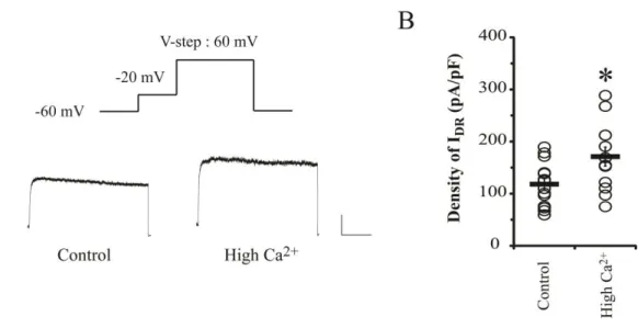
3. Results
3.1. Enhancement of IDR under the high Ca 2+
condition did not include any changes of kinetic properties in hippocampal neurons derived from DIV 6-9.
In this part, the mechanisms for regulation of IDR channels after inducing neuronal
overexcitability were investigated. The high Ca2+ (3.6 mM CaCl2) was used to induce
overexcitability by increasing the intracellular Ca2+ level. Figure 11 shows that the density of
IDR was significantly elevated by high Ca2+ application (Control= 112.62 ± 10.28 pA/pF, n=
13; high Ca2+= 171.05 ± 18.30 pA/pF, n= 12, p = 0.02), reflecting a possibility that
upregulating IDR channels may suppress the neuronal overexcitability via increasing K
+
outflux under high Ca2+ condition.
However, the activation kinetics of IDR channels were not changed under high Ca
2+
condition despite the enhancement of current density (Figure 12). The density as well as
increase rate of IDR also positively reflected the increase rates of command potentials. At +80
mv injection, significant changes of IDR were observed between control and high Ca
2+
treated
neurons (Control= 139.82 ± 15.62 pA/pF; High Ca2+= 226.79 ± 17.34 pA/pF, p= 0.02). The
increase rate of IDR was obtained by dividing the density of high Ca
2+
IDR with the control
value. This parameter was enhanced up to 60 % from -40 to 80 mV injection range compared
with the control group. Activation properties of IDR channels are shown in Figures 12 D and
E. Left-shifted pattern of activation curve by high Ca2+ according to slightly increased
voltage half (Vh) value of activation was observed but not significant (Vh of activation:
Control= 12.38 ± 2.62 mV; High Ca2+= 22.21 ± 4.4 mV, p= 0.1). This indicates that
upregulation of IDR channels under high Ca
2+
may not be dependent on their
56
Figure 11. IDR are increased by high Ca 2+
application in dissociated hippocampal neurons. A. Example traces of IDR after high Ca
2+
(3.6 mM CaCl2) treatment for 24 hrs.
Scale bars 500 pA, 100 ms. B. Individuals (circles) and averaged (square bars) densities of
57
Figure 12. Upregulating IDR by high Ca 2+
does not involve significant alteration of activation properties in hippocampal neurons. The activation properties were measured at
-60 to 80 mV with 20 or 40 mV steps after prepulse injection (-20 mV for 200 ms). A.
Example activation traces of IDR. Scale bars 500 pA, 100 ms. B. The density of IDR at each
command potential (-40 to 80 mV). C. The increase rate of IDR in high Ca
2+
58
compared with control group. The density of IDR was increased with the increment of
command potentials. D and E. Boltzmann fitted gating kinetics of IDR activation and its
voltage half (Vh) (thick gray dotted line in D). The significant change of the activation curves
59
3.2. Upregulation of IDR under high Ca 2+
condition is mediated with voltage-dependent Ca2+ channels.
A number of ion channels are regulated by Ca2+ signaling-mediated phosphorylation or
dephosphorylation. There are two major Ca2+ influx pathways opened by the depolarization
of membrane potentials; one is VDCC and the other is NMDAR. To confirm their
contribution to IDR upregulation, APV (100 μM) or nimodipine (10 μM ) with high Ca
2+
was applied to culture media for 24 hrs. As shown in Figure 13, Nimodipine, VDCCs antagonist,
critically blocked the increase of IDR by high Ca
2+
treatment, while APV treatment did not
show any effects on IDR alteration (APV = 151.55 ± 12.56 pA/pF, Nimodipine = 96.03 ± 9.34
pA/pF, P= 0.48, 0.008 respectively, compared with high Ca2+). In addition, each increase
pattern of IDR shown in APV- or nimodipine-treated neurons was respectively similar to the
pattern of high Ca2+- or non-treated neurons (Figure 14. B, at the 80 mV injection, APV=
192.53 ± 17.34, Nimodipine= 132.05 ± 15.06). However, effects of both antagonists on the
activation kinetics of IDR channels were not observed (Vh of activation in APV group= 10.11
± 1.91 and in Nimodipine group= 14.92 ± 1.63, p= 0.55 and 0.45 respectively). These results
indicate that Ca2+ influx through VDCCs may modulate the density of IDR channels in a
60
Figure 13. IDR upregulation to prevent neuronal overexcitation requires VDCCs. VDCC
antagonist nimodipine completely blocked the effect of high Ca2+ on density of IDR, but
NMDARs antagonist APV did not. A. Example traces of IDR after high Ca
2+
treatment with 100 μM APV or 10 μM nimodipine. Scale bars 500 pA, 100 ms. B. Square bars and open
circles indicate averaged and individual densities of IDR, respectively. Error bars represent
61
Figure 14. The blockade of IDR upregulation by nimodipine does not exhibit any
changes of IDR activation property. A. Example traces of IDR induced by injecting
command potentials from -40 mV to 80 mV. Scale bars 500 pA, 100 ms. B. Changes of IDR
densities according to each command potential. The increase pattern of IDR density in
62
IDR in APV-treated neurons resembled those of high Ca
2+
-treated neurons. C and D.
Boltzmann fitted activation curves of IDR channels and their Vh values. These activation
curves were not shifted and therefore, Vh values did not show any significant differences in