The Elbow Instability
경북의대 정형외과전인호∙김풍택
Biomechanics of elbow dislocation
Constraints to elbow instability
3 primary static constraints to elbow instability - the ulnohumeral articulation, MCL, LCL Secondary constraints - the radial head, the common flexor and extensor origins, the capsule Dynamic stabilizer - muscles (anconeus, triceps, brachialis…) that produce compressive forces.
Pathoanatomy
Disruption of the circle of soft tissue or bone from the lateral side and progresses to the medial. Stage 1 - the lateral collateral ligament is partially or completely disrupted
Stage 2 - additional disruption anteriorly and posteriorly
Stage 3A - all of the soft tissue around and including the posterior part of the MCL are disruption 3B - the entire medial collateral complex is disrupted
This circle of disruption (Horii circle)
Classification of elbow instability
According to 5 criteria(1) the articulation or articulations involved (the elbow or the radial head); (2) the direction of displacement (valgus, varus, anterior, or posterior rotator); (3) the degree of displacement (subluxation or dislocation);
(4) the timing (acute, chronic or recurrent); (5) the presence or absence of associated fractures
Acute Elbow Dislocation (Simple)
Appears to subluxate or dislocate → splint and AP&lat radiographs No subluxation → discharge / splint or sling
Reevaluation in 5~7 days: subluxation or incongruence → forearm pronation and evaluation of stability / stability is restored → hinged brace or cast-brace / elbow require an extension block of more than 30~40�→ surgical repair should be considered
Lateral pivot-shift maneuver
Patient placed supine on the table / The elbow is supinated, and a mild to moderate forced valgus stress is applied while the elbow is flexed past approximately 40�.
Management of acute dislocations
Acute Elbow Dislocation (Complex: Associated with Fracture)
The terrible-triad injury
Combination of an elbow dislocation and fractures of the radial head and coronoid process
Fractures of the coronoid process
Type I - small fleck of bone,
Type II - involves 50% of the height of the coronoid process or less,
Type III - more than 50% of the height of the coronoid process
Fig. 4.
Results and complications
The terrible triad is a grave and complex injury
Suggest that lesions of both the coronoid and the radial head need to be stabilized or reconstructed whenever possible
Hinged External Fixation for Unstable Elbows
Primary indication
(1) Persistent instability in association with an acute fracture-dislocation despite attempted ligament repair and fracture fixation or radial head replacement or both;
(2) Gross acute instability in a patient who is not a candidate for surgery;
(3) Delayed treatment (approximately 4 weeks or more after the time of injury) of a dislocated and stiff elbow
Relative indication - need to protect the stability and the fracture reduction during rehabilitation following surgical treatment of an unstable elbow
Recurrent Elbow Instability
LCL insufficiency is most commonly seen after elbow dislocation
Both the medial and the lateral collateral ligament complex are acutely disrupted with an elbow dislocation, the residual insufficiency most commonly involves the lateral collateral ligament complex for reasons that have not yet been elucidated
History & Exam
Instability, pain and discomfort, painful catching, slippage, or clicking with flexion and extension. Reduction in pronation.
Lateral pivot-shift maneuver, the posterolateral rotator drawer test, apprehension test
Medial Instability
Pitchers are prone to elbow injuries because of high and repetitive valgus stresses on the elbow. Recurrent instability after a simple elbow dislocation is rare
Etiology
The anterior bundle of the medial ulnar collateral ligament (MCL) of the elbow is the primary restraint and is often attenuated with time, leading to functional incompetence and ultimate failure.
Clinical Presentation
Pitchers with a history of medial elbow pain, reduced velocity, and loss of command may have an MCL injury in evolution.
Diagnosis
Physical examination and imaging can confirm the diagnosis.
Management
∙ Treatment begins with rest and activity modification. ∙ All medial elbow pain is not MCL injury.
∙ Surgery is considered only for talented athletes who wish to return to competitive play and may include elite scholastic and other collegiates and professionals.
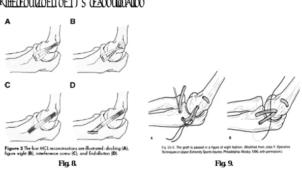
The technique for MCL reconstruction was first described in 1986. Many variations have been offered since then, which can result in predictable outcomes, allowing many to return to the same level of competitive play.
4 Different types of MCL reconstruction
Lateral Instability
Posterolateral rotatory instability of the elbow is the most common pattern of chronic lateral elbow instability.
Etiology
The primary lesion in posterolateral rotatory instability is injury or attenuation of the lateral ulnar collateral ligament.
Diagnosis
Posterolateral rotatory instability is diagnosed on the basis of careful history taking and specific physical examination techniques. (Table top relocation test, Pivot shift test)
Fig. 8. Fig. 9.
Treatment
Reconstruction of the LUCL with repair of the surrounding soft tissue structures is recommended in patients who have symptoms of recurrent lateral instability.
Open and arthroscopic reconstruction techniques have resulted in improvement of elbow function and satisfactory results in most patients, although mild limitation in terminal extension of the elbow is a common finding.
Rehabilitation
70~80�flexion with the forearm in full pronation for 10~14 days/ protected motion in a hinged brace for 2-6 weeks
After 6 weeks, removal of the hinged splint for sedentary activities.
At the end of additional 6 weeks, splint discontinued. Protection for a total 12 weeks after surgery After 12 weeks, activity is gradually resumed, with 100% activity allowed at 6 months
Results
No degenerative arthritis, radial head is intact → 90% have a satisfactory outcome with no subsequent subluxation
Radial head excision or degenerative arthritis → 67~75% of satisfactory result Fig. 12.
Summary of treatment principles
Reduction of acute dislocations and management after reduction
Closed reduction, forearm in neutral.3~5 days, place the elbow in a splint and start allowing movement unless the elbow tends to subluxate or dislocate
Careful examination and radiographs - every 5~7 days for the first three weeks
Management of acute fracture dislocation
Fix the bones internally and repair the ligaments so that early motion can be commenced
Repair of an acute ligament injury
Indicated in all fracture-dislocations requiring internal fixation of the radial head or the coronoid process or both.
Avulsion - repair directly to bone with heavy suture
Passing a heavy absorbable suture through the same course as that for a late ligament reconstruction and fixing it to the normal ligament attachment on the epicondyle and the ulna augments the repair