저작자표시-비영리-변경금지 2.0 대한민국 이용자는 아래의 조건을 따르는 경우에 한하여 자유롭게 l 이 저작물을 복제, 배포, 전송, 전시, 공연 및 방송할 수 있습니다. 다음과 같은 조건을 따라야 합니다: l 귀하는, 이 저작물의 재이용이나 배포의 경우, 이 저작물에 적용된 이용허락조건 을 명확하게 나타내어야 합니다. l 저작권자로부터 별도의 허가를 받으면 이러한 조건들은 적용되지 않습니다. 저작권법에 따른 이용자의 권리는 위의 내용에 의하여 영향을 받지 않습니다. 이것은 이용허락규약(Legal Code)을 이해하기 쉽게 요약한 것입니다. Disclaimer 저작자표시. 귀하는 원저작자를 표시하여야 합니다. 비영리. 귀하는 이 저작물을 영리 목적으로 이용할 수 없습니다. 변경금지. 귀하는 이 저작물을 개작, 변형 또는 가공할 수 없습니다.
Factors affecting recurrence of intrahepatic duct stone
and factors affecting development of cholangiocarcinoma
after initial treatment of intrahepatic duct stone
: 10-year analysis
by
Eunsoo Lim
Major in Medicine
Department of Medical Sciences
The Graduate School, Ajou University
and factors affecting development of cholangiocarcinoma
after initial treatment of intrahepatic duct stone
: 10-year analysis
by
Eunsoo Lim
A Dissertation Submitted to The Graduate School of
Ajou University in Partial Fulfillment of The Requirements
for The Degree of Master of Science in Medicine
Supervised by
Byung Moo Yoo, M.D., Ph.D.
Major in Medicine
Department of Medical Sciences
The Graduate School, Ajou University
This certifies that the dissertation
ofEunsoo Lim is approved.
SUPERVISORY COMMITTEE
____________________________
Byung Moo Yoo
____________________________
Sung Jae Shin
____________________________
KeeMyung Lee
The Graduate School, Ajou University
June, 19
th, 2015
i - ABSTRACT –
Factors affecting recurrence of intrahepatic duct stone andfactors affecting development of cholangiocarcinoma after initial treatment of intrahepatic duct stone:
10-year analysis
Background Hepatolithiasis causes recurrent cholangitis, biliary cirrhosis, and is an important risk factor for cholangiocarcinoma, which has a grave prognosis. Nevertheless, its intractable nature and high recurrence rate complicates treatment. Therefore, we investigated factors affecting recurrence of hepatolithiasis and factors affecting development of cholangiocarcinoma after treatment.
Methods Medical records of total 159 patients in Ajou University hospital from 1995 to 2014 were reviewed and types of hepatolithiasis were classified according to Tsunoda classification. Factors affecting complete removal of hepatolithiasis, recurrence of hepatolithiasis and development of cholangiocarcinoma in hepatolithiasispatients were analyzed.
Results The 159 patients consisted of 58 men and 101 women, with a mean age of 61.73
years. Median follow-up period was 53.69 months. Percutaneous
transhepaticcholedochoscopic stone lithotripsy (PTCS)and surgery were performed in 112 cases and in 34 cases, respectively. Peroraltranspapillary endoscopic lithotripsy was also used in 3 patients and 10 patients were followed up without any treatment because of their comorbidities. Location of stones, treatment modality, Tsunoda classification, presence of liver atrophy, dilatation of stenosis during treatment and previous biliary surgery were not related to the possibility of complete stone removal in univariate and multivariate analysis. Recurrence rate was significantly different according to the location of stones, treatment modality, presence of liver atrophy, previous history of biliary surgery in univariate analysis. However, in multivariate analysis, previous history of biliary surgery was the only statistically significant factor. Patients who had undergone biliary surgery had 2.699 higher risk of recurrence than patients who had not. Cholangiocarcinoma was diagnosed in 7 (4.4%)
ii
patients. Four patients were diagnosed cholangiocarcinoma and
hepatolithiasissimultaneously, and in the other three patients, cholangiocarcinoma developed during the follow-up period after treatment.Location of stones, Tsunoda classifications, previous history of previous biliary tract surgery, liver atrophy and complete stone removal were not related to development of cholangiocarcinomain multivariate analysis.
ConclusionWe could not find the factors related to the complete stone removal in this study. Recurrence rate was significantly different according to the previous history of surgery. Patients who had undergone biliary surgery have 2.699 higher risk of recurrence than patients who had not. No factor was associated with occurrence of cholangiocarcinoma. Lastly, patients with a history of biliary tract surgery should be followed up more closely and carefully for recurrence than patients who do not.
iii
TABLE OF CONTENTS
ABSTRACT……… i
TABLE OF CONTENTS……… iii
LIST OF FIGURES……… iv
LIST OF TABLES……… v
I. INTRODUCTION……… 1
II. PATIENTS AND METHODS………2
A. PATIENTS……… 2 B. METHODS………2 C. STATISTICAL ANALYSIS……… 3 III. RESULTS……… 4 IV. DISCUSSION……… 17 V. CONCLUSION……… 22 REFERENCES……… 23 국문요약………24
iv
LIST OF FIGURES
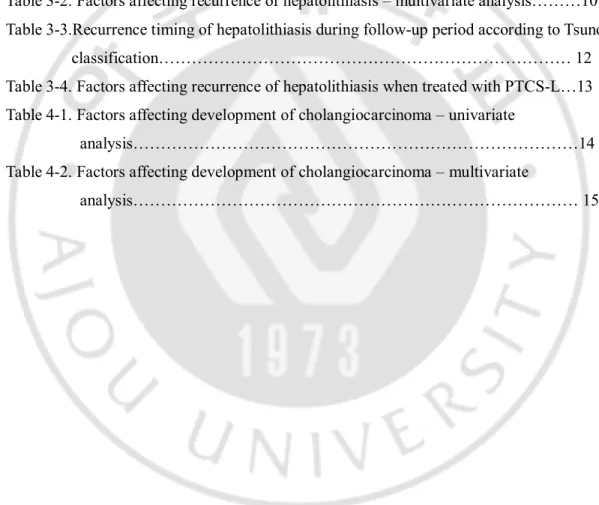
Fig 1. Recurrence timing of hepatolithiasis during follow-up period according to Tsunoda classification……… 12
v
LIST OF TABLES
Table 1.Clinical characteristicsof patients……… 4 Table 2-1. Factors affecting achievement of complete stone removal – univariate
analysis……… 7 Table2-2. Complete stone removal rate according to Tsunoda classification and treatment
modality……… 8 Table 3-1.Factors affecting recurrenceofhepatolithiasis– univariate analysis………… 9 Table 3-2. Factors affecting recurrence of hepatolithiasis – multivariate analysis………10 Table 3-3.Recurrence timing of hepatolithiasis during follow-up period according to Tsunoda
classification……… 12 Table 3-4. Factors affecting recurrence of hepatolithiasis when treated with PTCS-L…13 Table 4-1. Factors affecting development of cholangiocarcinoma – univariate
analysis………14 Table 4-2. Factors affecting development of cholangiocarcinoma – multivariate
1
I. INTRODUCTION
Hepatolithiasis refers to the presence of gallstones within intrahepatic duct. The incidence is much higher in East Asia, including Korea, compared with western countries. Hepatolithiasis causes recurrent cholangitis, liver abscesses, biliary cirrhosis, and atrophic changes of the affected liver. Also, it is an important risk factor for cholangiocarcinoma, which has a grave prognosis.(Cheon et al., 2009) Nevertheless, its intractable nature and high recurrence rate complicates treatment.Currently, the major two treatment modalities for the hepatolithiasis are surgery or percutaneous transhepaticcholangioscopic stone lithotripsy (PTCS).
Taken as a whole, residual or recurrent stones are noted in 18.6–29.8% of the patients.(Suzuki et al., 2014)When hepatolithiasis was treated with PTCS, complete stone removal was achieved in 77-85%, and cumulative stone recurrence rate ranged from 32.6% to 40.0%.(Chen et al., 2005; 황재철, 2012) Previous studies reported that liver atrophy, endoscopic treatment, bile duct strictures and residual stones are related to recurrence, but the results are not consistent.
Cholangiocarcinoma develops in 1.3–5.9% of hepatolithiasis patients.(Suzuki et al., 2014) Yet few incompatible risk factors affecting development of cholangiocarcinoma after treatment of hepatolithiasis are recognized including bile duct strictures.
Therefore, we retrospectively investigated the patients who were treated for hepatolithiasis and searched for the factors affecting complete stone removal, the factors affecting recurrence of hepatolithiasis and factors affecting development of cholangiocarcinoma.
2
II. PATIENTS AND METHODS
A. Patients
From 1995 to 2014, 511 patients who were radiologically (abdominal CT or ultrasonography) diagnosed with hepatolithiasis were enrolled. Among them, patients with insufficient medical records or imaging records and patients who had not been followed less than 6 months were excluded. Consequently, medical records of 159 patients in Ajou University hospital from 1995 to 2014 were reviewed retrospectively for this study.
B. Methods
According to medical records, basic clinical characteristics (age and sex), location of intrahepatic duct stones, treatment modality, Tsunoda classification, presence of atrophic changes of liver, whether dilatation for intrahepatic duct stricture during endoscopic procedures was done or not, previous history of biliary surgery, whether hepatolithiasis was removed completely or not, recurrence of hepatolithiasis, development of cholangiocarcinoma, and interval from the diagnosis of hepatolithiasis to occurrence of cholangiocarcinoma were analyzed.
Types of hepatolithiasis were classified according to Tsunoda classification.(Huang et al., 2003) Type I disease had no dilation or stricture of the intrahepatic bile ducts. Type II disease had diffuse dilation of the intrahepatic bile ducts and often had lesions associated with a common bile duct obstruction. Type III disease had unilateral solitary or multiple cystic dilation of the intrahepatic bile ducts, frequently accompanied by stricture of left or right intrahepatic bile ducts. Type IV disease had stricture and dilation of the intrahepatic bile ducts in bilateral hepatic lobes.
Liver atrophy was defined as presence of liver atrophy documented radiologically and also confirmed by radiologists. Intrahepatic duct stricture was defined only when stricture was noted during cholangioscopy or stricture was seen on ultrasonography or computed tomography. Recurrence of hepatolithiasis was defined as actual demonstration of one or more stones in ultrasonography or computed tomography during follow-up period after
3 complete removal of hepatolithiasis.
Variable treatment modalities were applied for the treatment of hepatolithiasis. PTCS ndsurgery were the major treatment modalities for the hepatolithiasis. In some patients, peroraltranspapillary endoscopic stone removal (POES) was performed.
PTCS was performed through percutaneous tract. Percutaneous transhepatic biliary drainage (PTBD) catheter was inserted at the same or the opposite side of hepatolithiasis. Two to three days after inserting PTBD, the PTBD tract was dilated up to 16-18Fr. And 1 week after dilating PTBD tract, hepatolithiasis was removed using choledochoscopy. Hepatolithiasis was removed with stone basket or fragmented with electrohydraulic lithotripter. Then the fragmented hepatolithiasis was removed with stone basket or pushed out to the duodenum by choledochoscopy. In cases with bile duct strictures, the bile duct was dilated with 6-10 mm balloon catheter.
Factors affecting complete removal of hepatolithiasis, recurrence of hepatolithiasisand development of cholangiocarcinoma in hepatolithiasis patients were analyzed.
C. Statistical analysis
Chi-square test was used for univariate analysis, and binary logistic regression was used for multivariate analysis. Relative risk was calculated with 95% confidence intervals. Survival curve using the Kaplan–Meier method was performed. P values were rounded up to the third digit after the decimal point. P values lower than 0.05 was considered statistically significant. Descriptive statistical values were rounded up to the second digit after the decimal point. The data was analyzed using SPSS 22.
4
RESULTS
1. Clinical characteristics
Total 159 patients consisted of 58 men and 101 women, with a mean age of 61.73 years. Mean follow-up period was 53.69 months and median follow-up period was 38 months. The stones were located in the left lobe in 82 (51.6%) patients, in the right lobe in 37 (23.3%) patients, and in both lobes in 40(25.2%) patients. The major two methods used to treat hepatolithiasis were PTCS in 112 (70.4%) cases and surgery in 34 (21.4%) cases, respectively. However, in 3 patients, peroraltranspapillary endoscopic lithotripsy was also used and 10 patients were followed up without any treatmentbecause of their comorbidities. The percentage and numbers of patients in each Tsunoda type were as follows: 10.1% (16 patients) in type I, 34.0% (54 patients) in type II, 32.1% (51 patients) in type III, and 23.9% (38 patients) in type IV. Liver atrophy was present in 15.1% (24 patients). Forty one (25.8%) patients had a history of previous biliary surgery. (Table 1) Fourteen patients underwent surgery due to hepatolithiasisbefore they were enrolled to this study, 16 patients due to choledochal cyst, and 11 patients due to stones or malignancy of common bile duct, respectively. In 70 patients, dilatation of bile duct stricture was performed during PTCS. Hepatolithiasis was completely removed in 93 (83.0%) patients treated with PTCS and 32 (94.1%) patients treated with surgery. Overall, complete stone removal was achieved in 80.5% of the patients.
Table 1.Clinical characteristicsof patients
Variable No. of patients (%)
Sex (M:F) 58(36.5)/101(63.5)
Age (mean, years) 61.73
Follow-up period (mean±SD, months) 53.69±44.82
Location of hepatolithiasis
5 Right 37(23.3) Both 40(25.2) Treatment modality PTCS 112(70.4) Surgery 34(21.4) POES 3(1.9) Observation 10(6.3) Tsunoda classification I 16(10.1) II 54(34.0) III 51(32.1) IV 38(23.9) Liver atrophy Atrophy (+) 24(15.1) Atrophy (-) 135(84.9)
Dilatation of stenosis during procedures
Dilatation (+) 70(44.0)
Dilatation (-) 47(29.6)
Unknown 42(26.4)
Previous biliary surgery
Surgery (-) 118(74.2)
Surgery (+) 41(25.8)
Achievement of complete stone removal
6 Incomplete removal 31(19.5) Recurrence Recurred 34(21.4) Not recurred 125(78.6) Development of cholangiocarcinoma Cholangiocarcionoma (+) 7(4.4) Cholangiocarcinoma (-) 152(95.6)
PTCS: percutaneous transhepaticcholedochoscopic stone removal POES: peroraltranspapillary endoscopic stone removal
2. Factors affecting complete stone removal
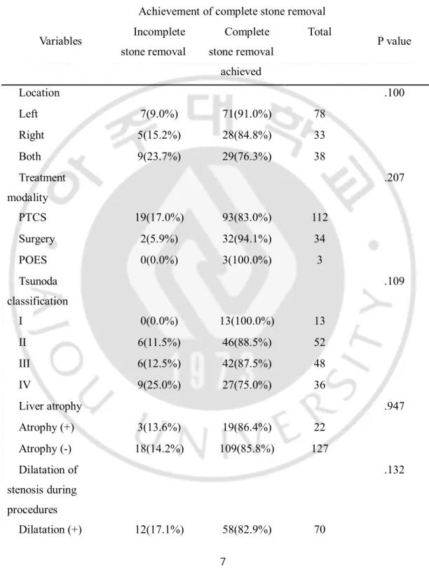
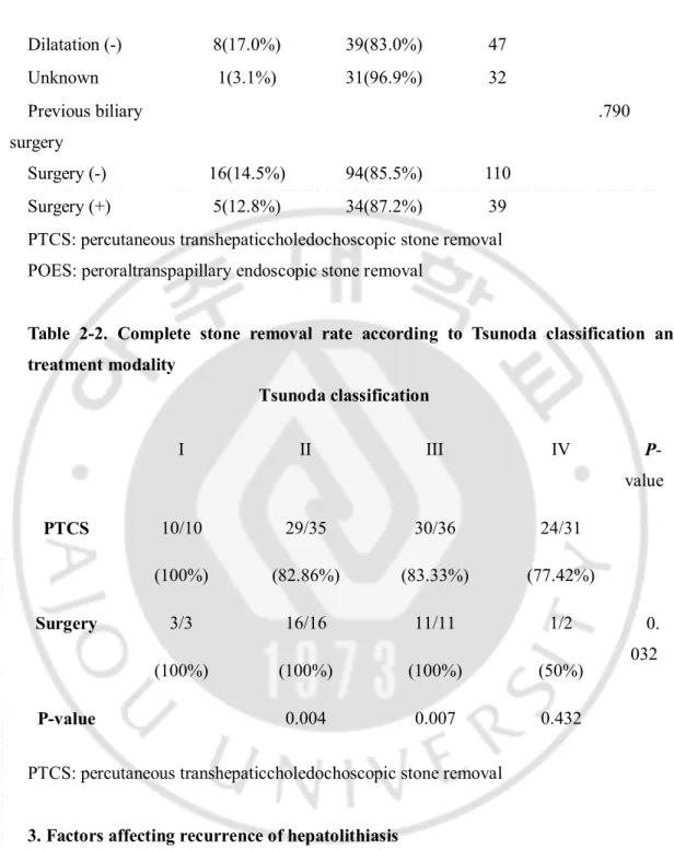
Hepatolithiasis were completely removed in 83.0% (93 patients) of patients treated with PTCS, and in 94.1% (32 patients) of patients treated with surgery. Location of stones, treatment modality, Tsunoda classification, presence of liver atrophy, dilatation of stenosis during procedure and previous biliary surgery were not related to complete stone removal in univariate analysis. (Table 2-1) Stones were completely removed in 100% of type I disease both in PTCS and surgery group. In type II disease, 83.0% of stones were removed completely in PTCS group, and 100% in surgery group. Complete stone removal rate was 83.33% in type III patients treated with PTCS and 100% in type III patients treated with surgery. Finally, stones were completely removed in 77.4% of type IV disease treated with PTCS, 50% of type IV disease treated with surgery. (Table 2-2) Complete stone removal rate was significantly different according to Tsunoda classification between PTCSgroup and surgery group. (p=0.032) Especially, surgery was superior in complete stone removal in both type II and III. The causes of incomplete stone removal were technical failure to approach and remove stones, patients’ refusal for further treatment, inadequate general condition to tolerate treatment, incidentally found stone in imaging study performed after treatment, and complications which precluded further treatment including bile duct injury and liver laceration.
7
Table 2-1. Factors affecting achievement of complete stone removal – univariate analysis
Variables
Achievement of complete stone removal
P value Incomplete stone removal Complete stone removal achieved Total Location .100 Left 7(9.0%) 71(91.0%) 78 Right 5(15.2%) 28(84.8%) 33 Both 9(23.7%) 29(76.3%) 38 Treatment modality .207 PTCS 19(17.0%) 93(83.0%) 112 Surgery 2(5.9%) 32(94.1%) 34 POES 0(0.0%) 3(100.0%) 3 Tsunoda classification .109 I 0(0.0%) 13(100.0%) 13 II 6(11.5%) 46(88.5%) 52 III 6(12.5%) 42(87.5%) 48 IV 9(25.0%) 27(75.0%) 36 Liver atrophy .947 Atrophy (+) 3(13.6%) 19(86.4%) 22 Atrophy (-) 18(14.2%) 109(85.8%) 127 Dilatation of stenosis during procedures .132 Dilatation (+) 12(17.1%) 58(82.9%) 70
8 Dilatation (-) 8(17.0%) 39(83.0%) 47 Unknown 1(3.1%) 31(96.9%) 32 Previous biliary surgery .790 Surgery (-) 16(14.5%) 94(85.5%) 110 Surgery (+) 5(12.8%) 34(87.2%) 39
PTCS: percutaneous transhepaticcholedochoscopic stone removal POES: peroraltranspapillary endoscopic stone removal
Table 2-2. Complete stone removal rate according to Tsunoda classification and treatment modality Tsunoda classification I II III IV P-value PTCS 10/10 (100%) 29/35 (82.86%) 30/36 (83.33%) 24/31 (77.42%) Surgery 3/3 (100%) 16/16 (100%) 11/11 (100%) 1/2 (50%) 0. 032 P-value 0.004 0.007 0.432
PTCS: percutaneous transhepaticcholedochoscopic stone removal 3. Factors affecting recurrence of hepatolithiasis
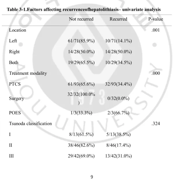
Except for 10 patients who were not treated, recurrence was detected in 34 (21.4%) patients. Recurrence rate was significantly different according to the location of stones, treatment modality, presence of liver atrophy and previous history of biliary surgery in
9
univariate analysis.(Table 3-1) In multivariate analysis, patients who had undergone biliary surgery have much higher risk of recurrence than patients who had not.(OR=2.699)(Table 3-2) Also, Kaplan-Meier curve which shows recurrence timing during follow-up period comparing each Tsunoda classification was calculated using log-rank test. (Table 3-3, Figure 1) Mean time to recurrence was shorter in type I disease, however, the result was not significantly different among each Tsunoda classification. Because recurrence was not observed in patients who were treated with surgery, 93 patients treated with PTCS were analyzed separately. (Table 3-4) Patients who underwent biliary tract surgery had a tendency toward higher risk of recurrence and other factors did not affect recurrence.
Table 3-1.Factors affecting recurrenceofhepatolithiasis– univariate analysis
Not recurred Recurred P-value
Location .001 Left 61/71(85.9%) 10/71(14.1%) Right 14/28(50.0%) 14/28(50.0%) Both 19/29(65.5%) 10/29(34.5%) Treatment modality .000 PTCS 61/93(65.6%) 32/93(34.4%) Surgery 32/32(100.0% ) 0/32(0.0%) POES 1/3(33.3%) 2/3(66.7%) Tsunoda classification .324 I 8/13(61.5%) 5/13(38.5%) II 38/46(82.6%) 8/46(17.4%) III 29/42(69.0%) 13/42(31.0%)
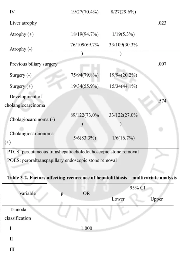
10 IV 19/27(70.4%) 8/27(29.6%) Liver atrophy .023 Atrophy (+) 18/19(94.7%) 1/19(5.3%) Atrophy (-) 76/109(69.7% ) 33/109(30.3% )
Previous biliary surgery .007
Surgery (-) 75/94(79.8%) 19/94(20.2%) Surgery (+) 19/34(55.9%) 15/34(44.1%) Development of cholangiocarcinoma .574 Cholagiocarcinoma (-) 89/122(73.0% ) 33/122(27.0% ) Cholangiocarcionoma (+) 5/6(83.3%) 1/6(16.7%)
PTCS: percutaneous transhepaticcholedochoscopic stone removal POES: peroraltranspapillary endoscopic stone removal
Table 3-2. Factors affecting recurrence of hepatolithiasis – multivariate analysis
Variable p OR 95% CI Lower Upper Tsunoda classification I 1.000 II III
11 IV Treatment modality .585 PTCS 1.000 Surgery .998 0.000 0.000 POES .300 3.686 0.313 43.454 Observation .999 0.000 0.000 Location .065 Left .313 0.571 0.193 1.694 Right .202 2.068 0.677 6.314 Both 1.000 Liver atrophy Atrophy (+) Atrophy (-) 1.000 Previous biliary surgery .037 Surgery (-) 1.000 Surgery (+) 2.699 1.061 6.863 Development of cholangiocarcinom a Cholangiocarcin oma (-) 1.000 Cholangiocarcin oma (+)
12
POES: peroraltranspapillary endoscopic stone removal
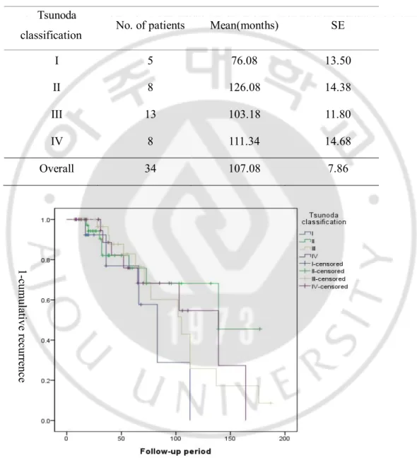
Table 3-3.Recurrence timing of hepatolithiasis during follow-up period according to Tsunoda classification
Tsunoda
classification No. of patients Mean(months) SE
I 5 76.08 13.50
II 8 126.08 14.38
III 13 103.18 11.80
IV 8 111.34 14.68
Overall 34 107.08 7.86
Fig 1.Recurrence timing of hepatolithiasis during follow-up period according to Tsunoda classification. 1-cu m ula tiv e r ec ur re nc e
13
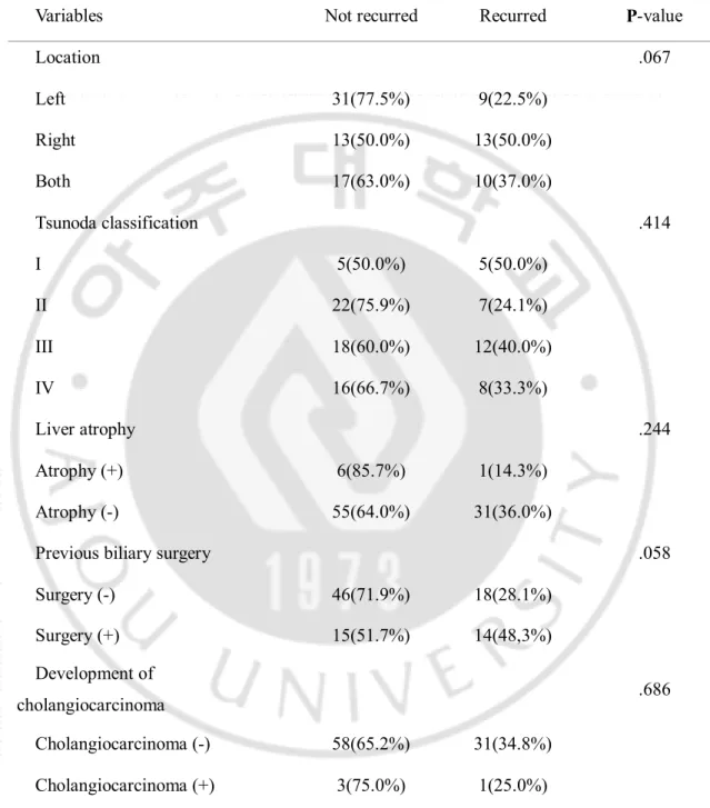
Table 3-4. Factors affecting recurrence of hepatolithiasis when treated with PTCS-L
Variables Not recurred Recurred P-value
Location .067 Left 31(77.5%) 9(22.5%) Right 13(50.0%) 13(50.0%) Both 17(63.0%) 10(37.0%) Tsunoda classification .414 I 5(50.0%) 5(50.0%) II 22(75.9%) 7(24.1%) III 18(60.0%) 12(40.0%) IV 16(66.7%) 8(33.3%) Liver atrophy .244 Atrophy (+) 6(85.7%) 1(14.3%) Atrophy (-) 55(64.0%) 31(36.0%)
Previous biliary surgery .058
Surgery (-) 46(71.9%) 18(28.1%) Surgery (+) 15(51.7%) 14(48,3%) Development of cholangiocarcinoma .686 Cholangiocarcinoma (-) 58(65.2%) 31(34.8%) Cholangiocarcinoma (+) 3(75.0%) 1(25.0%)
14
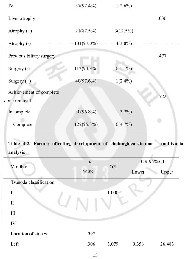
4. Factors affecting development of cholangiocarcinoma
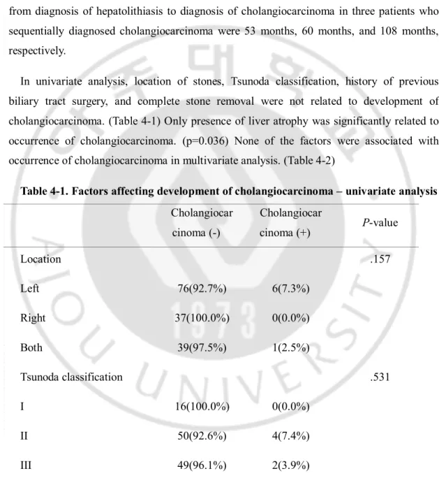
Cholangiocarcinoma was diagnosed in 7 (4.4%) patients. Four patients were diagnosed hepatolithiasis and cholangiocarcinomasimultaneously, and in the other three patients, cholangiocarcinoma developed during the follow-up period after treatment.The intervals from diagnosis of hepatolithiasis to diagnosis of cholangiocarcinoma in three patients who sequentially diagnosed cholangiocarcinoma were 53 months, 60 months, and 108 months, respectively.
In univariate analysis, location of stones, Tsunoda classification, history of previous biliary tract surgery, and complete stone removal were not related to development of cholangiocarcinoma. (Table 4-1) Only presence of liver atrophy was significantly related to occurrence of cholangiocarcinoma. (p=0.036) None of the factors were associated with occurrence of cholangiocarcinoma in multivariate analysis. (Table 4-2)
Table 4-1. Factors affecting development of cholangiocarcinoma – univariate analysis Cholangiocar cinoma (-) Cholangiocar cinoma (+) P-value Location .157 Left 76(92.7%) 6(7.3%) Right 37(100.0%) 0(0.0%) Both 39(97.5%) 1(2.5%) Tsunoda classification .531 I 16(100.0%) 0(0.0%) II 50(92.6%) 4(7.4%) III 49(96.1%) 2(3.9%)
15
IV 37(97.4%) 1(2.6%)
Liver atrophy .036
Atrophy (+) 21(87.5%) 3(12.5%)
Atrophy (-) 131(97.0%) 4(3.0%)
Previous biliary surgery .477
Surgery (-) 112(94,9%) 6(5.1%) Surgery (+) 40(97.6%) 1(2.4%) Achievement of complete stone removal .722 Incomplete 30(96.8%) 1(3.2%) Complete 122(95.3%) 6(4.7%)
Table 4-2. Factors affecting development of cholangiocarcinoma – multivariate analysis Varaible P-value OR OR 95% CI Lower Upper Tsunoda classification I 1.000 II III IV Location of stones .592 Left .306 3.079 0.358 26.483
16
Right .998 0.000 0.000
Both 1.000
Achievement of complete stone removal
Incomplete stone removal 1.000
Complete stone removal achieved
Previous biliary surgery
Surgery (-) 1.000
Surgery (+) Liver atrophy Atrophy (+)
17
DISCUSSION
The major two methods used to treat hepatolithiasis were PTCS and surgery. The general indications for endoscopic treatment are as follows; 1) when intrahepatic duct stones are located in both lobes 2) absence of liver atrophy 3) when patients have a history of previous biliary surgery4) when patient’s co-morbidities precludes surgery. (황재철, 2012)On the other hand, the indications for surgery are as follows; 1) presence of severe stricture of unilateral bile duct which contains stones 2) presence of liver atrophy and/or abscesses 3) presence of congenital anomaly of bile duct 4) when concomitant cholangiocarcinoma is suspected. (Chen et al., 2005; Cheon et al., 2009; 권형준김상걸, 2012; 황재철, 2012)
In 3 patients, only peroraltranspapillary endoscopic lithotripsy was used to remove hepatolithiasis. In these patients, hepatolithiasis was usually centrally located and removed at the same time while removing the common bile duct stone during peroraltranspapillary endoscopic lithotripsy. Cheon et al mentioned that peroraltranspapillary endoscopic lithotripsy is recommended for older patients, patients with bilateral multiple stones associated with bilateral stenosis of the intrahepatic duct accompanying uncompensated liver cirrhosis, and patients with localized intrahepatic calculi located not beyond the secondary branches. (Cheon et al., 2009)
In this study, of 159 patients, 149 patients went through treatment including surgery or endoscopic treatment and stones were completely removed in 85.9% of patients. The result is similar to other studies in which stones were completely removed in 77 to 85% of patients. (Chen et al., 2005; 황재철, 2012) According to treatment modality, 83.0% of patient treated with PTCS and 94.1% of patient treated with surgery achieved complete stone removal and this difference was not statistically significant in our study. On the other hand, Lee’s study similarly analyzed complete stone removal rate according to the treatment method in 168 patients, and the study concluded that PTCS is superior in achieving complete stone removal compared with surgery. (Lee et al., 2001)However, since there were differences in ways of selecting treatment modality for hepatolithiasis in each study, it is hard to say that which way
18 is more superior by simply comparing the results.
One of the crucial reasons for incomplete stone removal during endoscopic treatment is acute angulation of bile duct. To prevent incomplete stone removal due toacute angulation of bile duct, proper selection of the percutaneous transhepatic biliary drainage (PTBD) puncture site is very important.(Lee et al., 2001; Huang et al., 2003)Because it is easier to pass the scope from the left intrahepatic duct to the right intrahepatic duct or the common bile duct, left approach is suggested for the bilateral intrahepatic stones and/or the common bile duct stones. When attempting a right approach, a posterior rather than anterior approach can avoid angulation of the anterior segment. (Huang et al., 2003) In this study, we usually made the PTBD tract at the opposite side of hepatolithiasis and in some patients at both sides of the liver.
The above-mentioned indications for endoscopic treatment include bilateral stones, absence of liver atrophy and history of previous biliary surgery. In this study, however, location of stones, Tsunoda classification, presence of liver atrophy and previous biliary surgery were not related to complete stoneremoval in univariate and multivariate analysis. Thus, endoscopic treatment can be applied to various situations than it was thought tobe, and when percutaneous transhepatic biliary drainage tract location is properly selected, endoscopic treatment is not inferior and sometimes, superior to surgery in removing hepatolithiasis completely. However, dilatation of stenosis during endoscopic procedure was performed in 47% patients, but this also did not affect complete stone removal.
Except for 10 patients who were not treated, recurrence was noted in 34 (21.4%) patients and this result was consistent with other studies. Cheon et al reported that the overall recurrence rate for hepatolithiasis and/or cholangitis in patients after successful treatment was 20%.(Cheon et al., 2009) The cumulative recurrence rate of hepatolithiasis treated by PTCS appeared to be a little bit higher, ranging from 32.6% to 59 in one study,(Chen et al., 2005; 황재철, 2012)and the average number of PTCSneeded for complete removal was from 4 to 5 according to Hwang’s study.(황재철, 2012)
19
In our study, recurrence rate was significantly different according to the location of stones, treatment modality, presence of liver atrophy and previous history of biliary surgery in univariate analysis. However, multivariate analysis showed that only patients with previous history of biliary surgery had 2.699 higher risk of recurrence.
Cheon et al stated that there was no statistically significant difference in the recurrence rates between treatment methods.(Cheon et al., 2009)The advantage of surgery in the treatment of hepatolithiasis is that the stones and associated pathologic bile ducts can be completely resected.(Huang et al., 2003) In a study which compared recurrence rate according to treatment methods, rate of complete stone removal was higher in surgery group, but recurrence rate was not statistically different between the two groups.(임창섭 et al., 2010) In our study, recurrence rate was significantly different according to treatment modality in univariate analysis, but not in multivariate analysis. All patients treated with surgery did not experience recurrence.
Intrahepatic duct strictures had been known to be one of the major causes of treatment failure and recurrence, especially when treated with PTCS. (Lee et al., 2001; Chen et al., 2005; Suzuki et al., 2014)Huang et al reported that the duration till recurrence was shorter in cases with duct strictures than in cases without strictures (11 years and 18 years, respectively)(황재철, 2012) In our study, however, recurrence rate was not related to Tsunoda classification, which reflects bile duct strictures. We also scrutinized recurrence timing during follow-up period according to Tsunoda classification and there was no significant difference in recurrence timing amongTsunoda classifications. Manners of defining bile duct stricture which were different between the two studies explain this discrepancy.
Liver atrophy was one of the significant factors affecting recurrence in univariate analysis, but not in multivariate analysis. However it was a significant risk factor for recurrent cholangitis in Toshio’s study. (Tsuyuguchi et al., 2014)
20
significant in univariate and multivariate analysis. Patients who had undergone biliary surgery have 2.699 higher risk of recurrence than patients who had not. Therefore, patients with a history of biliary tract surgery should be followed up more closely and carefully because the recurrence rate of hepatolithiasiswas higher in these patients.
Recurrence of hepatolithiasis was not related to the development of cholangiocarcinoma. The results were grossly similar when patients treated with PTCS were analyzed separately. Patients who underwent biliary tract surgery had a tendency toward higher risk of recurrence, and it was not statistically significant. All the other factors were not associated with recurrence, either. However, Lee’s study concluded that recurrence was significantly frequent in severe stricture group and in Tsunoda type III and IV.(Lee et al., 2001)
Cholangiocarcinoma was identified in 1.3-12% of the hepatolithiasispatient in several studies, and the mean time from the diagnosis of hepatolithiasis to the diagnosis of malignancy ranged from 5.1 years to 10.4 years. (Hwang, 2012; Kwon, 2012; Suzuki et al., 2014; Tsuyuguchi et al., 2014)Cholangiocarcinoma was diagnosed in 7 (4.4%) patients in our study. Four patients were diagnosed cholangiocarcinoma and hepatolithiasis at the same time, and the other three patients were diagnosed cholangiocarcinoma after they treated hepatolithiasis.
Many factors were known to be associated with cholangiocarcinoma in hepatolithiasis patients, but the results are conflicting. Cheon et al mentioned that liver atrophy, choledochoenterostomy, being 65 years or older, stone removal only as the initial treatment, and retained or recurrent stones among patients who were treated with PTCS were associated with cholangiocarcinoma. (Chen et al., 2005; 황재철, 2012)
However, location of stones, Tsunoda classification, history of previous biliary tract surgery, and complete stone removal were not related to development of cholangiocarcinoma in univariate analysis. Only presence of liver atrophy was significantly related to occurrence of cholangiocarcinoma. (p=0.036) Multivariate analysis showed that no factor was associated with development of cholangiocarcinoma including liver atrophy. Our results
21
were supported by other studies which concluded that there was no proven significant risk factor(Tsuyuguchi et al., 2014) Toshio et al reported that even when there were residual
stones, there was no significant difference in the incidence of
cholangiocarcinoma.(Tsuyuguchi et al., 2014)
There are a few limitations to this study. First of all, this study essentially has a limitation because of its retrospective nature. Although we tried to review medical records thoroughly, sometimes indications for selecting each treatment modalities were unclear. Second, since the study was performed in a single medical center, the results might not be applicable to the general population. Third, many of patients had not been followed long enough. Therefore there were possibilities that diagnosis of cholangiocarcinoma or recurrence of hepatolithiasis might have been undetected in this study.
22
CONCLUSION
PTCS, surgery, and in a few selected cases, peroraltranspapillary endoscopic lithotripsy were used to treat hepatolithiasis. Overall, complete stone removal was achieved in 80.5% of the patients. Location of stones, treatment modality, Tsunoda classification, presence of liver atrophy, dilatation of stenosis during procedure and previous biliary surgery were not related to complete stone removal. Recurrence rate was significantly different according to the location of stones, treatment modality, presence of liver atrophy, and previous history of biliary surgery in univariate analysis. However, in multivariate analysis, previous history of biliary surgery was the only statistically significant factor ofrecurrence. The recurrence rate of hepatolithiasis after biliary surgery was higher than patients who had not. Cholangiocarcinoma was diagnosed in 7 (4.4%) patients. Only presence of liver atrophy was significantly related to occurrence of cholangiocarcinoma in univariate analysis. However, multivariate analysis showed that no factor was associated with development of cholangiocarcinoma including liver atrophy. In conclusion, patients with a history of biliary tract surgery should be followed up more closely and carefully for recurrence than patients who do not. Also, further prospective studies with a larger number of patients need to be conducted.
23
REFERENCES
1. 권형준김상걸: 간내담석의외과적치료. 대한췌담도학회지17: 19-27, 2012 2. 임창섭, 장진영, 이승은, 강미주, 김선회: 간내결석증의최근치료경험및장기치료성적분석. 한국간담췌외과학회지14: 37-45, 2010 3. 황재철: 간내담석의내과적치료. 대한췌담도학회지17: 12-18, 20124. Chen C, Huang M, Yang J, Yang C, Yeh Y, Wu H, Chou D, Yueh S, Nien C: Reappraisal of percutaneous transhepatic cholangioscopic lithotomy for primary hepatolithiasis. Surg Endosc 19: 505-509, 2005
5. Cheon YK, Cho YD, Moon JH, Lee JS, Shim CS: Evaluation of long-term results
and recurrent factors after operative and nonoperative treatment for hepatolithiasis. Surgery 146: 843-853, 2009
6. Huang MH, Chen CH, Yang JC, Yang CC, Yeh YH, Chou DA, Mo LR, Yueh SK,
Nien CK: Long-term outcome of percutaneous transhepatic cholangioscopic lithotomy for hepatolithiasis. Am J Gastroenterol 98: 2655-2662, 2003
7. Lee SK, Seo DW, Myung SJ, Park ET, Lim BC, Kim HJ, Yoo KS, Park HJ, Joo YH,
Kim MH, Min YI: Percutaneous transhepatic cholangioscopic treatment for hepatolithiasis: an evaluation of long-term results and risk factors for recurrence. Gastrointest Endosc 53: 318-323, 2001
8. Suzuki Y, Mori T, Yokoyama M, Nakazato T, Abe N, Nakanuma Y, Tsubouchi H,
Sugiyama M: Hepatolithiasis: analysis of Japanese nationwide surveys over a period of 40 years. J Hepatobiliary Pancreat Sci 21: 617-622, 2014
9. Tsuyuguchi T, Miyakawa K, Sugiyama H, Sakai Y, Nishikawa T, Sakamoto D,
Nakamura M, Yasui S, Mikata R, Yokosuka O: Ten-year long-term results after non-surgical management of hepatolithiasis, including cases with
24 - 국문요약–
간내 결석 환자에서 재발에 영향을 미치는 요인과
간내 결석 환자에서 담관암 발생의 위험 인자에 대한 연구
배경 간내 담석은 반복되는 담관염, 담즙성 경화를 일으키며, 예후가 매우 좋지 않은 것으로 알려진 담관암의 중요한 위험인자이다. 그럼에도 불구하고 간내 담석은 재발률이 높아 치료가 까다로운 것으로 알려져 있다. 이에 저자들은 간내 담석 환자에서 재발에 영향을 미치는 요인과, 담관암 발생의 위험 인자에 대해 조사하였다. 방법 아주대학교 병원에서 1995년부터 2014년까지 간내 담석을 진단받고 치료한 159명의 환자들의 의무기록을 검토하였다. 간내 담석은 Tsunoda 분류에 따라 분류되었다. 간내 결석 환자에서 결석의 완전 제거에 영향을 미치는 요인과 재발에 영향을 미치는 요인, 담관암 발생에 영향을 미치는 요인에 대해 분석하였다. 결과 합계 159명의 환자는 101명의 여자와 58명의 남자로 이루어져 있었으며 평균 나이는 61.73 세였다. 추적 관찰 기간의 중간값은 53.69 개월이었으며 경피 경간 담도 쇄석로 치료 받은 환자가 112명, 수술로 치료 받은 환자가 34명이었다. 경구경유두적 쇄석술이 3명에서 시행되었으며 10명의 환자는 동반 기저질환으로 인해 시술 없이 경과관찰 하였다. 담석의 위치, 치료 방법, Tsunoda 분류, 간위축의 동반 여부, 시술 중 협착에 대한 확장 시술 여부, 담도계 수술의 과거력은 담석의 완전제거와는 유의한 관련이 없었다. 재발률은 단변량 분석에서는 담석의 위치, 치료 방법, 간위축의 동반 여부, 담도계 수술의 과거력과 관련이 있었으나 다변량 분석에서는 담도계 수술의 과거력만이 재발과25 통계적으로 유의한 연관성이 있었다. 이전에 담도계 수술을 받았던 환자의 경우에는 그렇지 않은 경우에 비해 2.699배 재발할 위험이 높았다. 담관암은 7명 (4.4%)의 환자들에게서 진단이 되었으며 4명은 간내 담석과 동시에, 3명은 추적관찰 중 진단이 되었다.담석의 위치, Tsunoda 분류, 담도계 수술의 과거력, 간위축의 동반, 담석의 완전 제거는 담도암의 발생과 유의한 연관성은 보이지 않았다. 결론 본 연구에서는 간내 담석의 완전 제거와 관련된 인자는 발견되지 않았다. 재발률은 이전의 담도계 수술의 기왕력이 있던 환자에서 유의하게 높았다. 간내 담석 환자에서 담도암의 발생에 관련된 인자는 발견되지 않았다. 따라서 이전에 담도계 수술의 기왕력이 있는 간내 결석 환자는 재발 확률이 높으므로 그렇지 않은 사람들에 비해 보다 밀접한 관찰이 필요하겠다. 핵심어 : 간내담석; 재발성 간내 담석;담관암