INTRODUCTION
To perform a thorough decompression of the foramen during anterior cervical discectomy or corpectomy procedures, it is advantageous to mobilize the longus colli muscle laterally to
expose the lateral edge of the uncinate processes. This should be done carefully to avoid injury to the sympathetic trunk, as well as the vertebral artery. It has been reported that the distance between the medial border of the cervical sympathetic chain and the medial border of the longus colli muscle is 11.6 mm to 17.4 mm1-3 and that the distance between the medial border of vertebral artery and the medial border of the longus colli muscle is 8.0 mm to 11.6 mm, depending on the cervical level.4-6
While there have been reports concerning the inter-muscu-lar distances between the medial border of the longus colli muscles, as a reference for exposing disc spaces,1,2,5,7 to our knowledge, there are no reports concerning the extent to which one must elevate the longus colli muscle to expose the unci-nate processes. The purpose of the present study was to eluci-date the anatomic relationship between the longus colli mus-cle and the uncinate process of the cervical spine. Further, we
Surgical Anatomy of the Longus Colli Muscle and
Uncinate Process in the Cervical Spine
Moon Soo Park
1, Seong-Hwan Moon
2, Tae-Hwan Kim
1, Jae Keun Oh
3,
Hyung Joon Kim
1, Kun-Tae Park
1, and K. Daniel Riew
4Departments of 1Orthopaedic Surgery and 3Neurosurgery, Hallym University Sacred Heart Hospital, Hallym University College of Medicine,
Anyang;
2Department of Orthopaedic Surgery, Yonsei University College of Medicine, Seoul, Korea;
4Department of Orthopedic Surgery, Columbia University, The Spine Hospital at NY-Presbyterian/Allen Hospital, New York, NY, USA.
Purpose: There have been a few previous reports regarding the distances between the medial borders of the longus colli to expose
the disc space. However, to our knowledge, there are no reports concerning longus colli dissection to expose the uncinate pro-cesses. This study was undertaken to assess the surgical relationship between the longus colli muscle and the uncinate process in the cervical spine.
Materials and Methods: This study included 120 Korean patients randomly selected from 333 who had cervical spine MRIs and
CTs from January 2003 to October 2013. They consisted of 60 males and 60 females. Each group was subdivided into six groups by age from 20 to 70 years or more. We measured three parameters on MRIs from C3 to T1: left and right longus colli distance and inter-longus colli distance. We also measured three parameters on CT: left and right uncinate distance and inter-uncinate dis-tance.
Results: The longus colli distances, uncinate distances, and inter-uncinate distances increased from C3 to T1. The inter-longus
colli distances increased from C3 to C7. There was no difference in longus colli distances and uncinate distances between males and females. There was no difference in the six parameters for the different age groups.
Conclusion: Although approximate guidelines, we recommend the longus colli be dissected approximately 5 mm at C3–5, 6 mm
at C5–6, 7 mm at C6–7, and 8 mm at C7–T1 to expose the uncinate process to its lateral edge.
Key Words: Cervical spine, longus colli muscle, uncinate process, surgery
Yonsei Med J 2016 Jul;57(4):968-972
http://dx.doi.org/10.3349/ymj.2016.57.4.968 pISSN: 0513-5796 · eISSN: 1976-2437
Received: October 16, 2015 Revised: December 16, 2015 Accepted: December 16, 2015
Corresponding author: Dr. Moon Soo Park, Department of Orthopaedic Surgery,
Hallym University Sacred Heart Hospital, Hallym University College of Medicine, 22 Gwanpyeong-ro 170beon-gil, Dongan-gu, Anyang 14068, Korea.
Tel: 82-31-380-6000, Fax: 82-31-380-6008, E-mail: [email protected] •The authors have no financial conflicts of interest.
© Copyright: Yonsei University College of Medicine 2016
This is an Open Access article distributed under the terms of the Creative Com-mons Attribution Non-Commercial License (http://creativecomCom-mons.org/licenses/ by-nc/3.0) which permits unrestricted non-commercial use, distribution, and repro-duction in any medium, provided the original work is properly cited.
sought to determine the ease with which we could utilize MRI and CT scans to measure the extent to which the longus colli muscle needs to be dissected to expose the lateral border of the uncinate process.
MATERIALS AND METHODS
This study was approved by the Institutional Review Board (IRB) at Hallym University Sacred Heart Hospital (IRB num-ber: 2014-I002). This study involved 120 patients randomly se-lected from 333 Korean patients who had a cervical spine MRI and CT because of symptomatic cervical spine issues, including neck pain, pain radiating through the arm, or walking difficul-ty, from January 2003 to October 2013. There were 60 males and 60 females. They were subdivided into six groups accord-ing to their age: 20–29, 30–39, 40–49, 50–59, 60–69, and 70+ years. We excluded patients with a previous history of cervical trauma and any spinal operations for infective spondylitis, spi-nal tumor, or severe deformity. All MRIs were obtained using a 1.5-T superconductive imager (Intera, Koninklijke Philips Elec-tronics N.V., Amsterdam, the Netherlands) under the following settings: oblique T2-weighted fast spine-echo imaging (repeti-tion time/echo time 3500/148.58, thickness of slice 2 mm, field of view 249 mm, matrix size 512×247, number of excita-tion 3).
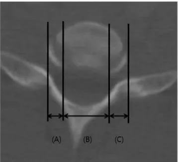
We measured three parameters on the MRI scans at C3–4, C4–5, C5–6, C6–7, and C7–T1 disc levels (Fig. 1). In addition, we measured three parameters on axial CT scans from C3–4
to C7–T1 (Fig. 2).
All statistical analyses were performed with SPSS software, version 13.0 for Windows (SPSS Inc., Chicago, IL, USA). Val-ues are expressed as mean valVal-ues with the standard deviation. Differences in continuous variables between the two groups were examined with a paired or unpaired t test. Differences in continuous variables between the different age groups were ex-amined with ANOVA test. Correlation of continuous variables between the two groups was examined with a Pearson corre-lation. It was considered significant when p was less than 0.05. In the preliminary study, all twenty patients were measured for intra-observer and inter-observer reliability. The intra-ob-server and inter-obintra-ob-server reliability were calculated using the reliability statistics by intraclass correlation (ICC). The ICC values were graded using previously described semiquantita-tive criteria: excellent for values in the 0.9–1.0 range, good for 0.7–0.89, fair/moderate for 0.50–0.69, low for 0.25–0.49, and poor for 0.0–0.24. Intra-observer reliability and inter-observer reliability for the uncinate process were good at 0.998 and 0.994, respectively, using ICC reliability statistics. Intra-ob-server reliability and inter-obIntra-ob-server reliability for the longus colli were good at 0.997 and 0.993, respectively, using ICC reli-ability statistics.
RESULTS
The mean values were 5.9±1.6 mm for the left longus colli dis-tance, 15.3±2.6 mm for the inter-longus colli disdis-tance, 6.5±1.9
Fig. 1. Distance of longus colli muscle on axial image of MRI: (A) the dis-tance from the lateral margin of the vertebral body to the medial margin of the right longus colli muscle (right longus colli distance), (B) the dis-tance from the medial margin of the right longus colli muscle to that of the left longus colli muscle (inter-longus colli distance), and (C) the distance from the medial margin of the left longus colli muscle to the lateral margin of the vertebral body (left longus colli distance).
Fig. 2. Distance of uncinate processes on axial image of CT: (A) the dis-tance from the lateral margin of the vertebral body to the medial margin of the right uncinate process (right uncinate distance), (B) the distance from the medial margin of the right uncinate process to that of the left un-cinate process (inter-unun-cinate distance), and (C) the distance from the medial margin of the left uncinate process to the lateral margin of the ver-tebral body (left uncinate distance).
mm for the right longus colli distance, 5.4±1.0 mm for the left uncinate distance, 17.0±2.3 mm for the inter-uncinate distance, and 5.4±1.1 mm for the right uncinate distance (Table 1). The longus colli distances, uncinate distances, and inter-uncinate distances increased from C3 to T1 (Table 1, Fig. 3). The inter-longus colli distances increased from C3 to C7 (Table 1). One hundred six patients were right-handed (88.3%) and fourteen were left-handed (11.7%). The right-handed patients had lon-ger longus colli distances on the right side than those on the left side at disc level C6–7 (p<0.05) (Table 1). The mean body mass index of the subjects was 21.05±5.3 kg/m2 (range: 13.5– 37.1 kg/m2).
To fully expose the uncinate processes, the longus colli mus-cles had to be dissected laterally 5.1±1.0 mm on the left and 5.5± 1.2 mm on the right at C3–4, 5.2±1.7 mm on the left and 5.6± 1.5 mm on the right at C4–5, 5.6±1.2 mm on the left and 6.0± 1.4 mm on the right at C5–6, 6.3±1.3 mm on the left and 7.3±
1.7 mm on the right at C6–7, and 7.2±1.6 mm on the left and 8.1±2.1 mm on the right at C7–T1 (Table 1).
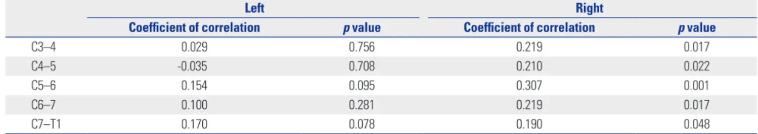
There was no difference in the longus colli distances and uncinate distances between males and females (Table 2). The inter-longus colli distances and inter-uncinate distances were larger in males than females, except the inter-longus colli dis-tances at C4–5 and C7–T1 (p<0.05) (Table 2). There were no differences in the longus colli distances, inter-longus colli dis-tances, uncinate disdis-tances, and inter-uncinate distances among the different age groups (Table 3). The longus colli distances were weakly correlated with uncinate distances from C3 to T1 on the right side only (p<0.05) (Table 4).
DISCUSSION
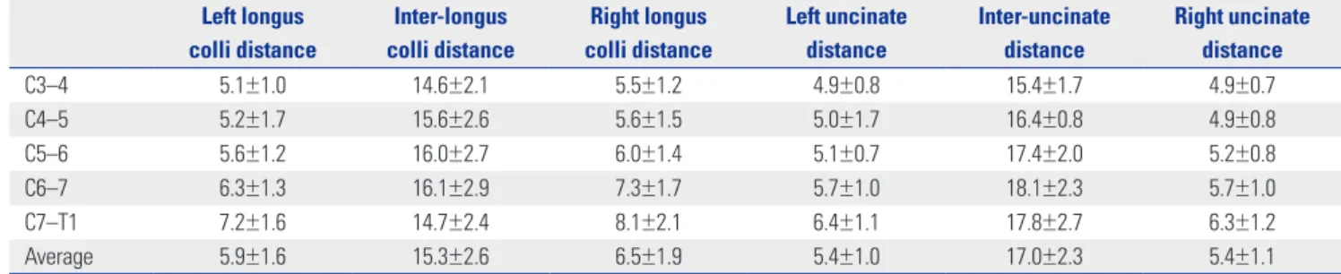
A number of studies have investigated the anatomical relation-Table 1. Distance of Longus Colli Muscles and Uncinate Processes According to Cervical Disc Levels (mm)
Left longus colli distance Inter-longus colli distance Right longus colli distance Left uncinate distance Inter-uncinate distance Right uncinate distance C3–4 5.1±1.0 14.6±2.1 5.5±1.2 4.9±0.8 15.4±1.7 4.9±0.7 C4–5 5.2±1.7 15.6±2.6 5.6±1.5 5.0±1.7 16.4±0.8 4.9±0.8 C5–6 5.6±1.2 16.0±2.7 6.0±1.4 5.1±0.7 17.4±2.0 5.2±0.8 C6–7 6.3±1.3 16.1±2.9 7.3±1.7 5.7±1.0 18.1±2.3 5.7±1.0 C7–T1 7.2±1.6 14.7±2.4 8.1±2.1 6.4±1.1 17.8±2.7 6.3±1.2 Average 5.9±1.6 15.3±2.6 6.5±1.9 5.4±1.0 17.0±2.3 5.4±1.1
Fig. 3. Distance of longus colli muscles and uncinate processes according to cervical disc levels (mm).
9 8 7 6 5 4 3 Distance C3–4 C4–5 C5–6 C6–7 C7–T1 Average Left uncinate distance Right uncinate distance
Left longus distance Right longus distance
Table 2. Distance of Longus Colli Muscles and Uncinate Processes According to Sex (mm) Left longus colli distance Inter-longus colli distance Right longus colli distance Left uncinate distance Inter-uncinate distance Right uncinate distance
Male Female Male Female Male Female Male Female Male Female Male Female
C3–4 5.3±1.0 5.0±1.1 15.5±2.0 13.7±1.9 5.7±1.3 5.4±1.2 5.1±0.7 4.8±0.8 16.1±1.5 14.8±1.6 5.0±0.8 4.9±0.7 C4–5 5.7±2.1 4.7±1.1 15.9±2.9 15.3±2.3 5.9±1.5 5.2±1.4 5.1±0.7 4.8±0.7 17.2±1.5 15.7±1.4 5.0±0.7 4.9±0.9 C5–6 5.8±1.0 5.4±1.3 16.7±2.6 15.3±2.6 6.2±1.5 5.9±1.2 5.2±0.7 5.0±0.7 18.3±1.8 16.5±1.8 5.3±0.8 5.2±0.9 C6–7 6.5±1.3 6.2±1.3 17.2±2.8 15.0±2.5 7.3±1.7 7.2±1.7 5.7±0.9 5.7±1.0 19.1±2.0 17.1±2.2 5.6±0.9 5.7±1.3 C7–T1 7.6±1.6 6.8±1.5 15.1±2.5 14.3±2.3 8.5±2.0 7.8±2.2 6.4±1.1 6.4±1.2 18.9±2.5 16.8±2.5 6.3±1.3 6.3±1.2 Average 6.1±1.6 5.6±1.4 16.0±2.6 14.7±2.4 6.7±1.9 6.2±1.8 5.4±0.9 5.3±1.0 17.8±2.1 16.1±2.1 5.4±0.9 5.3±1.1
ship between the longus colli muscle and the sympathetic chain, as well as the vertebral artery. Ebraheim, et al.2 found that the distance from the medial border of the sympathetic trunk to the medial border of the longus colli muscle was an av-erage of 10.6 mm in a study with 28 adult cadavers. Civelek, et al.3 found that the distance from the medial border of the sym-pathetic trunk to the medial border of the longus colli muscle was an average of 11.6±1.6 mm in a study with 30 cadavers. The distances from the medial border of the sympathetic trunk to the medial border of the longus colli muscle decreased from C3 to T1 in one study with 24 cadavers.1 In contrast, the dis-tance from the medial border of the vertebral artery to the medial border of the longus colli muscle gradually increased from C3 to C6 in a cadaveric study,5 as well as from C5 to C7 in a human CT study.6 Thorough knowledge of such anatomical relationships between the longus colli muscle and the sympa-thetic trunk, as well as the vertebral artery, can help reduce the risk of complications during anterior surgery. Similarly,
knowl-edge about the anatomical relationship between the uncinate process and the longus colli can be of benefit. This is especial-ly true when performing thorough decompressions of the fo-ramen in arthroplasty cases. However, we were unable to find any reports concerning the extent to which the longus colli must be mobilized to fully expose the uncinate processes out to its lateral margin during anterior cervical surgery.
We found that the longus colli distances, uncinate distances, and inter-uncinate distances increase from C3 to T1. To fully expose the uncinate processes, the longus colli muscles had to be dissected laterally 5.1±1.0 mm on the left and 5.5±1.2 mm on the right at C3–4, 5.2±1.7 mm on the left and 5.6±1.5 mm on the right at C4–5, 5.6±1.2 mm on the left and 6.0±1.4 mm on the right at C5–6, 6.3±1.3 mm on the left and 7.3±1.7 mm on the right at C6–7, and 7.2±1.6 mm on the left and 8.1±2.1 mm on the right at C7–T1. We suggest rounding off these numbers to make it easier to remember, such that the longus colli mus-cles are dissected laterally approximately 5 mm at C3–5, 6 mm at C5–6, 7 mm at C6–7, and 8 mm at C7–T1. The right side re-quired greater dissection than the left at all levels. There was no difference in the longus colli and uncinate distances be-tween males and females. Except for the inter-longus colli dis-tances at C4–5 and C7–T1, the inter-longus colli disdis-tances and inter-uncinate distances were larger in males than females. There were no differences in the longus colli distances, inter-longus colli distances, uncinate distances, and inter-uncinate distances among the different age groups. We were able to de-termine these values at all levels on all MRI and CT scans.
There are several reports concerning the lateral dimensions of the longus colli muscles using ultrasonography. The lateral dimension of the longus colli was reported to be 10.61±1.53 mm on the left side and 11.73±2.23 mm on the right side in 15 healthy subjects between 19 to 41 years old muscles measured by ultrasonography.8 In an ultrasonographic study of 20 pa-Table 5. Reports Concerning Widths of the Uncinate Process (mm)
Width Materials Pait, et al.7 6.0 at C3 6.1 at C4 5.3 at C5 5.8 at C6 6.7 at C7 6 cadaveric specimens Lu, et al.10 4.9±0.7 at C3 6.3±0.7 at C7 54 cadaveric specimens Ug˘ur, et al.11 5.0±0.8 at C3 5.0±0.9 at C4 5.1±0.8 at C5 5.1±1.0 at C6 5.3±1.1 at C7 49 cadaveric specimens
Table 3. Distance of Longus Colli Muscles and Uncinate Processes According to Age Group (mm) Left longus colli distance Inter-longus colli distance Right longus colli distance Left uncinate distance Inter-uncinate distance Right uncinate distance 20–29 6.1±1.7 14.5±2.3 7.1±2.0 5.6±1.3 16.5±2.3 5.4±1.3 30–39 5.9±1.5 14.9±2.2 7.1±2.0 5.6±1.0 17.0±2.3 5.7±1.3 40–49 6.1±1.4 14.6±2.4 6.8±1.9 5.6±0.9 16.0±1.9 5.8±1.0 50–59 5.7±1.9 15.8±2.7 6.3±1.6 5.4±1.1 17.0±2.1 5.4±1.0 60–69 5.7±1.5 16.2±2.6 5.9±1.8 5.1±0.8 17.6±2.7 5.1±0.8 70–79 5.9±1.4 16.3±2.8 5.9±1.6 5.2±0.9 18.0±2.1 5.1±0.9
Table 4. Correlation between the Distance of Longus Colli Muscles and Uncinate Processes According to Cervical Disc Levels
Left Right
Coefficient of correlation p value Coefficient of correlation p value
C3–4 0.029 0.756 0.219 0.017
C4–5 -0.035 0.708 0.210 0.022
C5–6 0.154 0.095 0.307 0.001
C6–7 0.100 0.281 0.219 0.017
tients with chronic neck pain and 20 healthy matched con-trols, the lateral dimensions of the longus colli were not differ-ent between the two groups.9 The lateral dimension of the longus colli in the patient group was 10.89±2.07 mm on the dominant side and 10.86±1.87 mm on the nondominant side versus 10.95±2.08 mm on the dominant side and 10.76±1.48 mm on the nondominant side in the controls.9 These values are higher than the ones we found. This is because the defini-tion of the longus colli distance in our study (i.e., the amount of longus colli muscle needed to be dissected to expose the uncinate process) is different from the ultrasonographic study, which measured the total width of the longus colli muscle. Our results are similar to the previous cadaveric studies in which the distances between the medial borders of longus colli mus-cles increased from C3 to C7.1,2,5,7
Our results showing increasing uncinate distances from C3 to T1 are similar to two previous studies (Table 5).10,11 Howev-er, another cadaveric study found no obvious pattern of in-creasing or dein-creasing distances from C3 to C7 (Table 5).7 This might be explained by the fact that they used different mea-suring methods: the width was measured from the medial to the lateral margins of the uncinate process at its base on the coronal plane,10,11 or from the medial to the lateral surfaces of the uncinate process at the mid-portion of the uncinate process on the coronal plane.7
As with any study, the present investigation has several lim-itations. First, the study was done in Koreans, and lengths may be different in other races. Second, the measurements were made in 120 cases and there may be rare variations in anato-my. There may also be individual variations based on body size such that one cannot blindly trust the numbers that we describe for any given patient. Our finding that CT and MRI scans could be utilized in all 120 cases points out the utility of using such studies to make these measurements prior to sur-gery. Despite these shortcomings, to our knowledge, this is the first report providing anatomic measurements that can be used as a guide for dissection of the longus colli muscle to ex-pose uncinate processes.
In conclusion, our results suggest that in most cases, one can safely dissect the longus colli muscle laterally approxi-mately 5 mm at C3–5, 6 mm at C5–6, 7 mm at C6–7, and 8 mm at C7–T1 to expose the uncinate process during anterior
cervi-cal surgery. It should be noted that these numbers should only serve as a guide and that individual measurements may differ due to anatomic variations. Therefore, it is recommended that the surgeon examine pre-operative CT or MRI prior to per-forming such dissection.
REFERENCES
1. Kiray A, Arman C, Naderi S, Güvencer M, Korman E. Surgical anatomy of the cervical sympathetic trunk. Clin Anat 2005;18: 179-85.
2. Ebraheim NA, Lu J, Yang H, Heck BE, Yeasting RA. Vulnerability of the sympathetic trunk during the anterior approach to the low-er clow-ervical spine. Spine (Phila Pa 1976) 2000;25:1603-6.
3. Civelek E, Karasu A, Cansever T, Hepgul K, Kiris T, Sabanci A, et al. Surgical anatomy of the cervical sympathetic trunk during an-terolateral approach to cervical spine. Eur Spine J 2008;17:991-5. 4. Kawashima M, Tanriover N, Rhoton AL Jr, Matsushima T. The
transverse process, intertransverse space, and vertebral artery in anterior approaches to the lower cervical spine. J Neurosurg 2003;98(2 Suppl):188-94.
5. Lu J, Ebraheim NA, Georgiadis GM, Yang H, Yeasting RA. Ana-tomic considerations of the vertebral artery: implications for an-terior decompression of the cervical spine. J Spinal Disord 1998; 11:233-6.
6. Hong JT, Park DK, Lee MJ, Kim SW, An HS. Anatomical variations of the vertebral artery segment in the lower cervical spine: analy-sis by three-dimensional computed tomography angiography. Spine (Phila Pa 1976) 2008;33:2422-6.
7. Pait TG, Killefer JA, Arnautovic KI. Surgical anatomy of the anteri-or cervical spine: the disc space, vertebral artery, and associated bony structures. Neurosurgery 1996;39:769-76.
8. Javanshir K, Mohseni-Bandpei MA, Rezasoltani A, Amiri M, Rah-gozar M. Ultrasonography of longus colli muscle: a reliability study on healthy subjects and patients with chronic neck pain. J Bodyw Mov Ther 2011;15:50-6.
9. Javanshir K, Rezasoltani A, Mohseni-Bandpei MA, Amiri M, Ortega-Santiago R, Fernández-de-LPeñas C. Ultrasound as-sessment of bilateral longus colli muscles in subjects with chronic bilateral neck pain. Am J Phys Med Rehabil 2011;90:293-301. 10. Lu J, Ebraheim NA, Yang H, Skie M, Yeasting RA. Cervical
unci-nate process: an anatomic study for anterior decompression of the cervical spine. Surg Radiol Anat 1998;20:249-52.
11. Ug˘ur HC, Uz A, Attar A, Tekdemir I, Egemen N, Elhan A. Anatom-ical projection of the cervAnatom-ical uncinate process in ventral, ventro-lateral, and posterior decompressive surgery. J Neurosurg 2000; 93(2 Suppl):248-51.