Utility of Single Shot Fast Spin Echo Technique in
Evaluating Pa n c reaticobiliary Diseases :
T 2 - weighted
I m a ge and Magnetic Resonance Cholangiopancre a to g ra p hy
1B young Wook Choi, M.D., Myeong-Jin Kim, M.D., Jae Bok Chung, M.D.2,
Heung Kyu Ko, M.D., Dong Joon Kim, M.D., Joo Hee Kim, M.D., Jae-Joon Chung, M.D., Hyung Sik Yoo, M.D., Jong Tae Lee, M.D.
Purpose : To evaluate the accuracy of T2-weighted imaging and MR
cholangiopancreatog-r a p hy using the single shot fast spin-echo technique focholangiopancreatog-r evaluating panccholangiopancreatog-reaticobiliacholangiopancreatog-ry dis-e a s dis-e.
Materials and Methods : B e t ween March and July 1997, axial and coronal T2-weighted
im-a g e s ( T E : 8 0-2 00 msec) im-and MR cholim-angiopim-ancreim-atogrim-ams(TE:800-1 2 00 msec) were ob-tained in two ways [single slab (thickness:30-50 mm) and multislice acquisition under chemical fat saturation] using SSFSE pulse sequencing in 131 cases of suspected pancreati-cobiliary disease. The accuracy of SSFSE MR imaging was assessed in 89 lesions of 74 pa-tients [male,48; female,26; age range, 3 0-86 (mean,59)years] confirmed surgicopathological-ly (50 lesions in 39 patients) and clinicalsurgicopathological-ly (39 lesions in 35 patients). Two radiologists re-v i ewed the MR images and diagnosis was determined by consensus.
Results : Correct diagnosis was confirmed in 84 of 89 lesions (94 %). Seven lesions we r e
falsely interpreted, false positive and false negative results accounting for two and five cas-e s, rcas-espcas-ectivcas-e l y. Two pancrcas-eatic canccas-ers wcas-ercas-e misdiagnoscas-ed as pancrcas-eatitis and a canccas-er of the proximal common bile duct(CBD) was interpreted as a distal CBD cancer. The sensitivi-ty of SSFSE MR imaging for malignancy was 93 %. One CBD stone revealed by endoscopic retrograde cholangiopancreatography (ERCP) was not detected on MR images. In contrast, a stone in the CBD seen on MR images was not apparent on subsequent ERC P. Sensitivity and specificity for calculous disease were 96 % and 99.7 %, respective l y. A benign stricture of the ampulla of Vater was falsely interpreted as normal, and correct diagnosis was possi-ble in two falsely diagnosed cases when MR images were rev i ewed retrospective l y.
Conclusion : The combination of T2-weighted and cholangiographic images using SSFSE is
an accurate method for diagnosing pancreaticobiliary diseases.
Index words :Magnetic resonance(MR), half-Fourier imaging
Bile ducts, MR
Bile ducts, stenosis or obstruction Pancreatic ducts, MR
Magnetic resonance (MR), rapid imaging
1Department of Diagnostic Radiology & Research institution of Radiological S c i e n c e
2Department of Internal Medicine, Division of Gastroenterology Yonsei University College of Medicine
이논문은보건의료기술연구개발사업연구비 ( H M P - 9 6 - M - 2 - 1 0 1 9 )의보조로이루어졌음 . Received March 15, 1999 ; Accepted June 1, 1999
Address reprint requests to : Myeong-Jin Kim, M.D., Department of Diagnostic Radiology, Severance Hospital, Yonsei University College of Medicine. #134, Shinchon-dong, Seodaemun-du Seoul 120-752, Korea.
The recently-introduced single shot fast spin echo (SS-FSE) or half-Fourier single shot turbo spin echo (HASTE) technique, using a single refocusing pulse and half-Fourier acquisition, shortens acquisition time to 2-2 0 seconds. For cholangiopancreatographic imaging of pancreaticobiliary diseases, most patients can hold their breath for this length of time. In terms of image-degra-dation due to motion during MR imaging, which is the major problem in MR imaging of pancreaticobiliary tracts, breath-hold MR cholangiopancreatography (MR-CP) using single shot half-Fourier acquisition provides good quality images free from motion artifact. Several investigators (1-6) have reported the superiority of this technique to previously-introduced sequences for MR-CP including two- or three-dimensional steady state free precession (SSFP)(7-11) and fast spin echo techniques ( 1 2-22) with or without the breath-hold technique. MR-CP using single shot half-Fourier acquisition can accu-rately depict normal structures and the presence and level of biliary obstruction without motion artifact and can, according to Regan et al. (4), be performed even in uncooperative and those who are ill.
T2-weighted images acquired using a relatively short echo time and without chemical fat saturation provide more detail of soft tissue than source images acquired for MIP (maximal intensity projection) reconstruction using a long echo time and chemical fat saturation. Although source images for MIP reconstruction have been reported by Yamashita et al. (6) to provide the best evaluation of stones in biliary trees, T2-weighted image can take the place of source images.
In many institutes, MRCP has been used to comple-ment endoscopic retrograde cholangiopancreatography (ERCP) or even routinely for the evaluation of pancre-aticobiliary diseases. Because of its advantages of nonin-vasiveness, virtually no procedure-related mortality or morbidity, and visualization of the duct proximal to complete obstruction -especially when therapy is not a n t i c i p a t ed-MRCP rather than ERCP is recommended. T2-weighted images without fat saturation can be ob-tained in a relatively short period of time using a SSFSE sequence in addition to MRCP. The two techniques in combination are believed to provide a more accurate di-agnosis as well as suggesting the next step in didi-agnosis and treatment. In patients with pancreaticobiliary dis-eases we evaluated the diagnostic accuracy of MRCP and T2-weighted images acquired using a SSFSE se-q u e n c e .
Materials and Methods
Between March and July 1997, T2 weighted images and MR cholangiopancreatograms were obtained using SSFSE pulse sequence in 131 cases of suspected pancreaticobil-iary disease. The accuracy of SSFSE MR imaging was as-sessed in 89 lesions of 74 patients [men,48; women, 2 6 ; age range,30-86 (mean age,59)years] confirmed surgico-pathologically (50 lesions in 39 patients) or clinically by ultrasonography, CT scanning, or direct cholangiogra-phy including ERCP (39 lesions in 35 patients, Table 1). The Remaining 47 patients were excluded due to insuffi-cient additional information or too short a period of fol-low-up. Using a 1.5-T system (Horizon, GE, Milwaukee, Wis., USA) and a phased-array torso coil, MR images were obtained. For coronal localizer images, single-shot fast spin-echo with a slice thickness of 8-10 mm and a s-lice gap of 0-2 mm with effective TE of 85-95 m s e c , phase encoding of 192, frequency encoding of 256, and receiver bandwidth of 31.3 kHz were used. Axial and coronal T2-weighted images were obtained using the fol-Table 1. Clinically Diagnosed Cases (n=35)
Diagnostic Modalities Diseases Total CT, US (n=7) Bile duct cancer(1)
GB cancer(1)
Extrahepatic duct stone(3) 1 1 Ihtrahepatic duct stone(3)
GB stone(2)
Recurrent pyogenic cholangitis(1) CT, ERCP (n=9) Bile duct cancer(4)
Pancreas cancer(2)
Extrahepatic duct stone(1) 1 0 Intrahepatic duct stone(1)
pancreatic duct stone(2) CT (n=6) Bile duct cancer(2)
GB cancer(1) 6 Pancreas cancer(1)
Extrahepatic duct stone(2) US (n=5) Bile duct cancer(1)
Pancreas cancer(1)
Liver malignancy(1) 5 Serous cystadenoma of pancreas(1) Adenomyomatosis of GB(1) ERCP (n=2) Ampullary cancer(1)
Extrahepatic duct stone(1) 2 CT, US, ERCP (n=2) Bile duct cancer(1)
Mucinous tumor of pancreas(1) 2 US, ERCP (n=1) Bile duct cancer(1) 1 US, PTC (n=1) Bile duct cancer(1) 1 PTC (n=1) Intrahepatic duct stone(1) 1
T o t a l 3 9
lowing parameters; TE,80-2 00 msec; matrix size,256× 256; thickness, 5 mm without gap. FOV was applied be-tween 24 cm and 36 cm according to a patient’s body habitus and expected extent of pathology based on the image for localization. Between eight and fourteen im-ages were acquired within 30 seconds, with 80 % of these within 20 seconds. MRCP was performed in two ways, single slab and multislice acquisition under chemi-cal fat saturation. Single slab image was obtained in the coronal plane with a TE of 800-1 2 00 msec, thickness of 3 0-50 mm, FOV of 28 cm, and matrix size of 256×2 5 6 . Acquisition time never exceeded two seconds. Multislice acquisition employed the same parameters as single slab acquisition except for a thickness of 3-5 mm without gap. For the reconstruction of maximal intensity projec-tion (MIP) images, multislice images were transferred to a workstation. MIP images were reconstructed in their an-terior-posterior perspective and rotated for up to 45 grees towards the left and right with an interval of 15 de-grees. No antiperistatic or paramagnetic agent was used.
Using clinical information, two abdominal radiologists read the MR images and a diagnosis was determined by consensus. Calculi were considered present when a round, oval, or multiangular signal void was seen within the lumen of bile ducts. To calculate diagnostic accuracy for calculous disease, five locations - namely the intra-hepatic, extraintra-hepatic, cystic, and pancreatic duct, and the gallbladder(total 74×5=370 locations) -were checked
for the presence of a stone. Malignancy was diagnosed when a definite mass was visible with or without nar-rowing or obstruction. To determine the accuracy with which malignancy was diagnosed, its origin was record-ed in four categories: the bile duct, pancreas, gallbladder, and ‘o t h e r’. Direct extension into adjacent organs was not included as a separate diagnosis. In cases of stenosis without a definite visible mass, abrupt narrowing, asym-metry, shouldering, or a combination of these findings was used to determine the cause of obstruction. Because many patients were ‘m i s s i n g’and thus had not been fol-lowed up, or because no confirmative diagnosis was available in clinically equivocal cases, pancreatitis was not included in this study. Mucinous neoplasm of the pancreas was classified as a miscellaneous disease rather than a malignancy. Mirizzi syndrome and recurrent pyo-genic cholangiohepatitis were considered as separate di-agnoses in addition to a diagnosis of calculi. Other mis-cellaneous diseases were diagnosed using the same crite-ria as those used in conventional CT and direct cholan-g i o cholan-g r a p h y .
R e s u l t s
Using MR cholangiography and T2 weighted images, diagnosis was correct in 84 of 89 lesions (94 %). Seven lesions were falsely interpreted, two cases being false positive and five false negative. Among the former, one
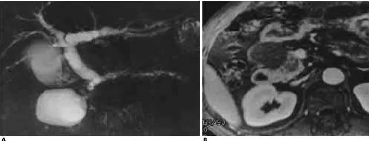
A B
Fig. 1. Pancreas cancer with pseudocyst
A. Single projection image shows a well demarcated large cystic mass on the right of the pancreas head portion. Diffuse dilatation
of pancreas duct and common bile duct is noted.
B . Gadolinium-enhanced T1-weighted axial image shows no definite mass lesion in the pancreas head except for a suspicious
poor-ly enhancing portion. One month later, jaundice was aggravated acutepoor-ly and follow-up CT scan shows a definite poorpoor-ly enhanced bulging mass in the head of the pancreas. Previously noted suspicious poorly enhancing portion of the pancreas head was postu-lated as the missing mass lesion retrospectively.
case was thought to be malignant and in the other a cal-culus was thought to be present, while among the latter, three malignant and two benign cases were misinter-p r e t e d .
Among 20 bile duct cancers, one case of proximal common duct cancer was misdiagnosed on the basis of MR images as distal common duct cancer and was counted as both a false positive for the distal CBD and a false negative for the proximal lesion. Two of nine pan-creatic cancers (sensitivity 78 %) were misdiagnosed simply as pancreatitis. On the basis of MR images, one of these cases was initially thought to be pancreatitis with a pseudocyst but without a definite mass lesion (Fig. 1). Retrospective review of MR images, however, revealed a suspicious lesion with low signal intensity and without bulging contour or ductal abnormality in the head of the pancreas. The other cases was falsely di-agnosed because of its small size (11 mm), the absence of mass effect and associated ductal abnormality seen on MRCP and T2-weighted images using SSFSE se-quencing. Ampullary carcinoma in six patients (sensitiv-ity 100 %) was correctly diagnosed on the basis of MR images (Fig. 2). Six cases of gallbladder carcinoma, all of which were correctly diagnosed (sensitivity 100 %), had visible masses with definite invasion of adjacent organs, especially the liver. One case of duodenal malignancy and three of liver malignancy were correctly diagnosed. The sensitivity of SSFSE MR imaging for malignancy was 93%(42/45) (Table 1).
Of the 89 lesions, 25 lesions (28 %) were calculous, and among these, extrahepatic duct stones were found in seven (28 %) (Fig. 3). Two cases of extrahepatic duct stone were misdiagnosed, one as false positive and one as false negative (Fig. 4). Intrahepatic duct stones were correctly diagnosed in nine patients. Four of these had CBD stones simultaneously, and in four others stones were present in the left hepatic duct (Fig. 5). In the other case, stones were located in the right hepatic duct, main-ly in the posterior segment. In three of four patients w i t h
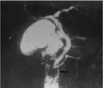
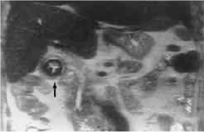
Fig. 2. Ampullary tumor.
Note a distal round dark-signaled obstructive mass (arrow) in M R C P .
A B
Fig. 3. Distal common bile duct stone and gallstones.
A . A rectangular-shaped calculus (arrow) is surrounded by bright signal of bile in the distal common bile duct on single projection
i m a g e .
B. T2-weighted axial images show multiple small stones (arrows) in the dependent portion of the gallbladder, which was not
stones in the left hepatic duct, recurrent pyogenic cho langitis with associated ductal dilatation and parenchy-mal atrophy of adjacent liver was diagnosed. Three of four cases of recurrent pyogenic cholangitis were con-firmed and surgically treated; the other patient under-went both CT scanning and ultrasonography. Six pa-tients with GB stones and one with a cystic duct stone were correctly diagnosed. Five of six patients with GB stones had other disease entities concurrently: a CBD s t o n e in three cases, Mirizzi syndrome in one (Fig. 6), and hilar cholangiocarcinoma in one. All these five
cas-es were correctly interpreted. Pancreatic duct stone was correctly diagnosed in two patients, one of whom show-ed a round signal void lesion, 1 cm in diameter, in the head portion of the main pancreatic duct and another 0 .7 cm signal void lesion in the body portion with dilata-tion of the main and side branches of the pancreatic duct. The other patient showed irregular tortuous dilatation of the pancreatic duct with a 1 cm round signal void le-sion in the head portion and multiple filling defects measuring 3-6 mm. Sensitivity and specificity for calcu-lous disease were 96 %(24/25) and 99.7 %(344/345),
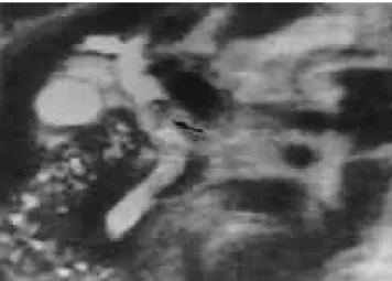
re-Fig. 4. False positive of choledocholithiasis. A sharply
demar-cated polygonal-shaped signal void lesion (arrow) is noted in the lumen of the mid portion of the common bile duct on T2-weighted coronal image. No calculus was demonstrated in the previous ERCP and no further evaluation was performed for c o n f i r m a t i o n .
Table 2.Malignant Biliary or Pancreatic Ductal Obstruction (n=45)
on MRCP
Correct Diagnosis Incorrect Diagnosis Bile duct cancer 1 9 2 * Pancreas cancer 7 2 Ampullary cancer 6 -GB cancer 6 -Duodenal Cancer 1 -Liver malignancy 3 -* : One case is a false positive.
Table 3. Calculous Disease (n=25) on MRCP
Correct Diagnosis Incorrect Diagnosis Extrahepatic duct 6 2 * Intrahepatic duct 9 -GB, cytic duct 7 -Pancreatic duct 2 -* : One case is a false positive.
A B
Fig. 5. Recurrent pyogenic cholangitis with left intrahepatic duct stone.
A . Single projection MR image clearly shows multiple variable-sized signal void lesions (arrows) in the dilated left intrahepatic
duct. However, surrounding liver parenchyma cannot be evaluated.
B. In contrast to the single projection image, T2-weighted coronal image shows atrophied surrounding liver parenchyma (arrows)
with increased signal intensity compared with normal parenchyma of the right lobe of the liver. Recurrent pyogenic cholangitis was confirmed by left lobectomy.
spectively, and diagnostic accuracy was 99. 5 % ( 3 6 8 / 370) (Table 3).
Twelve other miscellaneous diseases and their fre-quency of diagnostic accuracy are shown in Table 4. A benign stricture of the ampulla of Vater was falsely in-terpreted as normal. A subtle narrowing of the ampulla of Vater was neglected at the time of interpretation of MR images. The others were correctly interpreted on the basis of the same criteria applied in CT scanning and conventional cholangiopancreatography (Fig. 7).
D i s c u s s i o n
For the diagnosis of various pancreaticobiliary dis-eases, different diagnostic modalities such as computed
tomography and sonography have been employed in ra-diological fields and ERCP in internal medicine. ERCP has been regarded as ‘the gold standard’because duc-tal imaging is essential for the accurate diagnosis of dis-eases of the pancreatic and biliary tract, in which ductal structure plays a crucial role. However, the risk of pro-cedure-related complications of ERCP has been report-ed to be 1 %-5 % (23-25), an outcome mainly relatreport-ed to the invasiveness of the procedure. Since Wallner et al. (7) originally described MR cholangiography, many in-vestigators have used MRCP, with various pulse se-quences and advanced techniques, for the diagnosis of pancreaticobiliary diseases. Fast spin-echo techniques have been applied successfully to MRCP (17-22) and have shown a high signal-to-noise and contrast-to-noise
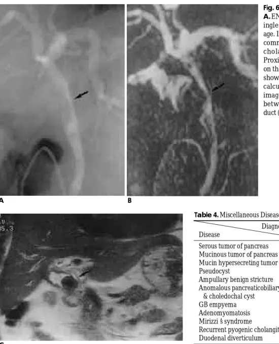
Fig. 6. Mirizzi syndrome
A. ENBD Cholangiogram, B . M R C P -
s-ingle projection, C . T2-weighted im-age. Irregular indentation on proximal common bile duct (arrow) is seen on cholangiographic images in A. B. Proximal CBD cancer was suggested on the ENBD cholangiogram. MRCP showed a round dark signal of a large calculus in the GB neck. C. Coronal image demonstates the relationship between the calculus and common duct (arrow).
A
C
B
Table 4. Miscellaneous Diseases (n=19)
D i a g n o s i s C o r r e c t I n c o r r e c t D i s e a s e
Serous tumor of pancreas 2 -Mucinous tumor of pancreas 1 -Mucin hypersecreting tumor of pancreas 3 -P s e u d o c y s t 1 -Ampullary benign stricture 1 1 Anomalous pancreaticobiliary duct union
& choledochal cyst 1 -GB empyema 1 -A d e n o m y o m a t o s i s 2 -M i r i z z i’s syndrome 1 -Recurrent pyogenic cholangitis 4 -Duodenal diverticulum 1
-ratio, reduced susceptibility effects, and reduced motion artifact compared with the initially introduced MRCP technique using gradient-echo sequences (7-10). With fast spin-echo techniques, three-dimensional acquisition with respiratory triggering has the advantage of yielding better-quality MIP reconstruction due to a more isotrop-ic voxel size (19, 20). More recently, half-Fourier single-shot rapid acquisition with a relaxation enhancement (RARE) sequence has been applied to MRCP, and this offers ultrafast acquisition times, low artifact suscepti-bility, and sequential thin slice capability (1-4).
Regan et al. (4) reported that the presence and level of obstruction was accurately depicted on 100 % and 87% of occasions, respectively. A little later, Ichikawa et al. (2) reported that single-shot hybrid RARE could provide consistently higher quality MRCP than FSE and the contrast-enhanced Fourier acquired steady-state tech-nique (CE-FAST) because sequential images by single-shot hybrid RARE minimized respiratory, bowel, and cardiac motion artifacts. Most recently, in a large study of 300 patients, MRCP with the half-Fourier RARE tech-nique was reported by Fulcher et al. (5) to be 100 % ac-curate in determining the presence of pancreaticobiliary disease, the presence and level of biliary obstruction, and obstruction due to bile duct calculi. In the same re-port, the accuracy of MRCP and MR imaging in deter-mining the presence and level of malignant obstruction was said to be 98.2 %. The accuracy they reported in di-agnosing pancreaticobiliary diseases using MRCP with FSE or HASTE was higher than in any previous reports. Among our patients, only two cases of calculi were mismatched with the results of ERCP, which was
per-formed before or after MR imaging. In a retrospective review of MR images of a false negative case of CBD stone side by side with ERCP, neither the signal void structure in the CBD lumen nor the dilated CBD were present in MR images. MR images showed only multi-ple stones in the patient’s left hepatic duct. The possi-bility that an intraheptic duct stone spontaneously moved to the CBD at some point during the interval be-tween the time of MR imaging and ERCP, which was performed 22 days after MR imaging, could not be ex-cluded. Three cases of malignancy were falsely inter-preted. Among these, one case of distal CBD cancer was correctly diagnosed after the retrospective review of MR images, but in two cases of pancreatic cancer, this review did not lead to correct diagnosis. The fact that pancreatic cancers were not detected in MR images sug-gested that the slightly different signal intensity of the suspicious lesion from that of surrounding normal parenchyma of the pancreas, was not enough to accu-rately diagnose or even detect a pancreatic mass which was not accompanied by ductal abnormality or mass ef-fect. Additional contrast-enhanced cross-sectional im-ages would have been necessary for correct diagnosis of these cases. In diagnosing malignancy, the small differ-ence in the accuracy of MRCP between our results and those of Fulcher et al. (5) can be explained by the fact that they used conventional cross-sectional MR images including contrast-enhanced T1-weighted images in ad-dition to MRCP in diagnosing suspicious malignancy. Conventional cross-sectional MR imaging with T1-weighted imaging and contrast enhancement does not, however, allow a definite conclusion regarding its rou-Fig. 7. Adenomyomatosis of gallbladder
T2-weighted coronal image. Multiple intramural high signal lesions from Rokitansky-Aschoff sinuses (arrows) are well demonstrated in the thickened GB wall.
Fig. 8. Gallstone-Mercedez Benz sign.
T2-weighted coronal MR image clearly depicts a large gall-stone showing the typical ‘M e r c e d e s - B e n z’sign (arrow).
tine use for suspicious pancreatic malignancy.
We added relatively short TE multislice imaging using SSFSE sequencing without fat suppression as in the case of conventional fast spin-echo T2-weighted imaging. This is in contrast to long TE multislice imaging with fat suppression used for MIP reconstruction. For tissue characterization, T2-weighted imaging with relatively short TE and without fat suppression was more infor-mative than source imaging for MIP, and small stones were detected no less accurately. Small stones in the gallbladder or biliary duct which were not detected or incompletely seen on MIP or single projection images, were clearly visualized on T2-weighted axial or coronal images. In view of the limitations of MRCP, short-T2 material in the lumen of bile ducts cannot be easily dif-ferentiated from a true stone or other obstructive le-sions. This is a consequence of water imaging with only bright or dark signals permitted, which resembles a di-rect cholangiogram in that a lesion is shown only as a fill-ing defect regardless of its nature. For example, Mirizzi syndrome (Fig. 6) was dramatically depicted on T2-weighted images without fat suppression, showing ir-regular GB wall thickening with a dark calcified stone impacted in the neck of the gallbladder and compressed mid-common duct without evidence of malignancy in surrounding tissue. MIP and multislice source images showed only bright lumen of CBD and GB, with focal narrowing and round filling defect, respectively. T2-weighted images showed more detailed characteristics of the stone, such as the Mercedes-Benz sign, suggesting the presence of a cholesterol stone in another case (Fig. 8). CNR is superior in MRCP with fat suppression, but CNR is not so critically decreased in T2-weighted im-ages without fat suppression that the lesion can be de-tected and characterized. Additionally, T2-weighted im-ages could be acquired in a reasonably short period of time, usually less than 20 seconds using SSFSE sequenc-ing, during which time most patients were able to hold their breath. T2-weighted images showed more detailed characteristics of stone and provided more information with which definite masses could be demonstrated and the extension of most malignancies evaluated. T2-weighted images were helpful for determining subse-quent diagnostic steps, such as further MR imaging with contrast enhancement or angiography with a more confident impression of malignancy, as well as endo-scopic biopsy for tissue confirmation in cases of suspect-ed malignancy without possible delay for other diagnos-tic outcomes. In addition, the use of T2-weighted
im-ages facilitates decision making with regard to the deter-mination of stone management, medical treatment, sur-gical removal, or endoscopic extraction.
However, small side branches of the pancreatic duct, if not dilated, could not be clearly evaluated with all SS-FSE sequences, though some studies (5, 13) have shown that MRCP can detect small ducts and stones as small as 2mm in diameter. With advance in MR equipment and technology, improvements in spatial resolution are ex-p e c t e d . In our study, ex-postsurgical biliary-enteric anatom-ic change (26) or susceptibility artifact from surganatom-ical m a-terial was not the cause of false interpretation of MRCP and T2-weighted images. In fact, only one case of CBD stone seen on MR images, which was mismatched with a negative result in ERCP, might be explained by certain reported pitfalls mimicking choledocholithiasis (27). In this case, unfortunately, no further attempt was made to confirm any pathologic condition or artifact (Fig. 4).
In spite of the high accuracy of MRCP in detecting the presence and level of biliary obstruction by calculi, it cannot easily replace ERCP because of the crucial ad-vantage this has as the means of providing simultaneous treatment at the time of diagnosis. Another advantage of ERCP is its role in facilitating biopsy in an equivocal case, particularly in ampullary lesions. In terms of non-invasiveness, however, MRCP can replace ERCP if a therapeutic procedure is not anticipated, and procedure-related mortality and morbidity can thus be avoided. Soto et al. (12) reported that MRCP played an important role in the care of patients in whom ERCP was unsuc-cessful, or incomplete, or when technical difficulties could be anticipated.
In conclusion, MR cholangiography with MIP or a sin-gle projection technique is an accurate method for diag-nosing various pancreaticobiliary diseases with high specificity and sensitivity while bearing in mind the need for a therapeutic procedure and resultant cost-ef-fectiveness. Additional T2-weighted images that can be acquired in a reasonably short time with MRCP would be informative in determining the next step of diagnosis or treatment.
R e f e r e n c e s
1. Reuther G, Kiefer B, Tuchmann A, Pesendorfer FX. Imaging find-ings of pancreaticobiliary duct diseases with single-shot MR cholangiopancreatography. A J R 1 9 9 7 ; 1 6 8 : 4 5 3 - 4 5 9
2. Ichikawa T, Nitatori T, Hachiya J, Mizutani Y. Breath-held MR cholangiopancreatography with half-averaged single shot hybrid rapid acquisition with relaxation enhancement sequence:
compar-ison of fast GRE and SE sequences. J Comput Assist Tomogr 1 9 9 6 ; 2 0 : 7 9 8 - 8 0 2
3. Miyazaki T, Yamashita Y, Tsuchigame T, Yamamoto H, Urata J, Takahashi M. MR cholangiopancreatography using HASTE(half-Fourier acquisition single-shot turbo spin-echo) sequences. A J R 1 9 9 6 ; 1 6 6 : 1 2 9 7 - 1 3 0 3
4. Regan F, Smith D, Khazan R, Bohlman M, Schultze-Haakh H, Campion J, et al. MR cholangiography in biliary obstruction using half-Fourier acquisition. J Comput Assist Tomogr 1 9 9 6 ; 2 0 : 6 2 7 - 6 3 2 5. Fulcher AS, Turner MA, Capps GW, Zfass AM, Baker KM.
Half-Fourier RARE MR cholangiopancreatography: Experience in 300 subjects. R a d i o l o g y 1998; 207:21-32
6. Yamashita Y, Abe Y, Tang Y, Urata J, Sumi S, Takahashi M. In vitro and clinical studies of image acquisition in breath-hold MR cholangiopancreatography: single-shot projection technique ver-sus multislice technique. A J R 1 9 9 7 ; 1 6 8 : 1 4 4 9 - 1 4 5 4
7. Wallner BK, Schumacher KA, Weidenmaier W, et al. Dilated bil-iary tract: evaluation with MR cholangiography with a T2-weight-ed contrast-enhancT2-weight-ed fast sequence. R a d i o l o g y 1 9 9 1 ; 1 8 1 : 8 0 5 - 8 0 8 8. Morimoto K, Shimoi M, Shirakawa T, et al. Biliary obstruction:
e-valuation with three-dimensional MR cholangiography. R a d i o l o g y 1 9 9 2 ; 1 8 3 : 5 7 8 - 5 8 0
9. Ishizaki Y, Wakayama T, Okada Y, et al. Magnetic resonance cholangiography for evaluation of obstructive jaundice. Am J
G a s t r o e n t e r o l 1 9 9 3 ; 8 8 : 2 0 7 2 - 2 0 7 7
1 0 . Hall-Craggs M, Allen CM, Owens CM, et al. MR cholangiography: clinical evaluation in 40 cases. Radiology 1 9 9 3 ; 1 8 9 : 4 2 3 - 4 2 7 1 1 . Lee MG, Lee HJ, Kim MH, et al. Extrahepatic biliary diseases: 3D
MR cholangiopancreatography compared with endoscopic retro-grade cholangiopancreatography. Radiology 1 9 9 7 ; 2 0 2 : 6 6 3 - 6 6 9 1 2 . Soto JA, Yucel EK, Barish MA, Chuttani R, Ferrucci JR. MR
cholangiopancreatography after unsuccessful or incomplete ER-CP. R a d i o l o g y 1 9 9 6 ; 1 9 9 : 9 1 - 9 8
1 3 . Becker CD, Grossholz M, Becker M, Mentha G, Peyer R, Terrier F. Choledocholithiasis and bile duct stenosis: diagnostic accuracy of MR cholangiopancreatography. R a d i o l o g y 1 9 9 7 ; 2 0 5 : 5 2 3 - 5 3 0 1 4 . Barish MA, Soto JA, Yucel EK. Magnetic resonance
cholangiopan-creatography of the biliary ducts: techniques, clinical applications, and limitations. J Magn Reson Imaging 1 9 9 6 ; 8 : 3 0 2 - 3 1 1
1 5 . Taourel P, Bret PM, Reinhold C, Barkun AN, Atri M. Anatomic variants of the biliary tree: diagnosis with MR cholangiopancre-atography. R a d i o l o g y 1 9 9 6 ; 1 9 9 : 5 2 1 - 5 2 7
1 6 . Guibaud L, Bret PM, Reinhold C, Atri M, Barkun AN. Bile duct obstruction and choledocholithiasis: diagnosis with MR cholan-giography. R a d i o l o g y 1 9 9 5 ; 1 9 7 : 1 0 9 - 1 1 5
1 7 . Reinhold C, Guibaud L, Genin G, Bret PM. MR cholangiopancre-atography: comparison between two-dimensional fast spin-echo and three-dimensional gradient-echo. J Magn Reson Imaging 1 9 9 5 ; 5 : 3 7 9 - 3 8 4
1 8 . Soto JA, Barish MA, Yucel EK, Ferrucci JT. MR cholangiopancre-atography: findings on 3D fast spin-echo imaging. A J R 1 9 9 5 ; 1 6 5 : 1 3 9 7 - 1 4 0 1
1 9 . Barishi MA, Yucel EK, Soto JA, Chuttani R, Ferrucci JT. MR cholangiopancreatography: efficacy of three-dimensional turbo spin-echo technique. A J R 1 9 9 5 ; 1 6 5 : 2 9 5 - 3 0 0
2 0 . Soto JA, Barish MA, Yucel EK, et al. Pancreatic duct: MR cholan-giopancreatography with a three-dimensional fast spin-echo tech-nique. R a d i o l o g y 1 9 9 5 ; 1 9 6 : 4 5 9 - 4 6 4
2 1 . Takehara Y, Ichijo K, Tooyama N, et al. Breath-hold MR cholan-giopancreatography with a long-echo-train fast spin-echo sequence and a surface coil in chronic pancratitis. R a d i o l o g y 1 9 9 4 ; 1 9 2 : 7 3 - 7 8 2 2 . Low RN, Sigeti JS, Francis IR, et al. Evaluation of malignant biliary
obstruction: efficacy of fast multiplanar spoiled gradient-recalled MR imaging vs spin-echo MR imaging, CT, and cholangiography.
A J R 1 9 9 4 ; 1 6 2 : 3 1 5 - 3 2 3
2 3 . Bilbao MK, Dotter CT, Lee TG, Katon RM. Complications of en-doscopic retrograde cholangiopancreatography(ERCP): a stucy of 10,000 cases. G a s t r o e n t e r o l o g y 1976; 70: 314-320
2 4 . Hamilton I, Lintott DJ, Rothwell J, Axon ATR. Acute pancreatitis following endoscopic retrograde cholangiopancreatography. C l i n
R a d i o l 1983; 34:543-546
2 5 . Thoeni RF, Fel SC, Goldberg HI. CT detection of asymptomatic pancreatitis following ERCP. Gastrointerst Radiol 1990; 15:291-295 2 6 . Pavone P, Laghi A, Catalano C, et al. MR cholangiography in the
examination of patients with biliary-enteric anastomoses. A J R 1997; 169:807-811
2 7 . David V, Reinhold C, Hochman M, et al. Pitfalls in the interpreta-tion of MR cholangiopancreatography. AJR 1 9 9 8 ; 1 7 0 : 1 0 5 5 - 1 0 5 9