저작자표시-비영리-변경금지 2.0 대한민국 이용자는 아래의 조건을 따르는 경우에 한하여 자유롭게 l 이 저작물을 복제, 배포, 전송, 전시, 공연 및 방송할 수 있습니다. 다음과 같은 조건을 따라야 합니다: l 귀하는, 이 저작물의 재이용이나 배포의 경우, 이 저작물에 적용된 이용허락조건 을 명확하게 나타내어야 합니다. l 저작권자로부터 별도의 허가를 받으면 이러한 조건들은 적용되지 않습니다. 저작권법에 따른 이용자의 권리는 위의 내용에 의하여 영향을 받지 않습니다. 이것은 이용허락규약(Legal Code)을 이해하기 쉽게 요약한 것입니다. Disclaimer 저작자표시. 귀하는 원저작자를 표시하여야 합니다. 비영리. 귀하는 이 저작물을 영리 목적으로 이용할 수 없습니다. 변경금지. 귀하는 이 저작물을 개작, 변형 또는 가공할 수 없습니다.
Management of Clinical T1N0M0 stage
Esophageal Cancer
Andrew Jihoon Yang
Department of Medicine
The Graduate School, Yonsei University
[UCI]I804:11046-000000522769
[UCI]I804:11046-000000522769
Management of Clinical T1N0M0 stage
Esophageal Cancer
Directed by Professor Chang Geol Lee
The Master's Thesis
submitted to the Department of Medicine,
the Graduate School of Yonsei University
in partial fulfillment of the requirements for the degree of Master of
Medical Science
Andrew Jihoon Yang
This certifies that the Master's Thesis of
Andrew Jihoon Yang is approved.
---
Thesis Supervisor : Chang Geol Lee
---
Thesis Committee Member#1 : Yong Chan Lee
---
Thesis Committee Member#2 : Dae Joon Kim
The Graduate School
Yonsei University
ACKNOWLEDGEMENTS
First of all, I would like to appreciate Professor Chang Geol Lee for
providing me such inspiring advice and support for preparing this thesis
and setting a good example for me by devoting himself to patients suffering
from esophageal cancer. Also I would like to express my gratitude to
professor Yong Chan Lee and Dae Joon Kim for giving me advices which
were essential for completing this article. Moreover, I would like to
appreciate to professor Seo Hee Choi, Hwa Kyung Byun, Hyun Ju kim, Jin
hyun Choi, Sang Kil Lee for always offering the great instructions with
careful concern. Any absence of help from those mentioned above would
have made my work impossible. I hereby once again thank everyone for
their aids in my research.
<TABLE OF CONTENTS>
ABSTRACT ··· 1
I. INTRODUCTION ··· 3
II. MATERIALS AND METHODS ··· 4
1. Patient selection ··· 4
2. Treatment··· 4
3. Evaluation of response ··· 6
4. Statistical analysis ··· 7
III. RESULTS ··· 7
1. Patient characteristics ··· 7
2. Treatment outcomes ··· 9
3. Patterns of failure ··· 14
4. Complication ··· 15
IV. DISCUSSION ··· 17
V. CONCLUSION ··· 21
REFERENCES ··· 22
APPENDICES ··· 23
ABSTRACT(IN KOREAN) ··· 24
PUBLICATION LIST ··· 26
LIST OF FIGURES
Fig 1. Treatment flow of all patients (N = 179) ··· 8
Fig 2. Comparison of Kaplan–Meier overall survival curves; (a) overall
survival (OS) and recurrence-free survival (RFS) of all patients, (b) OS and
RFS of patients with cT1a lesions after endoscopic resection, (c) OS and (d)
RFS of patients with cT1b lesions
according to the treatment group ··· 11
Fig 3. Flow of treatment outcomes
according to each treatment group ··· 14
LIST OF TABLES
Table 1. Patients’ characteristics ··· 9
Table 2. Patterns of failure according to the treatment group ·· 15
Table 3. Acute and late complication rates according to the treatment group in
patients with T1b lesions ··· 17
1
ABSTRACT
Management of Clinical T1N0M0 stage Esophageal Cancer
Andrew Jihoon Yang
Department of Medicine
The Graduate School, Yonsei University
(Directed by Professor Chang Geol Lee)
Purpose: Endoscopic resection is employed as a standard treatment for
stage T1a esophageal cancer, and esophagectomy or radical radiation
therapy (RT) are used for stage T1b lesion. The purpose of this study was
to compare treatment outcomes of each modality with clinical stage T1
esophageal cancer.
Materials and Methods: A total of 179 patients with clinical T1N0M0
stage esophageal cancer treated during 2006 and 2016 were
retrospectively evaluated. Sixty-two patients with clinical T1a stage
underwent endoscopic resection. Among 117 patients with clinical T1b
stage, 82 patients underwent esophagectomy, and 35 patients received
chemoradiotherapy or RT. We compared overall survival (OS) and
recurrence-free survival (RFS) rates by each treatment modality.
Results: Median follow-up time was 32 months (range, 1 - 120 months).
The 5-year OS and RFS rate for patients with stage T1a receiving
endoscopic resection was 100% and 85%, respectively. For patients with
stage T1b, the 5-year OS and RFS rate was 78% and 77% for the
esophagectomy group and 80% and 44% for the RT alone group and
96% and 80% for the chemoradiation group, respectively. The
esophagectomy group showed significantly higher RFS than RT alone
group (p=0.04). There was no significant difference in RFS between the
esophagectomy and the chemoradiation group (p=0.922). Grade 3 or
higher treatment-related complication occurred in 4 patients who
received esophagectomy.
2
Conclusion: The endoscopic resection alone appeared as adequate
treatment for patients with T1a stage esophageal cancer. Definitive
chemoradiation was comparable to esophagectomy in survival outcome
without serious complication for T1b stage esophageal cancer.
___________________________________________________________
Key words : esophageal cancer, endoscopic therapy, esophagectomy,
radiation therapy, chemoradiation
3
Management of Clinical T1N0M0 stage Esophageal Cancer
Andrew Jihoon Yang
Department of Medicine
The Graduate School, Yonsei University
(Directed by Professor Chang Geol Lee)
I. INTRODUCTION
Esophageal cancer is one of the most common and aggressive cancers worldwide. It is a frequently diagnosed cancer, with approximately 460,000 new cases and 400,000 deaths worldwide in 2012 (1). The incidence of this disease has increased dramatically during the past 3 decades (2). With this increasing incidence, the overall prognosis for patients with esophageal cancer was still poor, with a 5-year survival rate of 14% (3). However, with increasing use of endoscopic surveillance and early detection, early-stage cancers are being diagnosed more. Early-stage esophageal cancer (T1N0M0) means lesions limited to the mucosa (T1a) or submucosa (T1b) and comprises approximately 20% of all initial diagnoses (4). These early-stage cancers present the best opportunity for a cure at diagnosis to some extent that it can affect the overall prognosis of all esophageal cancer. The 5-year survival can be as high as 85%; a stark contrast to 10% when diagnosed at advanced stage (5).
Esophagectomy has been the standard treatment for T1b esophageal cancer, although with some debate. Recently, there has been increasing interest in endoscopic therapies for this stage lesion. These include ablative techniques such as photodynamic therapy (PDT) and endoscopic mucosal resection (EMR) (6-7). Radiotherapy (RT) can also be used for superficial esophageal cancer, and favorable outcomes with this noninvasive treatment also have been reported (8).
4
Moreover, RT has undergone notable advances during the past decade. Three-dimensional (3D) conformal RT and intensity-modulated radiation therapy (IMRT) based on computed tomography (CT) images, and multileaf collimator (MLC) have contributed to the delivery of high doses accurately to the target, sparing normal tissues. However, the role of definitive RT for superficial esophageal cancer compared to other treatment modalities as esophagectomy and endoscopic therapies has not been elucidated yet.
In our institution, we perform esophagectomy, endoscopic therapies, or RT in T1 stage esophageal cancers as physicians’ decision after the multidisciplinary discussion. The purpose of this study is to compare the outcomes of each treatment modalities while looking at the experience of the single institution for the treatment of early-stage esophageal cancers.
II. MATERIALS AND METHODS
1. Patients selection
We retrospectively identified 208 patients who received treatments for clinical T1N0M0 stage esophageal cancer between February 2006 and December 2016 in Severance Hospital. Among them, 27 patients with other malignancies and 2 patients with involvement of esophagogastric junction were excluded, and a total of 179 patients were included in the final analysis. Data were collected using the patients’ medical and operative records, and a departmental database.
2. Treatment
Before the treatment decision, endoscopic ultrasonography (EUS) was performed in all patients to evaluate the depth of cancer invasion. Depth of tumor infiltration for clinical T categorization was determined based on the deepest invasion depth by EUS. Based on the American Joint Committee on Cancer (AJCC) 7th edition, depth of invasion was categorized as ‘T1a’ if the
5
tumor invaded lamina propria or muscularis mucosae and ‘T1b’ if there was a submucosal invasion. Careful attention to ultrasound image provides presence of abnormal or enlarged lymph nodes likely to harbor cancer, and occasionally signs of distant spread, such as lesion in surrounding organs. In addition nodal status and distant metastasis were evaluated by CT and PET-CT.
Treatment decision was made fundamentally according to the National Comprehensive Cancer Network (NCCN) guidelines with these stages after multidisciplinary discussion. Physicians preferentially considered endoscopy therapy for T1a stage tumors and esophagectomy for T1b stage tumors. RT was performed for patients who had difficulty in surgery due to the location of the tumor, old age, or comorbidities, and patients who refused surgery. Patients who were initially diagnosed as having clinical T1a stage esophageal cancer but diagnosed as pathologic T1b stage after endoscopic therapy and received complete surgery or RT were included in this study. Those were analyzed separately from them who were finally diagnosed as pT1a.
A. Endoscopy therapy
Endoscopic resection was conducted by EMR consist of EMR using a transparent cap procedure (EMR-C), EMR with precutting (EMR-P) and endoscopic submucosal dissection (ESD) with various knives such as hook knife, IT knife-2 or dual knife. The detailed methods of endoscopic resections are described in our institution’s previous paper (9).
B. Radiotherapy
The total dose was determined by radiation oncologist considering the patient’s condition (e.g., age, performance status, and coexisting disease) and tolerance dose of adjacent normal tissues (e.g., spinal cord, lungs, and heart). RT was delivered 5 days a week at 1.8-2.1 Gy (median 1.8 Gy) per fraction. The total dose was ranged from 50.4 to 64.0 Gy (median 63.0 Gy) in 28 to 35 fractions. The gross tumor volume (GTV) was defined primary and nodaldisease, and was drawn onto the axial planning CT images using the EUS-derived ab oral
6
distances and EUS/CT findings as references. The clinical target volume (CTV) was defined as GTV plus a 3-5 cm margin in the longitudinal direction and a 0.5-1 cm margin in the lateral and anteroposterior directions. The planning target volume (PTV) was defined as the CTV plus an additional 0.3-0.5 cm margin to account for setup error and internal organ motion. Selection for RT modality (3D conformal RT or IMRT) was made considering the tumor location and temporal change. The number of patients with 3D conformal RT was 82.9%, and the number of IMRT patients was 17.1%.
Concurrent chemoradiotherapy (CCRT) was performed for patients with the good general condition, rather than RT alone. For CCRT, two courses of chemotherapy were given during RT on days 1 to 5, at a 3-week interval. Chemotherapy consisted of 5-fluorouracil (700 mg/m2 of body-surface area per day by 120-hour infusion) and cisplatin (15 mg/m2 of body-surface area per day intravenously).
C. Esophagectomy
For patients with middle and lower esophageal cancers, a two-field lymph node dissection and intrathoracic esophagogastrostomy were performed using the whole stomach as a conduit, and anastomosis was performed with a 28 mm end-to-end anastomosis stapler. For patients with upper esophageal cancer, a three-field lymph node dissection was routinely performed. If an intrathoracic esophagogastrostomy was possible, an intrathoracic anastomosis was performed using the entire stomach. For all others, a cervical esophagogastrostomy with a gastric tube was performed. The stomach was positioned in the posterior mediastinum. The detailed methods are available in another previous report (10).
3. Evaluation of response
Patients who underwent endoscopy therapy or RT were followed by endoscopy with biopsies, CT, and EUS every 6 months after treatment for 3 years and then every 1 year thereafter. The opinion of experienced physicians who performed the endoscopy was also important to discriminate between true
7
recurrence and treatment-related changes. Complete response was defined when no residual tumor findings were seen by all exams. Partial response was defined when residual tumor findings were diagnosed by all exams.
Patients who underwent esophagectomy were followed by CT every 6 months after treatment for 3 years and then every 1 year thereafter. Endoscopic evaluation was performed every 1 year and when abnormalities were seen on follow-up CT. Patterns of failure were classified according to the recurrence sites as follows. Local recurrence was defined as recurrence in the esophagus, and regional recurrence was defined as recurrence in regional nodes. Distant recurrence was defined as recurrence at the any other sites.
4. Statistical analysis
The overall survival (OS) was defined as the time from initial diagnosis to death from any cause or the last follow-up. The recurrence-free survival (RFS) was measured from initial diagnosis to any recurrences or the last follow-up. Both OS and RFS rates were calculated using the Kaplan-Meier method, and the significance of differences in survival was assessed by the log-rank test. Cox regression was used for identification of independent prognostic factors using multivariate analysis. Furthermore, the chi-square test was used to compare categorical variables, and the t-test was used to compare continuous variables. A p-value < 0.05 was considered statistically significant. SPSS ver. 23 (IBM, Armonk, NY, USA) was used for all statistical analyses.
III. RESULTS
1. Patients
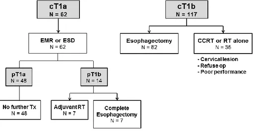
Among 179 patients who met inclusion criteria in this study, 62 patients were initially diagnosed with cT1a esophageal cancer, and the rest were diagnosed with cT1b esophageal cancer. As shown in Figure 1, 62 patients first received endoscopic resection (28: EMR, and 34: ESD). Further treatments were
8
performed according to the pathologic result of following endoscopic tests. A total of 14 patients were finally diagnosed with pT1b esophageal cancers, and half of them received adjuvant RT, and the rest underwent complete esophagectomy. Among 117 patients with cT1b esophageal cancers, 82 received esophagectomy and 35 received CCRT (N = 25) or RT alone (N = 10).
Figure 1. Treatment flow of all patients (N = 179)
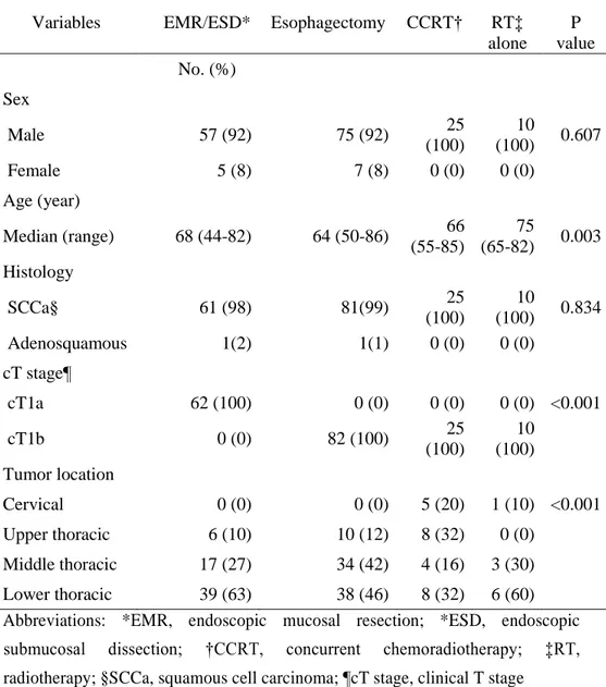
The baseline characteristics and cancer-related features of the 4 groups (‘EMR/ESD,’ ‘Esophagectomy,’ ‘CCRT,’ and ‘RT alone’ group) are summarized in Table 1. There was a significant difference in age and location according to the treatment method (p = 0.003, and <0.001, respectively). The median age (years) of each group was 68 in the endoscopy group, 64 in the esophagectomy group, 66 in the CCRT group, and 75 in the RT alone group. The median age of patients in the RT alone group was significantly higher than the rest. Significantly more numbers of tumors at the cervical or upper-thoracic area were included in the CCRT group than the others. As performing ‘laryngopharyngoesophagectomy’ for cervical location tumors might impair the quality of life subsequently, we selected CCRT or RT alone for those with upper-located lesions. Otherwise, no other significant difference was found in patients’ characteristics.
9
Table 1. Patients’ characteristics
Variables EMR/ESD* Esophagectomy CCRT†
RT‡ alone P value No. (%) Sex Male 57 (92) 75 (92) 25 (100) 10 (100) 0.607 Female 5 (8) 7 (8) 0 (0) 0 (0) Age (year) Median (range) 68 (44-82) 64 (50-86) 66 (55-85) 75 (65-82) 0.003 Histology SCCa§ 61 (98) 81(99) 25 (100) 10 (100) 0.834 Adenosquamous 1(2) 1(1) 0 (0) 0 (0) cT stage¶ cT1a 62 (100) 0 (0) 0 (0) 0 (0) <0.001 cT1b 0 (0) 82 (100) 25 (100) 10 (100) Tumor location Cervical 0 (0) 0 (0) 5 (20) 1 (10) <0.001 Upper thoracic 6 (10) 10 (12) 8 (32) 0 (0) Middle thoracic 17 (27) 34 (42) 4 (16) 3 (30) Lower thoracic 39 (63) 38 (46) 8 (32) 6 (60) Abbreviations: *EMR, endoscopic mucosal resection; *ESD, endoscopic submucosal dissection; †CCRT, concurrent chemoradiotherapy; ‡RT, radiotherapy; §SCCa, squamous cell carcinoma; ¶cT stage, clinical T stage
2. Treatment outcomes
10
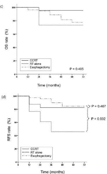
1-120 months). The 5-year OS and RFS rates of all patients were 86% and 77%, respectively [Figure 2A]. All patients with T1a stage lesions underwent endoscopic therapy (EMR/ESD). Except for the 14 patients who were diagnosed with pathologic T1b stage through endoscopic therapy, the 5-year OS and RFS rates of the EMR / ESD group were 100% and 85%, respectively [Fig. 2B]. Patients diagnosed with T1b stage were excluded from the outcome analysis of the EMR / ESD group because they had undergone salvage treament. Patients with T1b stage lesions underwent esophagectomy, CCRT, or RT. The 5-year OS and RFS rates were 78% and 77% for the esophagectomy group, 96% and 80% for the CCRT group, and 80% and 44% for the RT alone group, respectively [Figure 2C, 2D]. There was no significant difference in OS according to the treatment modality in patients with T1b stage (p = 0.405). The esophagectomy group showed significantly higher RFS than that of the RT alone group (p = 0.04). There was no significant difference in RFS between the esophagectomy and the CCRT group (p = 0.922). For OS, age, T stage, and treatment modality were significant factors in both univariate and multivariate analyses (p = 0.007, 0.056, and 0.026, respectively). For RFS, there were no significant factors in univariate and multivariate analyses.
12
Figure 2. Comparison of Kaplan–Meier overall survival curves; (a) overall survival (OS) and recurrence-free survival (RFS) of all patients, (b) OS and RFS of patients with cT1a lesions after endoscopic resection, (c) OS and (d) RFS of patients with cT1b lesions according to the treatment group
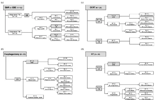
For the EMR/ESD group (N = 62), 48 patients were diagnosed with pT1a stage, and 14 patients were diagnosed with the pT1b stage. Of the 48 patients diagnosed with pT1a stage, 46 patients are alive without disease after no adjuvant
13
treatment, and 2 patients with local or regional recurrence are with the disease. Seven out of 14 patients with pT1b stage underwent adjuvant RT, and 7 underwent adjuvant surgery sequentially. Most patients except 2 with recurrence have been in complete response (CR) status and are alive without recurrence [Figure 3A].
For the esophagectomy group (N = 82), All esophageal cancers were completely removed through surgery. However, 3 of them underwent treatment-related death (sepsis after pneumonia or esophageal perforation). Otherwise, 71 patients were alive without any recurrence. Eight patients experienced recurrence (local = 1, regional = 4, distant = 3) and half of them died after disease progression [Figure 3B].
For the CCRT group (N = 25), 18 patients showed CR and 7 showed partial responses (PRs). Of the patients with CR, all except one who died of other medical cause are alive without disease. Of the patients with PR, none showed disease progression, and all except one who died of other medical cause are alive even without any additional treatment [Figure 3C].
For the RT alone group (N = 10), half of them showed CR and the other half showed PR. Of the patients with CR, only one regional recurrence developed and the rest were alive with or without the disease. Of the patient with PR, 3 recurrence (local = 1, regional = 1, and distant = 1) were noted and one with distant recurrence died of disease [Figure 3D]. An overview of outcomes in each treatment subgroup is seen in Figure 3.
14
Figure 3. Flow of treatment outcomes according to each treatment group
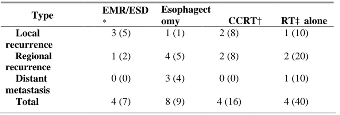
3. Patterns of failure
Table 2 lists the site of the first recurrence for patients in 4 subgroups. Twenty of the 179 patients had a recurrence. The group with the highest rate of recurrence was the RT alone group (7% in the EMR/ESD group, 9% in the esophagectomy group, 16% in the CCRT group, and 40% in the RT alone group). In the EMR/ESD group, although the overall rate of recurrence was low (N = 4), 3 (75%) were local recurrence, and 1 (25%) was a regional recurrence. In the esophagectomy group, regional recurrence was the most frequent recurrence pattern (50%), followed by distant (38%) and local recurrence (12%). In the CCRT group, half of the overall recurrences were local recurrence, and the rest were regional recurrence. There was no distant recurrence after CCRT. In the RT alone group, regional recurrence was in 50%, followed by local (25%) and distant recurrence (25%).
15
Table 2. Patterns of failure according to the treatment group Type EMR/ESD* Esophagect
omy CCRT† RT‡ alone Local recurrence 3 (5) 1 (1) 2 (8) 1 (10) Regional recurrence 1 (2) 4 (5) 2 (8) 2 (20) Distant metastasis 0 (0) 3 (4) 0 (0) 1 (10) Total 4 (7) 8 (9) 4 (16) 4 (40) Abbreviations: *EMR, endoscopic mucosal resection; *ESD, endoscopic submucosal resection; †CCRT, concurrent chemoradiotherapy; ‡RT, radiotherapy 4. Complication
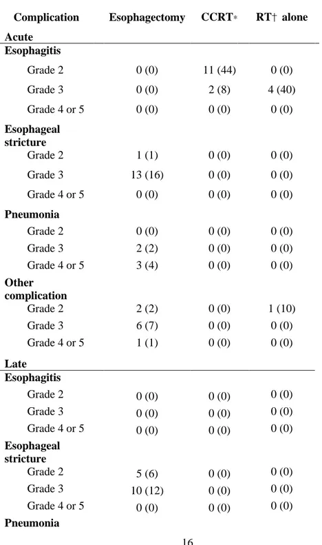
Table 3 lists the complication for patients in the 3 subgroups which included only T1b stage lesions. The overall rates of acute complications were 34%, 52%, and 50% in the esophagectomy, CCRT, and RT alone group, respectively. The most common acute complications were esophageal stricture (17%) in the esophagectomy group, esophagitis (52%) in the CCRT group, and esophagitis (40%) in the RT alone group. Grade 4 or higher complications were developed only in 4 patients (3%) of the esophagectomy group; Three patients suffered from pneumonia median 1 month after surgery and one patient experienced perforation 1 week after surgery. Three of them underwent treatment-related deaths after these complications. Otherwise, all except 2 in the CCRT group and 4 in the RT alone group (all manageable esophagitis during RT) were grade 2 complications. Late complications were only found in the esophagectomy group (24%). Most of them were an esophageal stricture, dysphagia, and regurgitation.
16
Table 3. Acute and late complication rates according to the treatment group in patients with T1b lesions
Complication Esophagectomy CCRT* RT† alone Acute Esophagitis Grade 2 0 (0) 11 (44) 0 (0) Grade 3 0 (0) 2 (8) 4 (40) Grade 4 or 5 0 (0) 0 (0) 0 (0) Esophageal stricture Grade 2 1 (1) 0 (0) 0 (0) Grade 3 13 (16) 0 (0) 0 (0) Grade 4 or 5 0 (0) 0 (0) 0 (0) Pneumonia Grade 2 0 (0) 0 (0) 0 (0) Grade 3 2 (2) 0 (0) 0 (0) Grade 4 or 5 3 (4) 0 (0) 0 (0) Other complication Grade 2 2 (2) 0 (0) 1 (10) Grade 3 6 (7) 0 (0) 0 (0) Grade 4 or 5 1 (1) 0 (0) 0 (0) Late Esophagitis Grade 2 0 (0) 0 (0) 0 (0) Grade 3 0 (0) 0 (0) 0 (0) Grade 4 or 5 0 (0) 0 (0) 0 (0) Esophageal stricture Grade 2 5 (6) 0 (0) 0 (0) Grade 3 10 (12) 0 (0) 0 (0) Grade 4 or 5 0 (0) 0 (0) 0 (0) Pneumonia
17 Grade 2 0 (0) 0 (0) 0 (0) Grade 3 0 (0) 0 (0) 0 (0) Grade 4 or 5 0 (0) 0 (0) 0 (0) Other complication Grade 2 4 (5) 0 (0) 0 (0) Grade 3 1 (1) 0 (0) 0 (0) Grade 4 or 5 0 (0) 0 (0) 0 (0)
Abbreviations: *CCRT, concurrent chemoradiotherapy; †RT, radiotherapy
IV. DISCUSSION
In this study, we compared the outcome of endoscopic therapy, esophagectomy, CCRT, and RT in patients with early-stage esophageal cancer. First, we have shown that patients with T1a esophageal cancer had a good prognosis with endoscopic eradication therapies. Second, esophagectomy did not improve OS compared to other noninvasive therapies in patients with T1b esophageal cancer. Esophagectomy prolonged RFS significantly compared to RT alone, however, there was no notable difference compared to CCRT. The outcomes of CCRT and RT should be assessed further, given that more patients with old age or poor performance status were treated with either CCRT or RT than surgery.
Traditionally, the ‘gold standard’ approach to the management of early-stage esophageal cancer has been esophagectomy, either alone or in combination with chemotherapy and/or RT. Although shown to improve survival, this approach has been associated with significant risks as surgical complications (e.g., anastomotic leaks, infections, pneumonia, or stenosis), or even mortality. Recent efforts to improve outcomes from early-stage esophageal cancer treatment, where the primary goal is ‘cure,’ have focussed on the development of less invasive, endoscopic treatments designed to destroy or remove cancerous lesions in the esophageal wall while sparing healthy tissue. These treatments were used
18
either alone or in combination with other techniques, without compromising oncologic outcomes. As in our results, grade IV or higher complications including pneumonia and esophageal perforation were only observed in the esophagectomy group, although any severe late complication did not appear in patients receiving CCRT or RT.
Currently, the National Cancer Institute recommends surgery as the standard treatment for Ib stage esophageal cancer, and this recommendation is based on the observation that T1b esophageal squamous cell carcinoma has a high incidence of lymph node metastasis (11). In our study, the patterns of failure data showed that the overall recurrence rate of patients with T1a stage was as good as only 7%. In the esophagectomy group, local recurrence was 1%, regional recurrence was 5%, and distant metastasis was 4%, which means surgical resection was good for local control. In the CCRT group, local and regional recurrence were all in 8% and 8%, respectively, with no distant metastasis. In the RT alone group, the regional recurrence rate was high as 20%, and local and distant metastasis rates were 10% and 10%, respectively. Comparing the CCRT group with the RT alone group, the absence of distant metastasis in the CCRT group indicates that the combination of chemotherapy might be effective enough for systemic control. Considering that patients with T1b lesions showed 7.6% of regional recurrence rate, we probably should include the regional lymph nodes in the RT field, not simply irradiating primary mass. Chemotherapy for systemic control also should be considered.
In patients with T1a stage, 5-year OS and RFS were 100% and 85%, respectively. In patients with T1b stage, there was no significant difference in outcome between the patients with esophagectomy and the patients with CCRT. This suggests that CCRT is sufficiently comparable to esophagectomy even after considering possible selection bias, which more patients with the poor condition were included in the CCRT or RT alone group.
There was one paper which demonstrated the superior outcome of esophagectomy in patients with cT1 esophageal cancer 11. Matsumoto et al. compared outcomes of 29 patients with radical esophagectomy and 38 patients
19
with definitive RT. Although there was no difference in the OS rate, the RFS was significantly higher in the surgery group than in the radiation group (5-year RFS 62% vs. 39%, p = 0.005). Especially, significantly higher RFS rate could be fulfilled in patients with tumors than invade the submucosa. However, before concluding that surgery is the most appropriate treatment for cT1 esophageal cancer, we should be noted that the authors did not distinguish patients with RT only or patients with CCRT. Considering that the RT alone group showed the inferior result in our paper, the actual results of Matsumoto’s paper are considered to be similar to the data of ours. The outcome of CCRT was superior to that of RT alone in esophageal cancer according to the previous prospective randomized trial (12). Therefore, we can propose that CCRT should be in an equal position rather than a simple alterative treatment, based on the results of those studies and other favorable data of RT (13-14).
When we performed multivariate analysis in this study, the factors significantly associated with OS were age, T stage, and treatment modality. T stage was a significant factor in several existing data, and treatment modality could also be a significant factor because there was a different baseline characteristic according to treatment modality related to T stage (15). Conversely, there might be no significant difference in OS according to treatment modality when patients with the same stage were analyzed. Other studies have shown that T stage, age, facility, and follow-up frequency in early-stage esophageal cancer are significantly associated with outcome (15-16). Most of them were similar to ours; this means that our cohort represents a representative group of patients with early-stage esophageal cancer who are not specific. Moreover, our study was a single institutional study which had no difference in follow-up schedules or facility according to the treatment group.
There are several limitations of this study. First, there could be a selection bias from the retrospective nature of this study. Although the main purpose of this study was to compare each treatment modality in the early-stage esophageal cancer, there was a trend to perform esophagectomy for patients with T1b stage unless patients did not refuse surgery. A definitive CCRT was
20
performed because of the difficulty of operation when the lesion was located at the cerivical level. Also, if the patient had any medical morbidity or poor performance status, physicians decided to perform CCRT or RT. Physician’s preference or differences in patient characteristics could affect the treatment outcomes unexpectedly. Second, because the study was based on a clinical stage, patients who were diagnosed with pathologic T1a stage after esophagectomy were also included in the esophagectomy group. We conclude that this comparison is reasonable because there will be patients in the T1a stage that are not identifiable in either the CCRT group or the RT alone group. However, this may result in overestimating the overall outcome of T1b stage patients. Third, this paper could not analyze the effect of RT technique development in the treatment outcome. Recently, our institution has been treating many patients with esophageal cancer using IMRT. This study included patients from the past, most of whom were treated with 3D-CRT (3D-CRT 82.8% vs. IMRT 17.2%). We could not find any difference according to RT modalities from these small number of patients. Lastly, we performed response evaluation using biopsy for patients who underwent definitive CCRT or RT. However, endoscopic biopsy after CCRT/RT sometimes shows low sensitivity/specificity in detection of recurred carcinoma. This might have resulted in a bias in the outcome.
Nevertheless, this study has a strength in that it analyzed the outcome of patients with early esophageal cancer who were treated with various treatment modalities based on medical records of single institution. Those patients were included in this study according to the strict eligibility criteria, compared to previous studies, and it enhanced the homogeneity of this study. In our institution, many patients with esophageal cancer have been referred to the multidisciplinary clinic and decided which was the most optimal treatment based on institutional policy. In addition, unlike existing papers, the more precise comparative study could be done by comparing the patient subgroups. To overcome the limitations by small numbers of each treatment to perform more structured comparative study, the Korea Radiation Oncology Group (KROG) group has the plan to perform a multi-institutional retrospective study on the optimal treatment policy in
21 early-stage esophageal cancer.
V. CONCLUSION
In conclusion, we assessed the clinical outcome of clinical T1N0M0 stage esophageal cancer patients according to each different treatment modality. In patients with T1a stage, endoscopic therapy was shown to be a satisfactory treatment. In patients with T1b stage, CCRT and esophagectomy showed a comparably good outcome although RT alone showed an inferior outcome than others. However, esophagectomy induced some patients with ≥grade 4 complications and favorable patients were included more than other groups, which might draw selection bias. Considering those results, CCRT should be considered as alternative treatments of esophagectomy for the treatment of T1b stage patients. To clarify our findings, further studies with randomized controlled design or larger cohort are warranted.
22
1. Zeng Y, Liang W, Liu J, He J. Endoscopic Treatment Versus Esophagectomy for Early-Stage Esophageal Cancer: a Population-Based Study Using Propensity Score Matching. Journal of gastrointestinal surgery : official journal of the Society for Surgery of the Alimentary Tract. 2017;21(12):1977-83.
2. Pennathur A, Farkas A, Krasinskas AM, Ferson PF, Gooding WE, Gibson MK, et al. Esophagectomy for T1 esophageal cancer: outcomes in 100 patients and implications for endoscopic therapy. The Annals of thoracic surgery. 2009;87(4):1048-54; discussion 54-5.
3. Enzinger PC, Mayer RJ. Esophageal cancer. The New England journal of medicine. 2003;349(23):2241-52.
4. Rice TW, Rusch VW, Ishwaran H, Blackstone EH, Worldwide Esophageal Cancer C. Cancer of the esophagus and esophagogastric junction: data-driven staging for the seventh edition of the American Joint Committee on Cancer/International Union Against Cancer Cancer Staging Manuals. Cancer. 2010;116(16):3763-73.
5. Wang S, Huang Y, Xie J, Zhuge L, Shao L, Xiang J, et al. Does delayed esophagectomy after endoscopic resection affect outcomes in patients with stage T1 esophageal cancer? A propensity score-based analysis. Surgical endoscopy. 2017.
6. Rice TW, Blackstone EH, Goldblum JR, DeCamp MM, Murthy SC, Falk GW, et al. Superficial adenocarcinoma of the esophagus. The Journal of thoracic and cardiovascular surgery. 2001;122(6):1077-90.
7. Pennathur A, Landreneau RJ, Luketich JD. Surgical aspects of the patient with high-grade dysplasia. Seminars in thoracic and cardiovascular surgery. 2005;17(4):326-32.
8. Cao C, Luo J, Gao L, Xu G, Yi J, Huang X, et al. Definitive radiotherapy for cervical esophageal cancer. Head & neck. 2015;37(2):151-5.
9. Lee HJ, Lee H, Park JC, Shin SK, Lee SK, Lee YC. Treatment Strategy after Endoscopic Resection of Superficial Esophageal Squamous Cell Carcinoma: A Single Institution Experience. Gut and liver. 2015;9(6):714-9.
23
thoracoscopic lymphadenectomy along the recurrent laryngeal nerves in radical esophagectomy for esophageal squamous carcinoma. Surgical endoscopy. 2014;28(6):1866-73.
11. Matsumoto S, Takayama T, Tamamoto T, Wakatsuki K, Enomoto K, Tanaka T, et al. A comparison of surgery and radiation therapy for cT1 esophageal squamous cell carcinoma. Diseases of the esophagus : official journal of the International Society for Diseases of the Esophagus. 2011;24(6):411-7.
12. Herskovic A, Martz K, al-Sarraf M, Leichman L, Brindle J, Vaitkevicius V, et al. Combined chemotherapy and radiotherapy compared with radiotherapy alone in patients with cancer of the esophagus. The New England journal of medicine. 1992;326(24):1593-8.
13. Nemoto K, Yamada S, Hareyama M, Nagakura H, Hirokawa Y. Radiation therapy for superficial esophageal cancer: a comparison of radiotherapy methods. International journal of radiation oncology, biology, physics. 2001;50(3):639-44. 14. Sasaki T, Nakamura K, Shioyama Y, Toh Y, Okamura K, Ohura H, et al. Treatment outcomes of radiotherapy for patients with stage I esophageal cancer: a single institute experience. American journal of clinical oncology. 2007;30(5):514-9.
15. Wani S, Drahos J, Cook MB, Rastogi A, Bansal A, Yen R, et al. Comparison of endoscopic therapies and surgical resection in patients with early esophageal cancer: a population-based study. Gastrointestinal endoscopy. 2014;79(2):224-32.e1.
16. Moreno AC, Verma V, Hofstetter WL, Lin SH. Patterns of Care and Treatment Outcomes of Elderly Patients with Stage I Esophageal Cancer: Analysis of the National Cancer Data Base. Journal of thoracic oncology : official publication of the International Association for the Study of Lung Cancer. 2017;12(7):1152-60.
24
ABSTRACT(IN KOREAN)
식도암 임상병기 T1N0M0 환자들의 치료
<지도교수 이 창 걸>
연세대학교 대학원 의학과
양 지 훈
목적: 내시경 절제는 T1a 병기 식도암에 대한 표준 치료법으로
사용되며, 식도 절제술 또는 근치적 방사선 치료 요법은
T1b병기 식도암에 사용됩니다. 이 연구의 목적은 임상 병기
T1 식도암에서 각 치료법의 결과를 비교하는 것 입니다.
대상 및 방법: 본 연구는 2006년부터 2016년까지 치료 된 임상
T1N0M0 단계 식도암 환자 총 179 명을 후향적으로
평가했습니다. 임상 T1a 병기를 가진 62 명의 환자는 내시경
절제술을 받았습니다. 임상 T1b 병기를 가진 117 명의 환자 중
82 명의 환자는 식도 절제술을 받았으며 35 명의 환자는 화학
방사선 치료 또는 방사선 치료를 받았다. 각 치료법 별로 전체
생존율과 재발 없는 생존율을 비교했습니다.
결과: 추적 관찰 기간의 중앙값은 32 개월 (1 ~ 120
개월)이었습니다. 내시경 절제술을 받는 병기 T1a를 가진
환자의 5 년 생존율 및 재발 없는 생존율은 각각 100 % 및
85 %였습니다. 병기 T1b를 가진 환자의 경우, 5 년 생존율 및
재발 없는 생존율은 식도 절제술 그룹의 경우 78 % 및 77 %,
방사선 치료 단독 그룹의 경우 80 % 및 44 %, 항암 방사선
치료 그룹의 경우 96 % 및 80 %였습니다. 식도 절제술 그룹은
방사선 단독 그룹보다 유의미하게 높은 재발 없는 생존율을
나타냈습니다 (p = 0.04). 식도 절제술과 항암 방사선 요법 그룹
간에는 재발 없는 생존율에 유의미한 차이가 없었습니다 (p =
25