unusual circumstances such as uveitic cataracts or some patients with trauma. This study provides support for that dosing choice since postoperative inflammation was adequately controlled in even our youngest enrollees. Infants less than 7 months of age were routinely left aphakic. Although it is true that some surgeons in the Infant Aphakia Treatment Study (IATS) used steroid drops more frequently than four times per day, the rate of inflammatory complications was not lower in those dosed more than four times per day.2We can make no statement about the use of difluprednate more frequently than four times per day, but our study demonstrated good control of postoperative inflammation at that dosage and no increase in adverse events when compared with prednisolone acetate, the current standard.
The centers in our study were chosen, in part, because they were experienced and routinely successful at checking intraocular pressure in infants and small children. The Icare (Finland) rebound tonometer has become a popular device among pediatric cataract surgeons in the USA for measuring IOP in this age group without sedation pediatric cataract surgeons in the USA and it was used in this study.3Both pre-surgery IOP and post-operative IOP readings were taken in a clinical area outside of the operating room. Our study IOP readings were not done under general anesthesia.
Since the IATS recommended that most infants under 7 months of age be left aphakic and treated with a contact lens, infants treated in this manner were enrolled and randomized in our study.4Extended-wear silicone contact lenses or daily-wear rigid gas permeable contact lenses were used. With these materials (0% water content), we found no adverse events related to placing the drops on the eye while the contact lens was being worn. It is likely that the package insert advising against the instillation of topical difluprednate while wearing contact lens is for high water content contact lenses that are not available in the powers needed to correct aphakic infants.
Drug choice and dosing in infants and young children after cataract surgery will remain a personal choice of the surgeon. Our study provides evidence that difluprednate can be safely used at QID dosing in children aged 0–3 years.
Conflict of interest
The authors declare no conflict of interest.
References
1 Wilson ME, O'Halloran H, VanderVeen D, Roarty J, Plager DA, Markwardt K et al. Difluprednate versus prednisolone acetate for inflammation following
cataract surgery in pediatric patients: a randomized safety and efficacy study. Eye (Lond) 2016; 30(9):
1187–1194.
2 Lambert SR, Plager DA, Buckley EG, Wilson ME, DuBois L et al. Infant Aphakia Treatment Study Group. The Infant Aphakia Treatment Study: further on intra- and postoperative complications in the intraocular lens group. J AAPOS. 2015; 19 (2): 101–103.
3 Lambert SR, Buffenn AN, Chiang MF, Coats DK, Melia M, Simpson JL et al. Rebound tonometry in children. Ophthalmology 2013; 120: e21–e27.
4 Lambert SR, Lynn MJ, Hartmann EE, DuBois L, Drews-Botsch CD et al. (Writing Committee) The Infant Aphakia Treatment Study Group. Comparison of contact lens and intraocular lens correction of monocular aphakia during infancy: a
randomized clinical trial of HOTV optotype acuity at age 4.5 years and clinicalfindings at age 5 years. JAMA Ophthalmol 2014; 132(6); 676–684.
ME Wilson, SR Lambert, DA Plager, D VanderVeen, J Roarty and H O’Halloran
Ophthalmology & Pediatrics, Storm Eye Institute, Medical University of South Carolina, Charleston, SC, USA
E-mail: [email protected]
Eye (2017) 31, 506–507; doi:10.1038/eye.2016.244; published online 4 November 2016
Sir,
Two-year outcomes of intravitreal bevacizumab for choroidal neovascularization associated with a dome-shaped macula in pathologic myopia
A dome-shaped macula (DSM) is an inward protrusion of the macula seen on optical coherence tomography (OCT) in highly myopic eyes, which wasfirst described in 2008 by Gaucher et al.1Since DSM appears to be a distinct feature of highly myopic eyes,2,3it could be suspected that choroidal neovascularization (CNV) that develops in myopic eyes with DSM may have different clinical features and may follow a course different from that of CNV found in myopic eyes without DSM. However, studies on the therapeutic outcome of CNV associated with DSM are limited. When searching PubMed using the keyword‘dome-shaped macula’, only one brief report that showed the visual outcome of myopic CNV with DSM after 1-year intravitreal ranibizumab treatment was identified.3The purpose of this study was to determine the 2-year visual and anatomical outcomes of intravitreal bevacizumab (IVB) treatment for myopic CNV with DSM features. The secondary objective was to compare patients with and without DSM, to investigate whether there was any difference in the clinical features and therapeutic outcome in these two groups after IVB treatment.
We retrospectively reviewed the medical records of 50 patients with myopic CNV who received IVB injections between 1 January 2009 and 30 April 2014. Inclusion criteria were as follows: (1) a refractive error≤ − 6.0 D or an axial length≥ 26.5 mm, (2) the presence of subfoveal, juxtafoveal, or extrafoveal CNV, (3) treatment-naive patients who were treated with at least one IVB, and (4) follow-up of 2 years or more after intravitreal injection. The study was approved by the institutional review board of Yonsei University College of Medicine. After the initial IVB at baseline, retreatments were applied pro re
Eye Correspondence
nata on the basis of changes in OCTfindings and/or changes in visual acuity. Statistical analysis was performed using SPSS version 20.0 (IBM, Chicago, IL, USA).
Out of 50 eyes with myopic CNV, 9 (18%) eyes showed DSM features. The demographics and
clinical characteristics of patients with and without DSM are summarized in Table 1. The eyes with myopic CNV associated with DSM received 4.00± 3.04
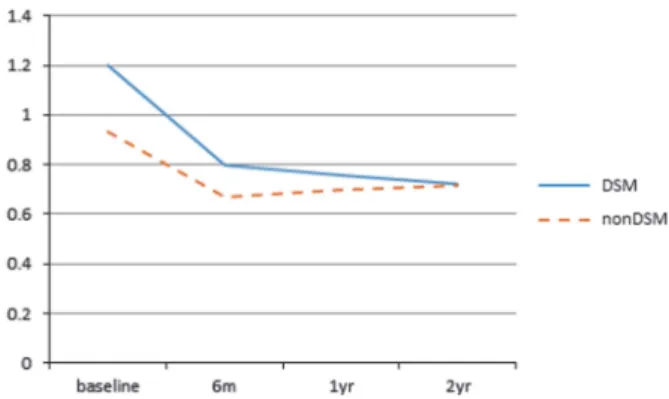
injections during 2 years, and the recurrence rate over the 2-year period was 33.3%. The mean logMAR best-corrected visual acuity in eyes with DSM significantly improved from 1.20 at baseline to 0.80 at 6 months, 0.76 at 1 year, and 0.72 at 2 years (P= 0.048, 0.034, 0.011, respectively). Both retinal and choroidal thickness decreased gradually over
2 years. There was no significant difference
between the DSM and the non-DSM group in terms of baseline characteristics and therapeutic outcomes, including number of injections and CNV
recurrence. Visual outcomes were also comparable between the two groups at 6 months, 1 year, and 2 years (Figure 1).
In conclusion, DSM features were present in 18% of myopic CNV patients, and after a 2-year treatment with
IVB, myopic CNV patients both with and without DSM showed equally significant visual benefits and
anatomical improvements. The development of CNV in patients with DSM may not be associated with DSM features, but with other more significant factors, such as myopia itself.
Conflict of interest
The authors declare no conflict of interest.
References
1 Gaucher D, Erginay A, Lecleire-Collet A, Haouchine B, Puech M, Cohen SY et al. Dome-shaped macula in eyes with myopic posterior staphyloma. Am J Ophthalmol 2008; 145(5): 909–914. 2 Cebeci Z, Kir N. Bilateral dome-shaped macula with serous
macular detachment in a child. Case Rep Ophthalmol Med 2015; 2015: 213968.
3 Ceklic L, Wolf-Schnurrbusch U, Gekkieva M, Wolf S. Visual acuity outcome in RADIANCE study patients with dome-shaped macular features. Ophthalmology 2014; 121(11): 2288–2289.
JH Lee, SC Lee, S Choi, HJ Koh, SS Kim and CS Lee Department of Ophthalmology, The Institute of Vision Research, Yonsei University College of Medicine, Seoul, Republic of Korea
E-mail: [email protected]
Eye (2017) 31, 507–508; doi:10.1038/eye.2016.249; published online 4 November 2016
Table 1 Demographics and clinical characteristics of patients with and without dome-shaped macula
DSM Non-DSM P-value
Number of patients (%) 9 (18%) 41 (82%)
Age (years) 59.00± 11.33 50.88± 15.19 0.138
Sex (male/female) 2/7 7/34 1.000
Refractive error (D) − 13.21 ± 4.02 − 10.74 ± 3.90 0.144 Baseline BCVA (logMAR) 1.20± 0.71 0.93± 0.66 0.281 2-year BCVA (logMAR) 0.72± 0.64 0.72± 0.66 0.974 CNV location
(subfoveal/non-subfoveal)
4/5 28/13 0.253
CNV area (mm2) 0.49± 0.35 0.46± 0.45 0.892
Subfoveal retinal thickness (μm)
Baseline 258.50± 106.80 332.58 ± 106.05 0.184 6 months 186.14± 78.07 253.10 ± 104.47 0.172 1-year 189.43± 75.52 261.67 ± 91.09 0.090 2-year 183.57± 71.28 271.46 ± 105.26 0.067 Subfoveal choroidal thickness (μm)
Baseline 66.44± 36.53 102.22± 73.33 0.174 6 months 54.13± 27.60 90.52± 57.12 0.055 1-year 49.67± 22.05 86.77± 57.55 0.051 2-year 49.56± 26.83 82.23± 66.10 0.188 CNV recurrence (%) 33.3 34.1 0.705 Number of injections 4.00± 3.04 4.90±3.48 0.475 Chorioretinal atrophy progression (%) 11.1 34.1 0.247
Abbreviations: BCVA, best-corrected visual acuity; CNV, choroidal neovascularization.
Figure 1 Visual outcome during the follow-up period after intravitreal bevacizumab treatment in patients with and without a dome-shaped macula. There was no difference between the 2 groups at 6 months, 1 year, and 2 years.
Eye
Correspondence 508