- ABSTRACT -
Collagenous Fibril Texture of the
Discoid Lateral Meniscus
Purpose: To provide the theoretical basis for treatment and the
understanding of the tear pattern of the discoid menisc us, we observed and analyzed the collagen orientation of discoid meniscus layer-by-layer.
Materials and Methods: Ten human complete type of discoid lateral menisc i
were obtained, of which collagen fibril orientation was observed in different region and layer utilizing polarizing filter microscope and scanning electr on microscope.
Results: The lateral discoid meniscus is divided into seven layers based on
collagen fibril orientation. The femoral surface of the discoid meniscus is covered by dense and well-arranged thick fibrils, resembling a bunched streak. The fibrils show a sagital isotropic arranged orientation. However, the tibial surface shows an irregular and anisotropically arranged orientation. In outer layer a meshwork of thin fibrils has been observed. The fibrils do not show a preferred orientation. The collagen fibrils in the in ner layer are orientated radially from the lateral side to the medial side, and perforating fibrils are also present underlying the meshwork fibrils. In central layer, the peripheral collagen fibrils display dense bundles running in circumferential pattern. However, a different pattern is revealed in the medial portion,
composed of thin loosely and irregularly arranged fibrils without a bundle formation.
Conclusion: The discoid meniscus can be divided into seven layers in a
symmetrical manner with the central layers as an axis, with an exception of the surface layer, which shows a difference between the femoral and the tibial surfaces. All layers have their own unique fibrillar orientations. The central layer seems to play a main role in resisting hoop tension since the peripheral portion has circumferential fashion of collagen fibers. Therefore, we can consider that preservation of the peripheral edge of the discoid meniscus would be better when resection of meniscus is mandatory.
TABLE OF CONTENTS
TITLE PAGE ---1 ABSTRACT ---2 TABLE OF CONTENTS ---4 LIST OF FIGURES--- 5 I. INTRODUCTION---6II. MATERIALS AND METHODS ---8
A. SIRIUS RED STAIN--- ---8
B. SCANNING ELECTRON MICROCOPE---9
III. RESULTS ---11
1. AXIAL SECTION---11
2. RADIAL SECTION---12
3. COLLAGEN ORIENTATION OF DIFFERENT ZONE IN CENTRAL LAYER---12 4. 3-D TEXTURE---13 IV. DISCUSSION---20 V. CONCLUSION ---26 BIBLIOGRAPH ---27 국문요약---31
LIST OF FIGURES
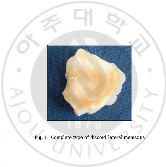
Fig. 1. Complete type of discoid lateral menisc us ---10
Fig. 2. Schematic drawing of the discoid meniscus ---13
Fig. 3. The scanning electron microscopic results---14
Fig. 4. The Sirius red stain results ---15
Fig. 5. Overall observation of the radial section ---16
Fig. 6. Collagen orientation of different segment in central layer ---17
Fig. 7. Synoptic drawing collagen orientation in the central layer ---18
I. INTRODUCTION
The meniscus plays the roles of load-bearing, shock absorption and stability for the human knee joint. These mechanical functions are determined not only by their anatomic characteristics but also by material properties of the meniscus. Particularly the dense and tightly woven collagen fibrils in the menisci can provide great elasticity and ability to withstand compression forces.2,3,5,7,15,22,27 Some studies by Kummer,20 Bullough et al.7 and Aspden et
al.3 depicted on normal meniscal microstructure regarding collagen fibrils
texture with different methods. Despite a few divergences, their concordant view is that the circumferential fibrils at the peripheral site can properly accommodate hoop tension from weight bearing. These orientations of collagen fibrils are believed to determine the tear pattern and treatment options. Therefore there is no doubt that the peripheral component of meniscus should be preserved as large as possible when partial meniscectomy is performed.8,11,19
The discoid lateral meniscus is the most common anomaly of the meniscus and more popular in Japanese and Korean than western countries.14,18,25 It is easy to be torn and occur self-degenerative changes due
to its typical shape and being caught in between the femur and the tibia.14,15,18,23,25 Tear patterns of discoid menisc us are more variable than
those of normal meniscus. Such as degenerative tear, horizontal tear and complex tear is common found in the discoid lateral meniscus, nevertheless
the common tear type of normal meniscus are longitudinal tear and flap tear. These tear patterns should be related to collagen fibril of discoid meniscus as in normal meniscus. Treatment for torn discoid meniscus is still on the debate. The same conception as the treatment of normal meniscal tear can hardly be applied to discoid meniscus because of its anomalous morphology. Presently, there are some advocates16,26 for performing total meniscectomy
in spite of reports that warn that early degenerative change of the articular cartilage could result from this operation. On the contrary the partial meniscectomy is recommended to preserve meniscal function as a desire that will prevent degenerative arthritis.1,14 However, this option cannot be
supported by either consentient clinical results or experimental data of load shearing of remained meniscus. In order to generate hoop tension, the discoid meniscus should retain collagen fibrils in circumferential fashion. To our knowledge, the c ollagen orientation of discoid meniscus has however never been reported so far.
This study observed and analyzed the collagen fibril orientation of discoid meniscus layer-by-layer, providing the theoretical basis for treatment and the understanding of the tear pattern of the discoid meniscus.
II. MATERIALS AND METHODS
Ten fresh obtained complete type of discoid lateral menisc i from ten patients with an average age of 37 years (range 17 to 62) were equally assigned into two groups, fixed in 10% formalin acid for three days (Fig. 1). First group was prepared to being observed under direct polarizing filter m icroscope (Eclipse E600, Nikon, Japan) view by Sirius red staining. Second group was prepared to being observed by scanning electron microscope (SEM; S-2400, Hitachi, Japan).
A. Sirius red stain
The discoid menisci in the first group were rinsed by water for 3-4 hours in order to remove residual formalin on the tissue, and then performed dehydration, clearing, paraffin infiltration using tissue processor (Shandon, Pittsburgh, PA). To be sectioned easily, the paraffin infiltrated tissues were carried out embedding procedure to form paraffin block in certain shape. The paraffin blocks were sectioned from the femoral surface to the tibial surface of the discoid meniscus utilizing sliding microtome (HM400R, Microm, Germany) in the thickness of 5μm. The folding slices were put into water bath (Jisco, Seoul, Korea) to plane down wrinkles, the plane slice was attached on the glass slide, subsequently put into 60℃ slide warmer (Jisco,
Seoul, Korea) for 1 hour to dissolve paraffin so that the slice is attached on the glass slide tightly. Before Sirius red staining, the slides were processed with xylene (Duksan, Ansan, Korea) to attain deparaffin and hydration. The hydrated slides were nuclear-stained by mayer’s hematoxylin (Merck, Darmstadt, Germany), and then stained by 0.1% Sirius red solution (0.1g siriusred (Polyscience, PA), 100ml picric acid (Sigma, MO)) for 1 hour. After the stained slides were differentiated with 50% acetic acid (Duksan, Ansan, Korea), respectively underwent twice dehydration with 95% and 100% alcohol, performed clearing with xylene and mounting procedures.
B . Scanning electron microscope
The rest of the five samples, as the second group, were subdivided into segments of radial and axial sections uniformly so as to permit a topographical classification. The different layers of the discoid meniscus segments were kept in 10% NaOH solution (Duksan, Ansan, Korea) at room temperature over a period of 5-6 days. Subsequently, the specimens were rinsed in distilled water for 1-2 days and were then saturated in 1% tannic acid (Duksan, Ansan, Korea) for 4-5 hours. After rinsing the specimens in distilled water for 24 hours, they were counterfixed in 1% OsO4 (Duksan,
Ansan, Korea) solution. The specimens were dehydrated in a series of graded concentrations of ethanol, freeze-cracked with a razor blade in liquid nitrogen and critical point-dried using liquid CO2. The dried specimens were
mounted on metal stubs with a double sticky tape, coated with gold. The obtained images were analyzed using image analysis program (Nikon, Tokyo, Japan).
III. RESULTS
1. Axial section
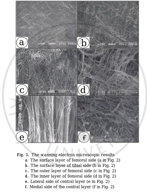
The polarizing filter microscope and SEM distinguish discoid meniscus into seven distinct layers in axial section by the arrangement of collagen fibril. The mid-portion of the meniscus is identified as the central layer, and three layers are symmetrically arranged in superoinferior directions with the central layer as a symmetric axis: surface, outer, inner and central layers (Fig. 2).
① Surface layer: The femoral surface of the meniscus is covered by dense and well-arranged thick fibrils, resembling a bunched streak. The fibrils show a sagital isotropic arranged orientation (Fig. 3a). However, the fibrils of the tibial surface show an irregularly and anisotropically arranged orientation (Fig. 3b).
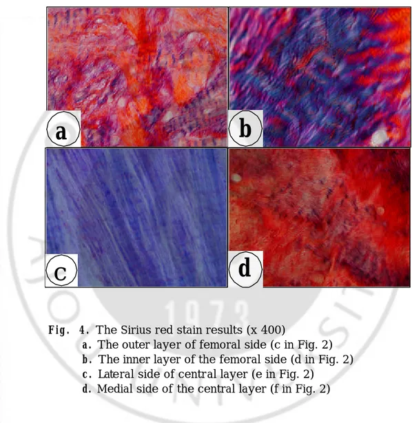
② Outer layer: Beneath the first layer a meshwork of thin fibrils have been observed. The fibrils do not show a preferred orientation (Fig. 3c, 4a).
③ Inner layer: The collagen fibrils are orientated radially from the lateral side to the medial side, and perforating fibrils are also present underlying the meshwork fibrils (Fig. 3d, 4b).
④ Central layer: The peripheral collagen fibrils that display dense bundles running in c ircumferential pattern are similar to normal meniscus (Fig. 3e, 4c). However, a different pattern is revealed in the medial portion,
composed of thin loosely and irregularly arranged fibrils without a bundle formation (Fig. 3f, 4d).
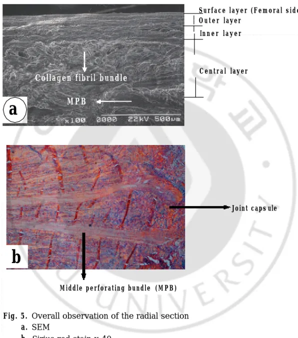
2. Radial section
Also we observed seven layers with differently arranged direction of collagen fibril. The central layer was showed arrangement of bundle collagen fibrils in its external circumference and thick middle perforating bundle (MPB) that was compact connection with the joint capsule in the medium portion. The central layer is the thickest occupying approximately 75% of full layer, every layers thickness of the femoral side is approximately equivalent to corresponding layers of the tibial side (Fig.5).
3. Collagen orientation of different zone in central layer
According to orientation of collagen fibrils in central layer, discoid meniscus is divided into four zones in overall: anterior, posterior and middle zone respectively, middle zone is subdivided into medial and lateral zone again. Both the anterior and posterior zones are coincident with meniscal attachment portion on bone. They show collagen fibrils with directly arranged orientation. Collagen fibrils in the medial middle zone display irregular arrangement, whereas collagen fibrils in the lateral middle zone represent circular arrangement occupying 20% of transverse diameter of the
discoid meniscus (Fig. 6, 7).
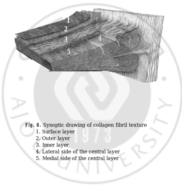
4. 3-D texture
In order to better understand layer arrangement of discoid meniscus from dimension, 3-D texture of full layers arrangement was drew with synoptic method based on above obtained images from polarizing filter microscope and SEM (Fig. 8).
.
Fig. 2. Schematic drawing of the discoid meniscus
d
f
e
Femoral sideb
c
Tibial sidea
Fig. 3. The scanning electron microscopic results a. The surface layer of femoral side (a in Fig. 2) b. The surface layer of tibial side (b in Fig. 2) c. The outer layer of femoral side (c in Fig. 2) d. The inner layer of femoral side (d in Fig. 2) e. Lateral side of central layer (e in Fig. 2) f. Medial side of the central layer (f in Fig. 2)
a
c
b
d
Fig. 4. The Sirius red stain results (x 400)
a. The outer layer of femoral side (c in Fig. 2) b. The inner layer of the femoral side (d in Fig. 2) c. Lateral side of central layer (e in Fig. 2)
d. Medial side of the central layer (f in Fig. 2)
a
b
d
Fig. 5. Overall observation of the radial section
a
. SEMb
. Sirius red stain x 40Surface layer (Femoral s i d e ) O u t e r layer
In n e r l a y e r
C e n t r a l layer
MPB
Collagen fibril
bundle
Joint caps ule
Middle perforating bundle (MPB)
b
a
F ig. 6. Collagen orientation of different zone in central layer (Sirius red stain
x40)
a. Anterior zone b. Posterior zone c. Lateral middle zone d. Medial middle zone
a
b
c
d
a
b
c
d
Fig. 7. Synoptic drawing collagen orientation in the central layer
According to orientation of collagen fibrils of central layer, the discoid lateral meniscus is divided into four zones in overall: anterior, posterior, lateral middle and medial middle zone respectively.
Anterior zone
Posterior zone
Medial
middle
Lateral
middle zone
Fig. 8. Synoptic drawing of collagen fibril texture
1. Surface layer 2. Outer layer 3. Inner layer
4. Lateral side of the central layer 5. Medial side of the central layer
1
2
3
4
IV. DISCUSSION
There have been many reports on normal meniscal microstructure regarding collagen fibrillar texture. A photoelastic analysis of the tensile trajectories undertaken by Kummer20 found that the collagen fibrils with supposedly
running in a radial direction in the internal circumference and in a circular direction in the external circumference displayed arcade-like arrangement. On the contrary, Bullough et al.7 and Aspden et al.3 demonstrated that
collagen fibrils are primarily orientated in a circular direction in both the internal and external circumferences, radial and perforating fibrils also are present. Petersen et al.22 utilized SEM and split line method to observe the
orientation of collagen fibrils layer-by-layer. It reveals three distinct layers in the meniscus cross section: the superficial network covered by a meshwork of thin fibrils, lamellar layer containing the bundles of collagen fibrils with a radial direction arrangement and intersection at various angles, central layer as the main portion of the meniscus collagen fibrils orientated in a circular manner.
Despite a few divergences, a view of agreement is that the circular fibrils at the peripheral site can accommodate properly to hoop tension from weight bearing. The circular fibers act in much the same way as metal hoops placed around a pressurized wooden barrel. The tension in the hoops keeps the wooden staves in place, thus the compression of the menisci by the tibia
and the femur generates outward forces that push the menisci out from between the bones. These hoop forces are transmitted to the tibia through the strong anterior and posterior attachments of the menisci. By animal experiment, Shrive24 has shown that the hoop tension is lost when a single
radial cut or tear extends to the capsular margin and that in terms of load-bearing, a single radial cut through the meniscus is equivalent to meniscectomy, thus the importance of the circular fibers is emphasized further. Either partial or complete excision of the menisci from the knees of dogs undertaken by King19 revealed that the hyaline cartilage of the articular
surfaces degenerated to a degree roughly proportional to the amount of meniscus excised. On the other hand, some studies8,11,13 have demonstrated
that the attempts at regeneration of menisci could be observed, however, a regenerated meniscus is frail, and they believed that the function of the regenerated meniscus in humans is probably insignificant. Once the collagen orientation is haphazard in the regenerated meniscus-like structure, the circumferential, or hoop-tension principle is not replicated. Therefore partial rather than total or subtotal meniscectomy always is preferable if possible.
The orientation of collagen fibrils is not only related to the principle of the treatment but also determines to some extent the characteristic patterns of meniscal tears.5,7,20 Beaupre et al.5 and Kummer20 found that the
occurrence of radial tears in the internal circumference and of circular bucket-handle tears in the external circumference is attributed to the arcade-like arrangement of the collagen fibrils. Nevertheless Bullough et al.7
considered that the most common longitudinal tear in clinical practice is germane to the circular collagen fibrils in deep layer, the radial fibrils rupture would lead to longitudinal tear, the circular fibrils rupture would lead to radial tear. It indicated that the meniscal tear is not only correlated to the mechanism of injury, but also depended on collagen fibril texture, so that the different segments would result in the different tear pattern.
The discoid meniscus is the most common type in meniscal anomaly that occurs more frequently laterally than medially. The incidence is higher in Korean and Japanese than in other populations.14,18,25 Watanabe et al.28 has
classified the discoid lateral meniscus into complete, incomplete, and Wrisberg-Ligament types based on the degree of coverage of the lateral tibial plateau and the presence or absence of the normal posterior meniscotibial attachment. Since Wrisberg-Ligament type discoid meniscus is similar to normal shape and is unstable, it is advocated total meniscectomy. This study investigates only complete type of discoid lateral meniscus.
Best of our knowledge, this is the first time that Sirius red staining method and SEM were used to observe collagen fibrils texture of discoid meniscus. Sirius red is specific for collagen, and the stained collagen with different arranged orientation are appeared in yellow, red/orange or green, according to their direction under polarizing filter light microscopy.4,17,29
By this study, the individual layers of discoid meniscus are distributed symmetrically from up to down based on central layer as an axial plane. The femoral surface of the discoid meniscus is covered by dense and
well-arranged thick fibrils, resembling a bunched streak. The fibrils show a sagital isotropic arranged orientation. However, the fibrils of the tibial surface show an irregularly and anisotropically arranged orientation. For such arrangement presentation on the femoral surface of the discoid meniscus, we can suppose that it is caused by global lateral condyle vigorously acting the rolling and gliding motion with various directions. In contrast the tibial surface of the discoid meniscus slipping on the flat articular tibial surface in limited range, on which collagen fibrils is irregular arrangement. For maintaining the meniscal shape, the role of a meshwork of thin fibrils in outer layer is more major than shock absorption. The inner layer looked as transition layer from the outer layer to central layer, which perhaps plays a connective role. The periphery of the central layer consists of the bundle collagen fibrils with circular arrangement. On contrast the arrangement of collagen fibrils in the medial portion is irregular without bundle fibrils. Thus it is believed that the periphery of the central layer is the most important portion so as to accommodate to hoop tension from weight bearing. The directly arranged collagen fibrils in anterior and posterior zone allow meniscus to attach on the tibia strongly. The MPB of central layer that allow discoid meniscus to insert at joint capsule tightly seems to play a role of stability.
Because discoid meniscus has anomalous morphology and awkward position, it is easy to cause tear and degenerative change.12,23 The
distribution of the tear pattern has been reported in a number of studies. Hayashi et al.14 investigated that the longitudinal tear is the most common
type of discoid meniscus damage. However, Smilie25 and Bin et al.6 stated
that horizontal cleavage is the most frequent type, which mostly belongs to degenerative tear and might be apparently invisible substantial tear. Moreover, a study by Ferrer-Roca and Vilalta9,10 indicated that the
degenerative horizontal cleavage most frequently occurs in the middle portion of the meniscus just as the portion of discoid meniscus containing middle perforating bundle. Regarded as poor vascularization in this portion, aging easily cause degenerative horizontal cleavage. Because discoid meniscus is thicker than the normal meniscus, furthermore, as knee joint acts flexion and extension, rotation and weight transmission, discoid meniscus is at the locking condition between the femur and tibia. MPB as an axis, superior and inferior layers undergo shear force of contrary direction, so it is easy to cause horizontal cleavage in the middle portion of the meniscus.
In clinical practice discoid meniscus is usually treated mainly according to symptom, tear degree, and type. However, the arragned orientation of collagen fibrils as a more important factor should be accurately acknowledged to determine range of excision precisely. Some studies16,26
showed satisfactory results after total meniscectomy. Nevertheless, total meniscectomy of a lateral non-discoid meniscus often leads to progressive osteoarthritis.21,30 Hayashi et al.14 advocated partial meniscectomies left 6-8
mm for incomplete and complete type, which not based on the orientation of collagen fibril. Araki et al.2 found the normal transverse diameter of the
13.1-30.0 mm. Ikeuchi16 studied 49 excised discoid lateral menisci and noted a
maximum thickness of 14 mm (minimum, 4 mm), Smillie25 examined 15
discoid and 30 normal menisci and found that the discoid menisci had a thicker-than normal central portion (especially the free margin), but the greater thickness did not extend to the periphery. Our results showed that the circular arrangement of collagen fibrils occupy 20% of transverse diameter of the lateral discoid meniscus, and referred to above literature results, therefore we considered that leaving 6 mm from the periphery seem to be desirable for discoid meniscectomy.
V. CONCLUSION
The discoid meniscus can be divided into seven layers in a symmetrical manner with the central layer as an axis, with an exception of the surface layer, which shows a difference between the femoral and the tibial surfaces. All layers have their own unique fibrillar orientations. The central layer seems to play a main role in resisting hoop tension since the peripheral portion has circumferential fashion of collagen fibers. Therefore, we can consider that preservation of the peripheral edge of the discoid meniscus would be better when resection of meniscus is mandatory.
BIBLIOGRAPHY
1 . Aichroth PM, Patel DV, Marx CL: Congenital discoid lateral meniscus in
children. A follow-up study and evaluation of management. J Bone Joint Surg Br 73(6):932-6, 1991
2. Araki Y, Yamamoto H, Nakamura H, Tsukaguchi I: MR diagnosis of discoid
lateral menisci of the knee. Eur J Radiol 18:92-5, 1994
3. Aspcen RM, Yarker YE, Hukins DW: Collagen orientations in the meniscus
of the knee joint. J Anat 140:371-80, 1985
4. Bancroft JD, Gamble M: Theory and practice of histological techniques.
Fifth edition pp 282, 2002
5. Beaupre A, Choukroun R, Guidouin R, Gerardin H, Cardou A: Knee menisci.
Correlation between microstructure and biomechanics. Clin Orthop 208:72-5, 1986
6. Bin SI, Kim JC, Kim JM, Park SS, Han YK: Correlation between type of
discoid lateral menisci and tear pattern. Knee Surg Sports Traumatol Arthrosc 10(4):218-22, 2002
7. Bullough PG, Munuera L, Murphy J, Weinstein AM: The strength of the
menisci of the knee as it relates to their fine structure. J Bone Joint Surg 52B:564-7, 1970
8. Cox JS, Cordell LD: The degenerative effects of medial meniscus tears in
9. Ferrer -Roca O, Vilalta C: Lesions of the meniscus. Part I: Macroscopic
and histologic findings. Clin Orthop 146:289-300, 1980
10. Ferrer -R o c a O , V i l a l t a C: Lesions of the meniscus. Part II: Horizontal
cleavages and lateral cysts. Clin Orthop 146:301-7, 1980
11. Fithian DC, Schmidt MB, Ratcliffe A, Mow VC: Human meniscus tensile
properties: Regional variation and biochemical correlation. Orthop Trans 14:205, 1989
12. Fritschy D, Gonseth D: Discoid lateral meiscus. Int Orthop. 15:145-7,
1991
13. Grood ES: Meniscal function. Adv Orthop Surg 7:193, 1984
14. Hayashi LK, Yamaga H, Miura T: Arthroscopic meniscectomy for discoid
lateral meniscus in children. J Bone Joint Surg Am 70(10):1495-500, 1988
15. Herwig J, Egner E, Budecke E: Chemical changes of human knee joint
menisci in various stage of degeneration. Ann Rheum Dis 43:635-40, 1984
16. Ikeuchi H: Arthroscopic treatment of the discoid meniscus. Technique
and long term result. Clin Orthop 167:19-28, 1982
17. Junqueira LC, Bignolas G, Brentani RR: Picrosirius staining plus
polarization microscopy, a specific method for collagen detection in tissue sections. Histochem J 11(4):447-55, 1979
18. Kim S J, Kim DW, Min BH: Discoid lateral meniscus associated with
1995
19. King D: The healing of semilunar cartilage. J Bone Joint Surg 18:333,
1936
20. Kummer B: Biomechanik des meniscus. Orthopade 23: 90-2, 1994
21. Manzione M, Pizzutillo DD, Peoples AB, Schweizer PA: Meniscectomy in
children. A long-term follow-up study. Am J sports Med 11:111-5, 1983
22. Petersen W, Tillmann B: Collagenous fibril texture of the human knee
joint menisci. Anat Embryol (Berl) 197(4):317-24, 1998
23. Rohren EM, Kosarek FJ, Helms CA: Discoid lateral meniscus and the
frequency of meniscal tears. Skeletal Radiol 30(6):316-20, 2001
24. S h r i v e N: The weight-bearing role of the menisci of the knee. In
Proceedings of the British Orthopaedic Research Sociey, J Bone Joint Surg 56B: 381, 1974
25. Smillie IS: The congenital discoid meniscus. J Bone Joint Surg. 30B:
671-82, 1948
26. Sugawara O, Miyatsu M, Yamashita I, Takemitsu, Onozawa T: Problems
with repeated arthroscopic surgery in the disc oid meniscus. Arthroscopy 7(1):68-71, 1991
27. Walker PS, Erkman MJ: The role of the menisci in force transmission
across the knee. Clin Orthop 109:184-92, 1975
28. Watanabe M, Takeda S, Ikeuchi H: Atlas of arthroscopy. Third edition.
Berlin, etc: Springer-Verlag, 1979
assessment of myocardial collagen with picrosirius red staining and circularly polarized light. Basic Res Cardiol 89(5):397-410, 1994
30. Zaman M, Leonard MA: Meniscectomy in children: Results in 59 knees.
- 국 문 요 약 –
외 측 원 판 형 연골의 교 원 섬유 배 열 구조
아 주 대 학 교 대 학 원 의 학 과
최 기 호
( 지도교수: 민 병 현 )
목 적: 외측 원판형 연골의 교원 섬유의 배열을 각 부위 및 층별로 관찰하고 분석함으로 써 원판형 연골 파열의 치료와 양상에 이론적 근거를 제공하려 한다. 대 상 및 방 법: 사람 슬관절에서 획득한 10 예의 완전형 외측 원판형 연골을 부위 및 층 별로 절편하고 Polarizing filter 광학현미경과 주사 전자 현미경을 사용하여 교원 섬유의 배열 방향을 관찰하고 분석하였다. 결 과: 외측 원판형 연골은 대퇴골 관절면으로부터 경골 관절면으로 교원 섬유의 배열 방 향에 따라 모두 7개의 층으로 나눌 수 있었다. 표면층의 대퇴골 관절면은 전방으로부터 후방으로 일치하게 정렬 된 가늘고 밀집된 교원 섬유로 줄 모양으로 주름을 형성하고 있 는 것을 관찰할 수 있었으나 경골 관절면은 배열이 매우 불규칙적인 교원 섬유로 덮어져 있었으며 일정한 모양을 이루지 않고 있었다. 외층은 가는 교원 섬유로 그물 모양의 배 열을 하고 있었고 방향성은 없었으며 천층의 바로 밑에 존재하는 내층은 첨단부로부터 변연부로 향하는 방사형으로 배열된 교원 섬유를 관찰할 수 있었으며 그 외에도 여러 섬 유들을 서로 이어주는 관통 섬유도 발견할 수 있었다. 중심층의 변연부 부분에서는 환형 으로 배열된 굵은 다발 형태의 교원 섬유를 볼 수 있었으며 첨단부 부분에서는 방향성없이 서로 엉켜있는 교원 섬유들이 다발을 형성하지 않은 상태로 존재하고 있었다. 결 론: 외측 원판형 연골은 대퇴골 관절면으로부터 경골 관절면으로 7개 층으로 나눌 수 있으며 대퇴골과 경골의 표면층을 제외하고 중심층을 축으로 하여 위 아래로 서로 대칭 되는 층으로 되어있다. 각 층의 교원 섬유는 모두 각자의 특징을 가지고 있으며 그 중에 서 중심층의 변연부에는 굵은 다발 형태를 가진 환형으로 배열된 교원 섬유가 존재하므 로 hoop tension에 잘 적응하는 기능을 수행함에 있어서 가장 중요한 부분으로 사료된 다. 이에 원판형 연골 파열의 치료에서 절제술을 시행할 때 변연부 부분을 환형으로 남 겨두는 것은 필수적이라고 사료된다. 핵 심 되 는 말: 원판형 연골, 교원 섬유, 주사 전자 현미경