Signet Ring Cell Mixed Histology
May Show More Aggressive
Behavior than Other Histologies in
Early Gastric Cancer
Cheal Wung Huh
Department of Medicine
Signet Ring Cell Mixed Histology
May Show More Aggressive
Behavior than Other Histologies in
Early Gastric Cancer
Cheal Wung Huh
Department of Medicine
Signet Ring Cell Mixed Histology
May Show More Aggressive
Behavior than Other Histologies in
Early Gastric Cancer
Directed by Professor Jie-Hyun Kim
The Master’s Thesis
submitted to the Department of Medicine,
the Graduate School of Yonsei University
in partial fulfillment of the requirements
for the degree of Master of Medical Science
Cheal Wung Huh
December 2011
This certifies that the Master’s Thesis
of Cheal Wung Huh is approved.
---
Thesis Supervisor : Jie-Hyun Kim
---
[Thesis Committee : Yong Chan Lee]
---
[Thesis Committee : Jae-Ho Cheong]
The Graduate School
Yonsei University
ACKNOWLEDGEMENTS
This thesis would not have been possible without the
support of many people. I am heartily thankful to my
supervisor, Jie-Hyun Kim, whose encouragement, guidance
and support from the initial to the final level enabled me to
develop an understanding of the subject. I owe my deepest
gratitude to my committee, Yong Chan Lee and Jae-Ho
Cheong without whose knowledge and assistance this study
would not have been successful. I am indebted to my many
of my colleagues to support me for accomplishing my thesis.
Lastly, I offer my regards and blessings to all of those who
supported me in any respect during the completion of the
project.
<TABLE OF CONTENTS>
ABSTRACT
···1
I. INTRODUCTION
···3
II. MATERIALS AND METHODS
···4
1. Patients
···4
2. Pathology
···5
3. Statistical analysis
···5
III. RESULTS
···6
IV. DISCUSSION
···14
V. CONCLUSION
···17
REFERENCES
···19
ABSTRACT(In Korean)
···22
LIST OF FIGURES
Figure 1. Cumulative survival curves for patients with
adenocarcinoma, signet ring cell carcinoma (SRC), and
mixed-SRC groups in early gastric cancer (SRC vs.
adenocarcinoma, P=0.002; mixed-SRC vs.adenocarcinoma,
P=0.623; mixed-SRC vs. SRC, P=0.010)
···13
LIST OF TABLES
Table 1. Comparisons of the clinicopathologic findings
among partly signet ring cell carcinoma (mixed-SRC), signet
ring cell carcinoma (SRC), and adenocarcinoma groups
···
7
Table 2. Comparisons of the clinicopathologic findings in
well/moderate differentiated adenocarcinoma
···
9
Table 3. Comparisons of the clinicopathological findings in
poorly differentiated adenocarcinoma
···
10
Table 4. Multivariate analysis of risk factors for lymph node
metastasis
···12
1
ABSTRACT
Signet Ring Cell Mixed Histology May Show More Aggressive
Behavior than Other Histologies in Early Gastric Cancer
Cheal Wung Huh
Department of Medicine
The Graduate School, Yonsei University
(Directed by Professor Jie-Hyun Kim)
OBJECTIVES: Signet ring cell carcinoma (SRC) of the stomach has been
known to have different microscopic and biologic characteristics compared to
non-SRC. Thus, a pathologic report has documented partly SRC component
with other main histologies in gastric cancer. However, the clinical
significance of SRC mixture has not been reported. The aim was to
investigate clinicopathologic features of mixed-SRC histology in early gastric
cancer (EGC).
METHODS: Between 1999 and 2005, 2208 patients were diagnosed with
EGC and underwent surgery at Severance and Gangnam Severance Hospital.
Among them, 156 patients were diagnosed with adenocarcinoma with partly
SRC (mixed-SRC group), 1,512 with only adenocarcinoma (adenocarcinoma
group), and 540 with SRC (SRC group). Clinicopathologic characteristics
2
RESULTS: The SRC group was more significantly associated with younger
age, female, mid-body location, mucosa-confined, depressed type, lower
lymph node metastasis (LNM), lower lymphovascular invasion, and a better
survival rate than the adenocarcinoma group. The mixed-SRC group was
more significantly associated with younger age, female, upper-body location,
and depressed type than the adenocarcinoma group, similar to the SRC group.
However, the mixed-SRC group showed more submucosal invasion, larger
size, and higher LNM than SRC and adenocarcinoma groups. Also, a
mixed-SRC component was one of the independent risk factors of LNM.
CONCLUSIONS: The mixed-SRC group displayed different
clinicopathologic characteristics from other groups. Mixed-SRC histology in
EGC showed more aggressive biologic characteristics than SRC and
adenocarcinoma. Thus, clinical considerations of mixed-SRC histology may
be helpful to decide on a specific cancer treatment.
---
Key words :
early gastric cancer, signet ring cell carcinoma, biologic characteristics3
Signet Ring Cell Mixed Histology May Show More Aggressive
Behavior than Other Histologies in Early Gastric Cancer
Cheal Wung Huh
Department of Medicine
The Graduate School, Yonsei University
(Directed by Professor Jie-Hyun Kim)
I. INTRODUCTION
Several published histological classifications in gastric cancer exist, such as the WHO, Lauren, Japanese, and Ming’s classifications.1-4
According to the
WHO classification, gastric cancer is categorized into papillary
adenocarcinoma, tubular adenocarcinoma, mucinous adenocarcinoma, and
signet ring cell carcinoma (SRC) based on the microscopic characteristics of
tumors. Any cell type that comprised more than 50% of the total was defined
to be the main histology of the tumor.1
Among them, SRC has been known to have different microscopic
characteristics as well as biologic characteristics compared with other types of
gastric adenocarcinoma.5-10 The incidence of SRC has been reported to vary
4
among women and young patients.6, 7, 10 Also, SRC has been known to have
different biologic characteristics between early stage and advanced stage
gastric cancer.5-9 In early gastric cancer (EGC), SRC has been reported to
have a better prognosis than non-SRC because of less lymph node metastasis
(LNM) and a more grossly depressed type, which is helpful for diagnosis.5-9, 11
However, in advanced gastric cancer (AGC), SRC has been characterized to
be a more grossly infiltrative type, with more peritoneal dissemination and a
similar or worse prognosis than non-SRC.5-8, 12-14 In this manner, SRC has
shown discriminative biologic characteristics compared to adenocarcinoma.
Therefore, pathologic reports documented partly SRC when a mixed-SRC
component existed in other main histologies. Generally, partly SRC is defined
as adenocarcinoma with a minor component less than 50% of SRC.1 However,
the clinical significance of mixed-SRC histology associated with gastric
adenocarcinoma has not been reported. The purpose of this study was to
investigate the clinicopathological features of mixed-SRC histology in EGC
compared to other histologies of gastric cancer.
II. MATERIALS AND METHODS
1. Patients
Between December 1999 and December 2005, 4,723 patients were
5
Gangnam Severance Hospital. In total, 2208 (47%) patients were diagnosed
with EGC. Among them, 156 (7.1%) were diagnosed with adenocarcinoma
with partly SRC (mixed-SRC group), 1,512 (68.4%) with only
adenocarcinoma (adenocarcinoma group), and 540 (24.5%) with SRC (SRC
group). Patient follow-up lasted until death or the cutoff date of December 1,
2010. The mean follow-up duration was 82 months (range, 0-133 months).
2. Pathology
Based on the WHO classification of gastric cancer, partly SRC was defined
as adenocarcinoma with a minor component (10-50%) of isolated carcinoma
cells containing mucin (mixed-SRC histology). Also, SRC was defined as a
predominant component (>50%) isolated carcinoma cells containing mucin
were present.1 Adenocarcinoma was categorized into well, moderate, and
poorly differentiated adenocarcinoma according to the WHO classification.1
The mixed-SRC group was compared to the SRC and adenocarcinoma groups
in terms of host, lesion, and biologic factors such as gender, age, tumor size,
tumor location, gross appearance, depth of invasion, LNM, lymphovascular
invasion (LVI), and perineural invasion (PNI). Patient information was
obtained from medical records and analyzed retrospectively.
3. Statistical Analysis
6
the chi-square test. The LNM rate was compared between groups after
stratifying the clinicopathologic characteristics. Risk factors affecting LNM
were evaluated by logistic regression analysis. The Kaplan-Meier method was
used to determine the cumulative survival rate, and the log-rank test was used
to analyze differences in the survival curve. A Cox proportional hazards
regression model for evaluating prognostic factors was applied for a
multivariable analysis. The accepted significant level was a P value < .05. All
statistical analyses were performed using the software SPSS version 12.0 for
Windows (SPSS Inc., Chicago, IL, USA).
III. RESULTS
1. Demographics and clinicopathologic characteristics among the mixed-SRC, SRC, and adenocarcinoma groups
Table 1 shows the clinicopathologic characteristics among the mixed-SRC,
SRC, and adenocarcinoma groups. The mixed type, defined according to the
Lauren classification, had a significantly higher proportion in the mixed-SRC
group. The clinicopathologic features of the SRC group were similar to those
previously reported.5-9, 11 That is, the SRC group was composed of a larger
proportion of younger patients and females than the adenocarcinoma group.
Furthermore, the SRC group had a larger ratio of mucosa-confined and
7
depressed gross type was more common, and the LNM rate and LVI were
significantly less in the SRC group than in the adenocarcinoma group (Table
1).
The host and lesion factors of the mixed-SRC group were similar to those of
the SRC group. The proportion of younger patients and females in the
mixed-SRC group was larger than in the adenocarcinoma group. Also, the
mixed-SRC group had a larger proportion of upper/mid-body located lesions
and the depressed gross type than the adenocarcinoma group. However, the
mixed-SRC group showed more submucosa invasion, larger size, and a higher
LNM rate than the adenocarcinoma group (P < .05) contrast to the SRC group
(Table 1).
TABLE 1 Comparisons of the clinicopathologic findings among partly signet ring cell carcinoma (mixed-SRC), signet ring cell carcinoma (SRC), and adenocarcinoma groups
Variables Adenocarcinoma (N=1512) (n, %) Mixed-SRC (N=156) (n, %) SRC (N=540) (n, %)
Age (years, mean) 59.1±10.6 51.7±12.0* 51.2±11.7**
Gender Male 1101 (72.8) 87 (55.8) 288 (53.3) Female 411 (27.2) 69 (44.2)* 252 (46.7)** Tumor size (mm, mean±SD) 23.2±15.6 26.5±16.1 24.0±15.4 Lauren classification Intestinal 506 (33.5) 5 (3.2) 8 (1.5) Diffuse 61 (4.0) 19 (12.2) 180 (33.3)** Mixed 47 (3.1) 22 (14.1)*, *** 6 (1.1) Unknown 898 (59.4) 110 (70.5) 346 (64.1) Depth of invasion Mucosa (T1a) 770 (50.9) 62 (39.7) 371 (68.7)** Submucosa (T1b) 742 (49.1) 94 (60.3)*, *** 169 (31.3) Location Upper 65 (4.3) 15 (9.6)*, *** 17 (3.1) Middle 681 (45.0) 84 (53.8) 328 (60.8)** Lower 766 (50.7) 57 (36.5) 195 (36.1) Operation Subtotal Total 1341 (88.7) 171 (11.3) 134 (85.9) 22 (14.1) 464 (85.9) 76 (14.1)
8 Macroscopic type Elevated 246 (16.2) 6 (3.8) 13 (2.4) Flat 450 (29.8) 35 (22.4) 201 (37.2) Depressed 816 (54.0) 115 (73.7)*, *** 326 (60.4)** Lymph node metastasis
(7th UICC) N0 N1 N2 N3
Lymph node metastasis
1362 (90.1) 128 (8.5) 20 (1.3) 2 (0.1) 126 (80.8) *, *** 26 (16.7) 5 (2.5) 0 (0) 508 (94.1) 26 (4.8) 6 (1.1) 0 (0) Positive 150 (9.9) 30 (19.2)*, *** 32 (5.9)** Negative 1362 (90.1) 126 (80.8) 508 (94.1) Lymphovascular invasion Present 133 (8.8) 12 (7.7)*** 21 (3.9)** Absent 1379 (91.2) 144 (92.3) 519 (96.1) Perineural invasion Present 5 (0.3) 1 (0.6) 3 (0.6) Absent 1507 (99.7) 155 (99.4) 537 (99.4)
*P<.05 compared to the adenocarcinoma group **P<.05 compared to the adenocarcinoma group
***P<.05 compared to the SRC group
2. Clinicopathologic characteristics between the mixed-SRC and
adenocarcinoma groups according to the main histological types
According to the main histological types, the mixed-SRC group was divided
into well/moderate differentiated adenocarcinoma with mixed-SRC histology
and poorly differentiated adenocarcinoma with mixed-SRC histology. Then,
the mixed-SRC group was compared to each adenocarcinoma group:
well/moderate adenocarcinoma with mixed-SRC histology vs. well/moderate
adenocarcinoma and poorly differentiated adenocarcinoma with mixed-SRC
histology vs. poorly differentiated adenocarcinoma.
9
be in the mixed-SRC group associated with a well/moderate differentiated
adenocarcinoma compared to those with a well/moderate differentiated
adenocarcinoma (Table 2). In addition, those in the mixed-SRC group with a
well/moderate differentiated adenocarcinoma showed more submucosa
invasion and larger tumor size than those with a well/moderate differentiated
adenocarcinoma (Table 2).
When the mixed-SRC group with a poorly differentiated adenocarcinoma
was compared to those with a poorly differentiated adenocarcinoma, younger
patients and females were more commonly associated with the mixed-SRC
group with a poorly differentiated adenocarcinoma (Table 3). Also, the
mixed-SRC group with a poorly differentiated adenocarcinoma had more
upper/mid-body located lesions and more depressed gross type than those in
the poorly differentiated adenocarcinoma group. The mixed-SRC group
showed a higher incidence of LNM than the poorly differentiated
adenocarcinoma group (Table 3).
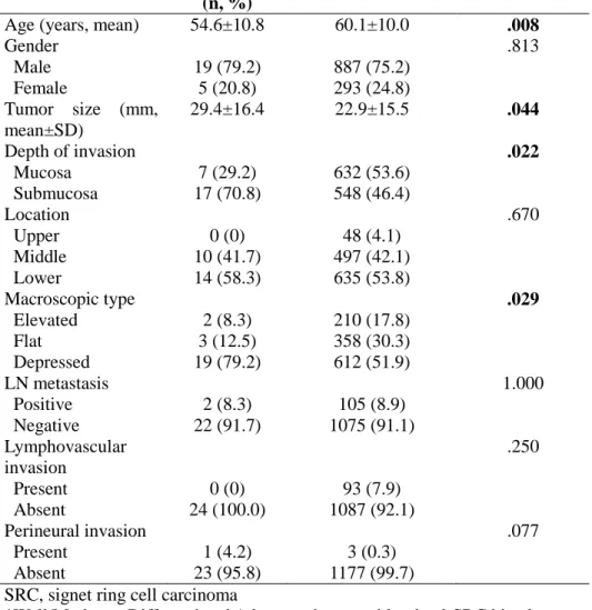
TABLE 2 Comparisons of the clinicopathologic findings in well/moderate
differentiated adenocarcinoma Variables Mixed-SRC* (N=24) Adenocarcinoma** (N=1180) (n, %) P value
10
(n, %)
Age (years, mean) 54.6±10.8 60.1±10.0 .008
Gender .813 Male 19 (79.2) 887 (75.2) Female 5 (20.8) 293 (24.8) Tumor size (mm, mean±SD) 29.4±16.4 22.9±15.5 .044 Depth of invasion .022 Mucosa 7 (29.2) 632 (53.6) Submucosa 17 (70.8) 548 (46.4) Location .670 Upper 0 (0) 48 (4.1) Middle 10 (41.7) 497 (42.1) Lower 14 (58.3) 635 (53.8) Macroscopic type .029 Elevated 2 (8.3) 210 (17.8) Flat 3 (12.5) 358 (30.3) Depressed 19 (79.2) 612 (51.9) LN metastasis 1.000 Positive 2 (8.3) 105 (8.9) Negative 22 (91.7) 1075 (91.1) Lymphovascular invasion .250 Present 0 (0) 93 (7.9) Absent 24 (100.0) 1087 (92.1) Perineural invasion .077 Present 1 (4.2) 3 (0.3) Absent 23 (95.8) 1177 (99.7)
SRC, signet ring cell carcinoma
*Well/Moderate Differentiated Adenocarcinoma with mixed-SRC histology **Well/Moderate Differentiated Adenocarcinoma
TABLE 3 Comparisons of the clinicopathological findings in poorly
differentiated adenocarcinoma Variables Mixed-SRC* (N=132) (n, %) Adenocarcinoma** (N=332) (n, %) P value
11 Gender .011 Male 68 (51.5) 214 (64.5) Female 64 (48.5) 118 (35.5) Tumor size (mm, mean±SD) 26.3±15.9 24.4±16.2 .238 Depth of invasion 1.000 Mucosa 55 (41.7) 138 (41.6) Submucosa 77 (58.3) 194 (58.4) Location .036 Upper 15 (11.4) 17 (5.1) Middle 74 (56.1) 183 (55.1) Lower 43 (32.6) 132 (39.8) Macroscopic type .011 Elevated 4 (3.0) 36 (10.8) Flat 32 (24.2) 92 (27.7) Depressed 96 (72.8) 204 (61.4) LN metastasis .048 Positive 28 (21.2) 45 (13.6) Negative 104 (78.8) 287 (86.4) Lymphovascular invasion .417 Present 12 (9.1) 40 (12.0) Absent 120 (90.9) 292 (88.0) Perineural invasion .592 Present 0 (0) 2 (0.6) Absent 132 (100.0) 330 (99.4)
SRC, signet ring cell carcinoma
*Poorly Differentiated Adenocarcinoma with mixed-SRC histology **Poorly Differentiated Adenocarcinoma
3. Multivariate analysis of LNM
Males, tumors of larger size, submucosal invasion, LVI, and mixed-SRC
12 analysis using logistic regression (Table 4).
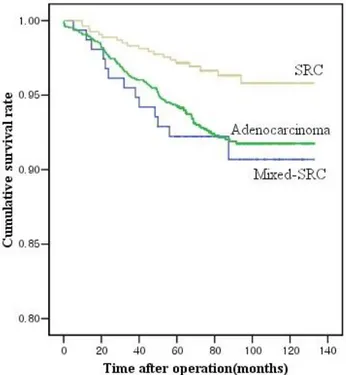
Kaplan-Meier analysis indicated that the patients with SRC had a higher
cumulative survival rate than those with an adenocarcinoma in EGC.
Although it was not statistically significant, the survival rates of mixed-SRC
group were lower than adenocarcinoma group (Fig. 1).
TABLE 4 Multivariate analysis of risk factors for lymph node metastasis
Factors Odds ratio (95% CI) P value
Gender (male vs. female) 1.65 (1.12-2.41) .010
Tumor size (≥20 mm vs. <20 mm) 2.60 (1.66-4.07) <.001
Macroscopic appearance 1.16 (0.67-2.03) .558
Depth of invasion (mucosa vs. submucosa) 8.23 (4.62-14.65) <.001
Lymphovascular invasion (present vs. absent) 8.98 (5.91-13.64) <.001
Histology (mixed-SRC vs. adenocarcinoma) 2.30 (1.38-3.83) .001
13
FIG. 1 Cumulative survival curves for patients with adenocarcinoma, signet
ring cell carcinoma (SRC), and mixed-SRC groups in early gastric cancer (SRC vs. adenocarcinoma, P=0.002; mixed-SRC vs. adenocarcinoma,
14
IV. DISCUSSION
The incidence of SRC in gastric cancer has been shown to have an
increasing tendency and reported to vary from 8.7% to 23.4%.5-9, 15 Earlier
studies showed a higher prevalence in females and younger patients and a
higher predominance for a mid-body tumor location in SRC compared to
non-SRC.5-10, 12
In EGC, several studies have reported that SRC occurred more frequently
with superficial spreading and depressed lesions compared to non-SRC and
had a significantly lower LNM than non-SRC. Therefore, the survival of
patients with SRC was better than that of patients with non-SRC.5-9 That is,
SRC in EGC has been known to be less aggressive than non-SRC. Thus, the
recent trend is to perform a less invasive treatment such as minimal invasive
surgery for the treatment of SRC in EGC.7, 9
In this study, SRC was higher in female and younger patients and had a
larger proportion of mucosa-confined and mid-body located lesions than
adenocarcinoma among patients with EGC. Also, the SRC group showed
more flat, depressed gross types, lower LNM, less LVI, and a better survival
rate than the adenocarcinoma group, similar to previous studies.
Because SRC showed different clinicopathologic and biologic characteristics
compared to non-SRC as stated above, a pathologic report has documented a
partly SRC component with other main histologies when SRC histology is
15
histology has not been reported. We analyzed the clinicopathologic and
biologic characteristics of mixed-SRC histology in EGC.
In our study, the host and lesion factors such as female gender, a younger age,
an upper/mid-body location, and a depressed gross type in the mixed-SRC
group were similar to those in the SRC group. However, the factors that
reflected more aggressive biologic characteristics in EGC, such as deeper
invasion depth, larger tumor size, positive LVI, and positive LNM, were more
significantly associated with the mixed-SRC group than with other histologies
in EGC. To investigate the influence of main histology, we examined
clinicopathologic features of mixed-SRC histology based on the main
histology, e.g., a well/moderate differentiated adenocarcinoma or a poorly
differentiated adenocarcinoma. Irrespective of the main histology, locally
aggressive biologic characteristics such as more submucosal invasion and
LNM were significantly associated with a mixed-SRC histology (mixed-SRC
group).
Mixed-SRC histology was one of the independent risk factors of LNM in
EGC. Also, the mixed-SRC group showed significantly lower survival rates
than SRC group and the tendency of lower survival rates than
adenocarcinoma group.
Zheng et al. reported that mixed-type carcinoma of the stomach showed
more aggressive characteristics such as deeper invasion, larger size, and more
16
They explained that the reason for the locally aggressive characteristics in
mixed-type gastric cancer was the increased expression of proteins such as
Ki-67, EMMPRIN, and VEGF, which are involved in the angiogenetic
process and cell proliferation in mixed-type gastric cancer.16 Also, Kozuki et al.
reported that mixed-type carcinoma had a larger proportion of LNM than
diffuse type carcinoma of the stomach.17 Park et al. suggested that mixed-type
gastric carcinoma frequently showed CpG island hypermethylation. So, the
mixed-type gastric carcinoma appeared to be more aggressive than the
intestinal or diffused type.18
In our study, we could not analyze the clinicopathologic characteristics of
mixed-type gastric cancer based on the Lauren classification because of some
unknown data for the Lauren classification. However, the mixed-type
carcinoma using the Lauren classification was more common in the
mixed-SRC group. Also, the mixed-SRC group displayed more aggressive
characteristics than other groups such as the adenocarcinoma and SRC groups.
Intestinal-type carcinoma based on the Lauren classification was more
common in the adenocarcinoma group, whereas diffuse-type carcinoma based
on the Lauren classification was more common in the SRC group. Therefore,
our study might demonstrate more consistent results compared to previous
studies.
As mentioned above, SRC in EGC has been recommended to be treated by
17
aggressive biologic characteristics.9 However, mixed-SRC histology showed
more aggressive characteristics than other histologies in EGC. The different
clinicopathologic characteristics of mixed-SRC histology may suggest that
one must document mixed-SRC histology with the main histology in a
pathologic report. Furthermore, aggressive characteristics such as deeper
invasion or more LNM may suggest that the clinical treatment approach of
mixed-SRC histology is different from SRC and non-SRC histologies in EGC.
A mixed-SRC histology in EGC may require a more aggressive treatment
approach than SRC and non-SRC histologies such as a poorly differentiated
adenocarcinoma or a well/moderate differentiated adenocarcinoma. For
example, mixed-SRC histology might be helpful for decisions on necessity of
additional surgery after endoscopic resection or range of LN dissection for
minimal invasive surgery. However, further research is necessary to
investigate the mechanisms involved to explain why a mixed-SRC histology
is more aggressive than others.
V. CONCLUSION
In conclusion, the mixed-SRC group displayed different clinicopathologic
characteristics from other groups. A mixed-SRC histology in EGC was more
aggressive than SRC, a well/moderate differentiated adenocarcinoma, and a
poorly differentiated adenocarcinoma. Thus, clinical considerations of
18 treatment.
19
References
1. Watanabe H JJ, Sobin LH. Histological typing od oesophageal and
gastric tumours. WHO international histological classification of
tumors. 2nd edBerlin: Springer-Verlag 1990.
2. Sugano H, Nakamura K, Kato Y. Pathological studies of human
gastric cancer. Acta Pathol Jpn 1982;32 Suppl 2:329-47.
3. Ming SC. Gastric carcinoma. A pathobiological classification. Cancer
1977;39:2475-85.
4. Lauren P. The Two Histological Main Types of Gastric Carcinoma:
Diffuse and So-Called Intestinal-Type Carcinoma. An Attempt at a
Histo-Clinical Classification. Acta Pathol Microbiol Scand
1965;64:31-49.
5. Zhang M, Zhu G, Zhang H, Gao H, Xue Y. Clinicopathologic features
of gastric carcinoma with signet ring cell histology. J Gastrointest
Surg 2010;14:601-6.
6. Chiu CT, Kuo CJ, Yeh TS, Hsu JT, Liu KH, Yeh CN, et al. Early
Signet Ring Cell Gastric Cancer. Dig Dis Sci 2010.
7. Kunisaki C, Shimada H, Nomura M, Matsuda G, Otsuka Y, Akiyama
H. Therapeutic strategy for signet ring cell carcinoma of the stomach.
Br J Surg 2004;91:1319-24.
20
Clinicopathological characteristics of signet ring cell carcinoma of the
stomach. ANZ J Surg 2004;74:1060-4.
9. Hyung WJ, Noh SH, Lee JH, Huh JJ, Lah KH, Choi SH, et al. Early
gastric carcinoma with signet ring cell histology. Cancer
2002;94:78-83.
10. Maehara Y, Sakaguchi Y, Moriguchi S, Orita H, Korenaga D,
Kohnoe S, et al. Signet ring cell carcinoma of the stomach. Cancer
1992;69:1645-50.
11. Otsuji E, Yamaguchi T, Sawai K, Takahashi T. Characterization of
signet ring cell carcinoma of the stomach. J Surg Oncol
1998;67:216-20.
12. Li C, Kim S, Lai JF, Hyung WJ, Choi WH, Choi SH, et al. Advanced
gastric carcinoma with signet ring cell histology. Oncology
2007;72:64-8.
13. Adachi Y, Yasuda K, Inomata M, Sato K, Shiraishi N, Kitano S.
Pathology and prognosis of gastric carcinoma: well versus poorly
differentiated type. Cancer 2000;89:1418-24.
14. Yamashiro K, Suzuki H, Nagayo T. Electron microscopic study of
signet-ring cells in diffuse carcinoma of the human stomach.
Virchows Arch A Pathol Anat Histol 1977;374:275-84.
15. Henson DE, Dittus C, Younes M, Nguyen H, Albores-Saavedra J.
21
carcinoma in the United States, 1973-2000: increase in the signet ring
cell type. Arch Pathol Lab Med 2004;128:765-70.
16. Zheng HC, Li XH, Hara T, Masuda S, Yang XH, Guan YF, et al.
Mixed-type gastric carcinomas exhibit more aggressive features and
indicate the histogenesis of carcinomas. Virchows Arch
2008;452:525-34.
17. Kozuki T, Yao T, Nakamura S, Matsumoto T, Tsuneyoshi M.
Differences in p53 and cadherin-catenin complex expression between
histological subtypes in diffusely infiltrating gastric carcinoma.
Histopathology 2002;41:56-64.
18. Park SY, Kook MC, Kim YW, Cho NY, Kim TY, Kang GH.
Mixed-type gastric cancer and its association with high-frequency
22
ABSTRACT(In Korean)
조기 위암에서 부분 반지 세포암의 임상적 의미
<지도교수 : 김지현>
연세대학교 대학원 의학과
허철웅
목적 : 위암에서 반지 세포암은 비반지세포암과는 다른 조직학적, 생 물 학 적 특 성 을 보 인 다 고 알 려 져 있 다 . 그 래 서 병 리 학 자 들 은 조직에서 부분적인 반지 세포가 보일 경우 병리결과에 이를 같이 보고 하고 있으나 임상적 중요성에 대해서는 알려진 바 없다. 따라서 저자들은 조기 위암에서의 부분적 반지 세포암의 특징과 임상양상을 조사하였다. 방법 : 1999년부터 2005년까지 세브란스, 강남세브란스병원에서 조기위암으로 수술받은 2208명을 대상으로 하였다. 그 중 156명이 부분 반지 세포를 동반한 선암(mixe d -SRC gr oup ), 1512 명이 선 암 ( a d e n o c a r c i n o m a g r o u p ) , 5 4 0 명 이 반 지 세 포 암 ( S R C group)이었다. 상기 세 그룹으로 나눈 뒤 분석을 시행하였다. 결과 : 반지 세포암군은 선암군에 비해 젊은 연령, 여성, 점막 국한, 함몰성 병변이 많았고 림프절 전이와 림프혈관 침범이 적었으며 예후가 더 좋았다. 부분 반지 세포암군은 젊은 연령, 여성, 함몰성 병변이 많은 점은 반지 세포암군과 유사하였으나 반지 세포암군이나 선암군보다 점막하층 침범, 림프절 전이가 유의미하게 많았고 종양 크 기 가 컸 다 . 또 한 부 분 반 지 세 포 는 림 프 절 전 이 에 독 림 적 인 인자였다.23 결론 : 부분 반지 세포암군은 비교군과는 다른 임상양상을 보임을 확인할 수 있었다. 조기위암에서 부분 반지 세포암은 반지 세포암이나 선암보다 더 공격적인 생물학적 특징을 보였다. 이러한 부분 반지 세 포 암 의 특 징 은 추 후 조 기 위 암 의 치 료 에 도 움 이 될 수 있 다 고 사료된다. --- 핵심되는 말 : 조기위암, 반지세포암, 생물학적 특징