16-1 / Z. Hou
• IMID 2009 DIGEST
Abstract
One-dimensional rare earth oxide luminescence nano materials have been prepared by a combination method of sol-gel process and electrospinning. Systematic studie s on optical properties indicate that electrospinning is a facile and novel route for development luminescen ce materials that are useful in fluorescent lamps an d field emission dispalys.
1. Introduction
One-dimensional (1D) nanomaterials including nanowires, nanofibers, nanotubes, and nanorods have
attracted great research interests 1, 2 because of their
specific and fascinating properties, such as high luminescence efficiency, superior mechanical toughness, metal insulator transition, and lowered threshold. 1D nanomaterials with different compositions have been developed using various methods including the chemical or physical vapour deposition, laser ablation, solution, arc discharge, vapour-phase transport process, and a template based
method. Compared to these, electrospinning 3 is an
effective and simple method for preparing nanofibers
from a rich variety of materials, 4–6 such as polymers,
inorganic and hybrid (organic-inorganic) compounds. On the other hand, the sol–gel process has been
proved as an efficient way to produce nanoparticles 7
and nanocoatings 8 of metal oxide. Sol-gel techniques
can be employed to prepare precursor solutions, which have been used to deposit coatings by spinning and
dipping process. 9 In the past few years, our group has
extended the application of sol-gel process combining with other methods to fabricate various kinds of optical materials, mainly luminescence and pigment materials with different forms (powder, core-shell structures, thin film, and patterning). 1D
nanomaterials fabricated by a combination method of
sol-gel process and electrospinning 10–12 have become
important for their exceptionally long length, uniform diameter, diverse composition and high surface, which can be applied in biomedical fields, reinforced composites, catalyst supports, sensors, electronic and optical devices, as well as sacrificial templates.
Among all the nanomaterials, rare earth compounds have been widely used in the fields of high-performance luminescent devices, catalysts, and other-functional materials based on their electronic, optical, and chemical characteristics arising from their 4f electrons. Furthermore, the rare earth compounds fabricated in the form of 1D nanostructure are expected to be highly functionalized materials as a result of both shape-specific and quantum confinement effects, acting as electrically, magnetically, and optically functional host materials
as well. Yttrium vanadate (YVO4), lanthanum
orthophosphate (LaPO4) and calcium tungstate (Ca
WO4) have been shown to be useful host materials for
rare earth ions to produce phosphors emitting a variety of colors, since high luminescence quantum yields are observed for the f–f transitions. Accordingly in this paper, we report the preparation of 1D fiberlike
YVO4:Eu3+, LaPO4:Ce3+, Tb3+ and CaWO4:Tb3+
phosphors through a combination method of sol-gel process and electrospinning, as well as their photoluminescent and cathodoluminescent properties.
2. Experimental
One-dimensional rare earth oxide phosphors were prepared by a method of sol-gel process and electrospinning. The stoichiometric amounts of origin
al materials (including Ln(NO3)3, Ca(NO3)2, NH4V
Rare earth oxide luminescence materials via
electrospinning: synthesis and characteristics
Zhiyao Hou1, 2 and Jun Lin1
1State Key Laboratory of Rare Earth Resource Utilization, Changchun Institute of
Applied Chemistry, Chinese Academy of Sciences, Changchun 130022, China TEL:86-431-85262031, e-mail: [email protected]
2College of Materials Science and Chemical Engineering, Harbin Engineering
University, Harbin 150001, China
16-1 / Z. Hou
IMID 2009 DIGEST •
O3, (NH4)H2PO4 and H26N6O41W12 · 18H2O) were
dissolved in deionized water and the pH value of the solution was kept between two and three by adding
HNO3. Then the solution was mixed with a
water-ethanol solution (final volume ratio of water to ethanol is 1:4) containing citric acid as a chelating agent for the metal ions. The molar ratio of metal ions to citric acid was 1:2. A certain amount of
polyvinylpyrrolidone (PVP, Mw = 1,300,000, Aldrich)
was added to adjust the viscoelastic behavior of the solution. The solution was stirred for 4 h to obtain a homogeneous hybrid sol for further electrospinning. The distance between the spinneret (a metallic needle) and collector (a grounded conductor), the high-voltage supply and the spinning rate (controlled by a syringe pump) were fixed at proper conditions. The as-prepared hybrid precursor samples were annealing to the desired temperature with the heating rate of 2 °C ·
min–1 and held there for 4 h in air.
3. Results and discussion
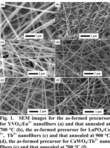
Fig. 1 shows the SEM micrographs of the
as-formed precursors for YVO4:Eu3+, LaPO4:Ce3+, Tb3+
and CaWO4:Tb3+ fiber-like samples, as well as tho
se samples annealed at high temperatures, respecti vely. From Fig. 1a, 1c and 1e, they can be seen that the precursor fibers are uniform with diamete rs ranging from 100 nm to 300 nm. In order to obtain pure inorganic fibers, a high temperature a nnealing is employed to remove the organic templ ates. After being calcined at high temperature for 4 h, the fibers shrink and become curvy due to the decomposition of PVP and crystallization of r are earth oxides. The obtained inorganic samples present fiber-like morphology and consist of fine
particles. The diameters of for YVO4:Eu3+ (Fig. 1
b), LaPO4:Ce3+, Tb3+ (Fig. 1d)and CaWO4:Tb3+ (Fig.
1f)nanofibers are 50–100 nm, 75–150 nm and 10 0–150 nm, respectively.
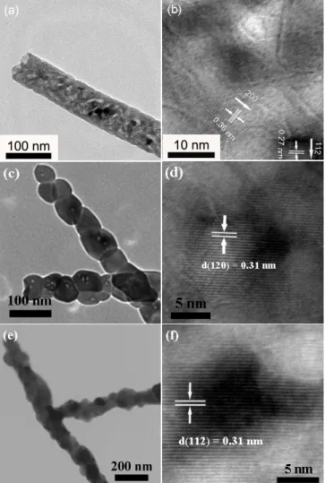
The TEM and HRTEM images of YVO4:Eu3+, L
aPO4:Ce3+, Tb3+and CaWO4:Tb3+ nanofibers are sho
wn in Fig. 2, respectively. From Fig. 2a, 2c and 2e, it can be seen that the nanofibers are further composed of fine and closed-linked nanoparticles. The typical HRTEM of nanofibers clearly show la ttice fringes with interplanar spacing of 0.36 nm and 0.27 nm, 0.31 nm, 0.31 nm that correspond t
o the (200) and (112) plane of YVO4, the (112)
plane of LaPO4 and the (112) plane of CaWO4,re
spectively.
Fig. 1. SEM images for the as-formed precursor for YVO4:Eu3+ nanofibers (a) and that annealed at 700 °C (b), the as-formed precursor for LaPO4:Ce 3+, Tb3+ nanofibers (c) and that annealed at 900 °C (d), the as-formed precursor for CaWO4:Tb3+ nano fibers (e) and that annealed at 700 °C (f).
The morphology and diameter of electrospun sa mples are dependent on a number of process para meters including the intrinsic properties of solutio n and the operational conditions. In order to obtai n uniform 1D morphology, searching for a balanc e point of various electrospinning parameters is v ery important.
Fig. 3 shows the photoluminescence excitation
and emission spectra of YVO4:Eu3+, LaPO4:Ce3+, T
b3+and CaWO
4:Tb3+ nanofibers, respectively. The E
xcitation spectrum of YVO4:Eu3+ (Fig. 2a) was ob
tained by monitoring the emission of Eu3+ 5D
0–7F2
transition at 618 nm. It can be seen clearly that the excitation spectrum consists of a broad inten
se band from 200 to 350 nm due to VO43– group
s, this confirms that the emission of Eu3+ occurs
via an energy transfers from the excited VO43–. E
xcitation into the VO43– group at 280 nm yields t
he emissions spectrum (Fig. 2b) corresponding the f–f transition at 618 nm. No emission from the
VO43– groups is observed with the excitation of 2
80 nm UV, suggesting that the energy transfer fro
m VO43– to Eu3+ is quite efficient. The excitation
16-1 / Z. Hou
• IMID 2009 DIGEST
d with the 543 nm emission (5D
4–7F5) of Tb3+ is
composed exclusively of the excitation bands of
Ce3+, which is similar to the excitation spectrum
of LaPO4:Ce3+. Excitation into the Ce3+ band at 2
78 nm (Fig. 2d) yields both the weak emission o
f Ce3+ (300-360 nm) and the strong emission of
Tb3+ (370-700 nm, 5D
4–7FJ, J = 3, 4, 5, 6), this i
ndicates that a energy transfer from Ce3+ to Tb3+
occurs in the nanofiber of LaPO4:Ce3+, Tb3+. The
excitation spectrum of CaWO4:Tb3+ (Fig. 2e) moni
tored with the 545 nm emission (5D
4–7F5) of Tb3+
consists of an intense broad band from 200 to 3 00 nm with a maximum at 249 nm, which is bad
ically identical to that of CaWO4. Excitation into
the WO42– group at 249 nm yields the emission s
pectrum (Fig. 2f) corresponding to the f–f transiti
on of Tb3+, which is dominated by the emission
of 5D
4–7F5 transition at 545 nm. Compared with t
he emission of Tb3+, the intrinsic emission from
Fig. 2. TEM and HRTEM micrographs of YVO43–: Eu3+, LaPO
4:Ce3+, Tb3+ and CaWO4:Tb3+ nanofi bers.
WO42– group is very weak, suggesting that an effi
cient energy transfer from WO42– group to Tb3+ h
as occurred. All the energy tranfer processes can be further established by time-resolved spectra.
Fig. 3. Excitation and emission spectra of YVO43–: Eu3+, LaPO
4:Ce3+, Tb3+ and CaWO4:Tb3+ nanofi bers.
Under low-voltage electron beam excitation, the
as-prepared YVO4:Eu3+, LaPO4:Ce3+, Tb3+and CaW
O4:Tb3+ nanofibers also exhibit their characteristic
emissions as the UV excitation, respectively. The representative cathodoluminescence spectra of thos
16-1 / Z. Hou
IMID 2009 DIGEST •
e nanofiber phosphors under the excitation of elec tron beam are shown in Fig. 4, which have inden tical shapes as the PL emission spectra. The CL emission intensity for those nanofiber phosphors i ncreased with the raising accelerating voltage and filament current. The increase in CL brightness c an be attributed to deeper penetration of electrons into the phosphors and the larger electron beam current density.
Fig. 4. Cathodoluminescence spectra of (a) YVO4 3–:Eu3+, (b) LaPO
4:Ce3+, Tb3+ and (c) CaWO4:T b3+ nanofibers.
4. Summary
In summary, YVO4:Eu3+, LaPO4:Ce3+, Tb3+and Ca
WO4:Tb3+ nanofibers have been successfully synth
esized by means of electrospinning technique in c onjunction with sol-gel process. Due to the energy
transfer processes from VO43– to Eu3+, from Ce3+
to Tb3+, and from WO
42– to Tb3+, the Eu3+ and
Tb3+ show their characteristic emissions up on exc
itation into the VO43– group, Ce3+ ion and WO42–
group, respectively. Under low-voltage electron b
eam excitation, the as-prepared YVO4:Eu3+, LaPO4:
Ce3+, Tb3+and CaWO
4:Tb3+ nanofibers also exhibit
their characteristic emissions as the UV excitation. Those studies indicate that electrospinning is a f acile and novel route for the development 1D lu minescent nanomaterials that are useful in many t ypes of color display fields.
5. References
1. M. Bockrath and W. Liang (eds.), Science, 291,
283 (2001).
2. Y. Xia and P. Yang (eds.), Adv. Mater., 15, 353
(2003).
3. A. Formhals, Process and apprartus for preparin
g artificial threads, US Patent no. 1, 975, 504(193
4).
4. S. Madhugiri and A. Dalton (eds.), J. Am. Che
m. Soc., 125, 14531(2003).
5. L. Yao and T. W. Haas (eds.), Chem. Mater., 1
5, 1860(2003).
6. H. Q. Hou and D. H. Reneker, Adv. Mater., 1
6, 69(2004).
7. P. Yang and M. K. Lu (eds.), J. Phys. Chem.
Solid, 63, 2047(2002).
8. X. A. Fu and S. Qutubuddin, Colloid Surf. A,
186, 245(2001).
9. J. W. Zhai amd L. Y. Zhang (eds.), Surf. Coat.
Technol., 138, 135(2001).
10. M. M. Bergshoef and G. J. Vancso, Adv. Mat
er., 11, 1362(1999).
11. H. Q. Liu and J. Kameka (eds.), Nano Lett.,
4, 671(2004).
12. C. L. Casper and J. S. Stephens (eds.), Macr