저작자표시-변경금지 2.0 대한민국 이용자는 아래의 조건을 따르는 경우에 한하여 자유롭게
l 이 저작물을 복제, 배포, 전송, 전시, 공연 및 방송할 수 있습니다. l 이 저작물을 영리 목적으로 이용할 수 있습니다.
다음과 같은 조건을 따라야 합니다:
l 귀하는, 이 저작물의 재이용이나 배포의 경우, 이 저작물에 적용된 이용허락조건 을 명확하게 나타내어야 합니다.
l 저작권자로부터 별도의 허가를 받으면 이러한 조건들은 적용되지 않습니다.
저작권법에 따른 이용자의 권리는 위의 내용에 의하여 영향을 받지 않습니다. 이것은 이용허락규약(Legal Code)을 이해하기 쉽게 요약한 것입니다.
Disclaimer
저작자표시. 귀하는 원저작자를 표시하여야 합니다.
변경금지. 귀하는 이 저작물을 개작, 변형 또는 가공할 수 없습니다.
A Dissertation for the Degree of Doctor of Philosophy
Development of Anti-Cancer
Therapeutic Strategy Using Synthetic Non-Viral Carriers for Gene and Drug Delivery in Targeted Cancers
February 2017
By
Nomundelger Gankhuyag
Department of Veterinary Medicine
Development of Anti-Cancer
Therapeutic Strategy Using Synthetic Non-Viral Carriers for Gene and Drug Delivery in Targeted Cancers
By Nomundelger Gankhuyag
Supervised by Professor Je-Yeol Cho, D.V.M., Ph.D.
A Dissertation Submitted to the Faculty of the Graduate School of Seoul National University in Partial Fulfillment of the Requirements for the
Degree of Doctor of Philosophy in Veterinary Medicine
October 2016
Supervisory Committee Approval of Thesis Submitted by December 2016
Chairman of Committee:
Vice Chairman of Committee:
Member of Committee:
Member of Committee:
Abstract
Development of Anti-Cancer Therapeutic Strategy Using Synthetic Non-Viral Carriers for Gene and Drug Delivery in Targeted
Cancers
(Supervisor: Je-Yeol Cho, D.V.M., Ph.D.) Nomundelger Gankhuyag
Department of Veterinary Medicine
Graduate School of Seoul National University
Despite significant advances in cancer therapy, cancer continues to be a leading cause of death in the world. One of the major hurdles in cancer therapy is abnormal cancer cell proliferation leading to metastasis.
Traditional cancer therapies are based on the fact that rapidly proliferating cancer cells are more sensitive to anti-cancer drugs, but unfortunately, these anti-cancer drugs show non-specific toxicity to proliferating normal cells too. Therefore, apart from the understanding the molecular basis of cancer, it is now necessary to explore and develop new anti-cancer therapeutic strategy by using non-viral carrier for gene and drug delivery. In first study, lithocholic acid (LCA), a potent anti-cancer drug, is converted to two forms of poly(ethyleneglycol) PEG conjugates, PEG-LCA(PL) and lactobionic acid (LBA) conjugated PEG-LCA (LPL). HepG2 cancer cells and LO2 normal cells were used for in vitro experiments. We further confirmed C6- loaded PL and C6-loaded LPL nanoparticles were injected into in H-ras model mice through IV administration.
In another study, we performed the gene therapeutic effect of short hairpin Rab25 RNA (shRab25) on 4-(Methylnitrosamino)-1-(3-pyridyl)-1- butanone (NNK)- induced lung tumorigenesis in female A/J mice. Mice were injected with single dose of NNK by intraperitoneal injection to induce the tumor. Eight weeks later, Glycerol proxylate triacrylate and spermine (GPT-SPE) which is used for non-viral cationic carrier that can deliver gene to lung via aerosol delivery. GPT-SPE/Rab25 using short hairpin RNA complexes delivered into tobacco-induced lung cancer model A/J mice through aerosol-only inhalation twice a week for 8 weeks. Aerosol mediated
gene delivery performs a noninvasive alternative for the targeting genes to lungs. Repeated aerosol delivery of GPT-SPE/Rab25 shRNA complexes greatly suppressed lung tumorigenesis. Those results strongly provide the novel and potential therapeutic outcome for liver and lung cancer treatment.
Key words: anti-cancer drug, apoptosis, hepatocyte-specific, nanoparticles, lithocholic acid, aerosol-delivered shRab25, lung cancer, gene therapy.
Student number: 2012-30770
Contents
Abstract ………..……..………..i
Contents……….………...iv
List of Figures………...vi
List of Tables………....….…...ix
List of Abbreviations………...x
General introduction……….…1
I. Galactosylated-poly(ethyleneglycol)-lithocholic acid selectively kills hepatoma cells, while sparing normal liver cells...5
Abstract……….………....6
Introduction ………..7
Material and methods ………..…….9
Results and Discussion ...………....15
II. Suppression of tobacco carcinogen-induced lung tumorigenesis by aerosol-delivered glycerol propoxylate triacrylate-spermine copolymer/short hairpin Rab25 RNA complexes in female A/J mice...33
Abstract………..….…34
Introduction ………..…….…… 35
Material and methods ………..……...38
Results ………..…..43
Discussion ...………..….58
References ……….…………63
Abstract in Korean .……….….73
List of Figures
Chapter I. Galactosylated-poly(ethyleneglycol)-lithocholic acid selectively kills hepatoma cells, while sparing normal liver cells
Figure 1.1H NMR spectra of LCA (a), LBA (b), PEG (c) and LPL (d).
Figure 2. FTIR spectrum of LPL. The characteristic IR peak of LBA, PEG and LCA are indicated by arrowheads.
Figure 3. Size and morphology of nanoparticles. The mean hydrodyna mic diameter of LPL(a), and PL(b) nanoparticles was measured by D LS. TEM images of LPL(c), and PL nanoparticles (d). Scale bar=200 nm.
Figure 4. Cell viability test with LPL and PL nanoparticles. LO2 and HepG2 cells were treated with LPL, PL, LCA, DMSO, and untreated group as a control, for 24 h. cell viability of the cells were analyzed by CCK-8 assay (n=3 independent experiments).
Figure 5. Fluorescent images of tissue sections to detect the cell specificity of LPL and PL nanoparticles. C6-loaded LPL or Pl was administered in mice (n=4) by IV injection. After 2 h, the mice were sacrificed and several organs were taken for tissue section to observe the distribution of the nanoparticles.
Figure 6. Effects of LPL and PL nanoparticles on the expression of apoptotic proteins in cells. LO2 and HepG2 cells were treated with LPL, PL, LCA, DMSO, and untreated group as a control, for 48 h. Whole-cell lysates were separated by SDS-PAGE and analyzed with specific antibodies on western blot.
Figure 7. Apoptosis detection by Annexin V-FITC analysis.
Chapter II. Suppression of tobacco carcinogen-induced lung tumorigenesis by aerosol-delivered GPT-SPE/shRab25 complex in female A/J mice
Figure 1. (A) Experimental design of tumor induction and treatment. Six- week-old mice (eight mice/group) were injected intraperitoneally with NNK(100mg/kg). At 22 weeks, mice were sacrificed and lungs were harvested for evaluation of lung tumors. (B) Body weight of mice from 14 to 21 weeks during aerosol delivery (n = 5, *p < 0.05 saline compared to NNK+GPT-SPE only, NNK+GPT-SPE/shScr, and NNK+GPT- SPE/shRab25). GPT-SPE glycerol propoxylate triacrylate-spermine;
shRab25, short hairpin Rab25 RNA.
Figure 2.Therapeutic efficiency of shRab25 through aerosol delivery in A/J mice. Therapeutic efficiency of shRab25 through aerosol delivery in A/J mice. (A) Lung tumor lesions. (B) Total tumor numbers (n = 5, ***p < 0.001 compared to NNK control, **p < 0.01 compared to NNK+GPT-SPE only
and NNK+GPT-SPE/shScr). (C) Total tumor volume (**p < 0.01 compared to NNK control and NNK+GPT-SPE/shScr). (D) Histopathological features (magnification, 100´and 200´).
Figure 3.Inhibition of cell proliferation by aerosol delivery of shRab25. (A) Western blot analysis of PCNA. (B) Quantification of PCNA expression.
Bands were analyzed by densitometer (n = 3, **p < 0.01 compared to NNK control, NNK+GPT-SPE only, and NNK+GPT-SPE/shScr. #p < 0.05 compared to saline control).
(C) PCNA immunohistochemistry analysis (magnification, 400´). (D) PCNA counting (n = 3, **p < 0.01compared to NNK control, ***p < 0.001 compared GPT-SPE only and GPT-SPE/shScr, ###p < 0.001 compared to saline control, ##p < 0.01 compared to saline control). PCNA, proliferating cell nuclear antigen.
Figure 4. Determination of apoptosis mechanism by aerosol delivery of shRab25. (A, C)
Western blot analysis. Statistical analysis of western blot (B) Rab25 (n = 3,
**p < 0.01 compared to NNK control). (D) Bcl-2 (n = 3, *p < 0.05 compared to NNK control, NNK+GPT-SPE only, NNK+GPT-SPE/shScr, #p<0.05 compared to saline control). (E) Bax (n = 3, ## p<0.01 compared to saline control). (F) Colorimetric TUNEL assay.
List of Tables
Chapter II. Suppression of tobacco carcinogen-induced lung tumorigenesis by aerosol-delivered GPT-SPE/shRab25 complex in female A/J mice
Table 1. Summary of tumor incidence in NNK treated A/J mice
**p < 0.01 compared to NNK group, #p < 0.05 compared to GPT-SPE only,
$$$ p < 0.001 compared to GPT-SPE/shScr. GPT-SPE, glycerol propoxylate triacrylate-spermine; NNK only, mice induced with NNK as a positive control; NNK+GTP-SPE only, mice induced with NNK and treated with GPT-SPE copolymer; NNK+GTP-SPE/shScr, mice induced with NNK and treated with GPTSPE/shScr complex; NNK+GTP-SPE/shRab25, mice induced with NNK and treated with GPT-SPE/shRab25 complex; shRab25, short hairpin Rab25 RNA; STD, standard deviation
List of Abbreviation
Bad Bcl-2-associated death promoter protein
Bax Bcl-2-associated X protein
Bcl-2 B-cell lymphoma 2
CCK8 Cell counting kit 8
C6 Coumarin 6
DAB 3,3¢-Diaminobenzidinetetrahydrocholoride
DCC N,N¢-dicyclohexylcarbodiimide
DLS Dynamic light scattering
DMEM Dulbecco’s modified Eagle medium
DMSO Dimethylsulfoxide
FBS Fetal bovine serum
FTIR Fourier transform infrared spectroscopy
GPT-SPE Glycerol propoxylate triacrylate and spermine
H&E Hematoxylin and Eosin
HRP Horseradish peroxidase
IHC Immunohistochemistry
i.p intraperitoneal
i.v intravenous
LBA Lactobionic acid
LCA Lithocholic acid
NHS N-hydroxysuccinimide
NNK 4-(Methylnitrosamino)-1-(3-pyridyl)-1-butanone
PBS Phosphate buffered saline
PCNA Proliferating cell nuclear antigen
PEG Polyethyleneglycol
PEI Polyethyleneimine
qPCR Quantitative PCR
RNAi RNA interference
ROS Reactive oxygen species
SDS-PAGE Sodium dodecyl sulfate-polyacrylamide gel electrophoresis
shRab25 shRNA construct targeting Rab25
shRNA Short hairpin RNA
TEM Transmission electron microscopy
TTBS Tris-buffered saline +Tween 20
General introduction
Hepatocellular carcinoma (HCC) has become one of the serious reason of liver cancer with low 5-year survival worldwide (Li et al., 2016). The liver is important organ in human body, is removing many toxins and chemical waste products from our blood . Therefore, lung cancer is the leading cause of c ancer deaths worldwide, especially due to tobacco smoking that causes nearly 6 million deaths per year. Despite significant advances in cancer therapy, cancer continues to be a leading cause of death in the world.
Multiple therapies have been proposed to target cancers still needed urgent therapy.
I tried to develop anti-cancer therapeutic strategy using polymeric vector based drug and gene delivery, those have been responsible for targeted cancers. Polycationic vector polymers have been attracted to increase application for gene and drug delivery last two decades (Cordeiro et al.). To make a nanoparticle formulation, drugs can be either encapsulated into a polymeric vehicle or chemically attached to PEG in a process called pegylation. The latter process also improves the pharmacokinetic and pharmacodynamic properties of drugs by increasing water solubility, reducing renal clearance and limiting toxicity (Harris and Chess, 2003).
Lithocholic acid is a naturally occurring secondary bile acid that is formed in the gut during bacterial degradation of the primary bile acids and metabolized in liver (Nicholson et al., 2012). Bile acids are steroidal amphipathic molecules that facilitate excretion, absorption, and transport of fats and sterols in the intestine and liver (Patil et al., 2013). Although some bile acid derivatives have been identified to exhibit potent cytotoxic properties, (Kozoni et al., 2000) recent researches suggest that LCA not only kills neuroblastoma cells selectively, but also displays anti-tumor effect in several types of cultured cancer cells at physiologically relevant concentrations (Goldberg et al., 2011). Due to versatile properties of LCA, i aimed to design a nanoparticle formulation of LCA as PEG conjugate and test the efficacy of the formulation for enhanced antitumor effect.
Furthermore, tobacco contains a variety of carcinogens, of whi ch NNK has been identified as the most potent lung carcinogen (Schu ller et al., 2003). It is noted that a single IP injection of NNK at a dose of 2 mg is enough to induce an average of 10–12 lung adenom as per A/J mouse (Hecht et al., 1989). Hence, A/J mouse is a suitabl e model to study the molecular mechanisms of NNK to induce the de velopment and progression of lung tumors. Based on these animal mo dels, although many antitumor agents have been identified to suppress the NNK-induced lung carcinogenesis to some extent, it is still neces
sary to develop advanced methods to treat the lung cancer at genetic level. It has been reported that chromosome instability, genetic mutatio n, and activation of oncogenes are occurred in NNK-induced lung tum ors due to disruption of the expression of several enzymes of various cellular signal pathways (Zheng and Takano, 2011).
Many scientists have therefore largely focused on RNA interfe rence (RNAi)-based knock down for cancer therapy. Usually, two kind of RNAi-based therapeutics are well-known: a vector-based short hairpin RNA (shRNA) based on a vector or a chemically synthesized double- stranded small interfering RNA (siRNA) (Rao et al., 2009). Aerosol- mediated gene delivery performs a noninvasive alternative for the targeting of genes to the lungs (Merlin JL, 2001).
Rab25 signaling pathway plays a role in the regulation of cell proliferation and apoptosis in cancer cells indicating that the Rab25 gene plays an important role in tumor occurrence and development (Agola et al., 2011). Rab25 belongs to the Rab-related small guanosine triphosphatase (GTPases) proteins; these proteins are considered key regulators of intracellular membrane trafficking involved in various diseases, including cancer (Subramani and Alahari, 2010; Tanos and Rodriguez-Boulan, 2008).
Several Rab GTPases, including Rab25, have been examined for being potential drivers in carcinogenesis (Luen Tang, 2010).
Glycerol proxylate triacrylate and spermine (GPT-SPE) which is used for non-viral cationic carrier that can deliver gene to lung via aerosol delivery. GPT-SPE has been known to good delivery efficiency of shRNA and have less toxicity than PEI-25K(Jiang et al., 2011). Cationic polymers are easy to be improved their molecular weight, surface charge, and other characterizations (Merdan et al., 2002). Gene therapy as a promising treatment strategy for cancer, aerosol delivery of therapeutic genes seems the most convenient way for lung cancer gene therapy because it is simple, non-invasive and direct method that makes instant access to lungs without any effect to other organs due to the anatomical structure and location of the lungs inside the body (Gautam et al., 2003).
Chapter I
Galactosylated-Poly(Ethyleneglycol)- Lithocholic Acid Selectively Kills
Hepatoma Cells, While Sparing Normal Liver Cells
(Published in 2015, Macromolecular Bioscience)
Abstract
Delivering drugs selectively to cancer cells but not to nearby normal cells is a major obstacle in drug therapy. Recent reports suggest that lithocholic acid (LCA) is a potent anti-cancer drug that selectively kills cancer cells, sparing normal cells. Although LCA holds great promise for killing cancer cells, it is usually inappropriate for in vivo use because of its low solubility, short shelf life, short circulating half-life, and rapid renal clearance from the body.
Conversely, the afore-mentioned defects can be overcome through a process called PEGylation. In this study, LCA was converted to two forms of PEG conjugates, viz., PEG-LCA and Galactose-PEG-LCA. The latter form contains a galactose ligand to target the hepatocytes. Both forms are self- assembled to form nanoparticle formulation, and they have high potency to kill HepG2 cancer cells, sparing normal LO2 cells, similar to the function of LCA as reported before. Besides, Galactose-PEG-LCA had high specificity to HepG2 cells in killing the cells. Western blot results confirmed that the cell death is occurred through apoptosis induced by LCA nanoparticles.
Further loading of anti-cancer drugs inside the core of LCA nanoparticles as drug delivery vehicles will be of great importance to evaluate the potential of this system.
Introduction
Despite significant advances in cancer therapy, cancer continues to be a leading cause of death in the world (Jemal et al., 2008). One of the major hurdles in cancer therapy is abnormal cancer cell proliferation leading to metastasis (Chaffer and Weinberg, 2011) Traditional cancer therapies are based on the fact that rapidly proliferating cancer cells are more sensitive to anti-cancer drugs, but unfortunately, these anti-cancer drugs show non- specific toxicity to proliferating normal cells too (Pasut and Veronese, 2009).
Therefore, apart from the understanding the molecular basis of cancer, it is now necessary to explore and develop new anti-cancer drugs which are much more selective to cancer cells.
Recently, lithocholic acid (LCA), a bile acid, has been reported to kill human neuroblastoma cells, while sparing normal human primary neurons due to the selective initiation of an extrinsic apoptotic programme of cell death in neuroblastoma cells (Goldberg et al., 2011).
It is now documented that the LCA inhibits the proliferation of prostate cancer cells without killing immortalized normal prostate epithelial cells, where the extrinsic pathway of apoptosis and cell death induced by LCA is partially dependent on the activation of caspase-8 and -3 (Goldberg et al., 2013).
Although LCA holds great promise for killing cancer cells, it is usually
inappropriate for in vivo use because of its low solubility, short circulating half-life, and rapid renal clearance from the body. Conversely, the afore- mentioned defects can be overcome by conjugation of poly(ethylene glycol) (PEG) with drugs through a process called PEGylation that has been identified as a promising strategy in drug delivery system to improve solubility and biocompatibility of the drugs and to facilitate the particle diffusion in biological environments (Cu and Saltzman, 2009; Pendri A, 1996; Vllasaliu et al., 2014)
Moreover, receptor-mediated cellular uptake of the drugs has received much attention in the field of drug delivery systems, (Leamon and Low, 2001;
Qian et al., 2002; Sudimack and Lee, 2000) as drugs can be targeted to the specific cells or tissue through the specific interaction of ligands with receptors of the cells (Wagner E. Curiel D, 1994) Accordingly, galactose is one of the most used ligands for targeting liver parenchymal cells because hepatocytes have large number of cell surface receptors called asialoglycoproteins that have high-affinity with galactose moiety (Wu and Wu, 1998)
The present study aims to develop a potent anticancer drug, lactobionic acid (LBA)-PEG-LCA (LPL), for targeted delivery that selectively kills hepatoma cells sparing normal liver cells.
Materials and methods
Chemicals and reagents
Lithocholic acid (LCA), lactobionic acid (LBA), N,N′- dicyclohexylcarbodiimide (DCC), N-hydroxysuccinimide (NHS), dimethylsulfoxide (DMSO), Coumarin 6 (C6) were purchased from Sigma- Aldrich (USA). Diamino polyethyleneglycol 6000 Da (PEG) was purchased from SunBio (Korea). All reagents were of analytical grade and used without further purification.
Anti-PARP and anti-Caspase-3, anti-Cleaved Caspase-3 antibodies were from Cell Signaling (Beverly, MA, USA). The other indicated anti-b-Actin, anti-Bad antibodies were from Santa Cruz Biotechnology (Santa Cruz, CA, USA).
Synthesis of LPL
LPL was synthesized in two-step reactions (Scheme 1). First, LBA (2 mM) was activated in DMSO (10 ml) with DCC (4 mM) in the presence of NHS (4 mM) at room temperature for 18 h. The activated LBA was then reacted with PEG (20 mM) at room temperature for 4 h. The reaction product was first dialyzed in DMSO using a dialysis membrane (Spectra/Por MWCO:
1,000) for 24 h to remove the unreacted LBA, and finally dialyzed against
water at 4 °C overnight. The first product, LBA-PEG (LP), was freeze dried.
To conjugate LCA with LP, LCA (50 mM) was activated in DMSO (10 ml) with DCC (100 mM) in the presence of NHS (100 mM) at room temperature for 18 h. The activated LCA was reacted with LP (10 mM) at room temperature for 4 h. The reaction product, LPL, was first dialyzed in DMSO using a dialysis membrane (MWCO: 1,000) for 24 h to remove unreacted LCA, and finally dialyzed against water at 4 °C overnight. LPL nanoparticles thus formed during dialysis step was collected, and washed with water to remove unreacted water soluble PEG portion. LPL was further freeze dried and stored at -80°C until use. Similarly, C6-loaded PL or LPL nanoparticles were prepared by dialysis method. C6 (0.4 mg) was mixed with PL or LPL (20 mg) in DMSO (5 ml). The mixture was dialyzed in water using Spectra/Por membrane (MWCO: 1,000) at 4 °C overnight. Due to hydrophobic nature of LCA, C6-loaded PL or LPL nanoparticles are formed during dialysis due to the transition from DMSO to hydrophilic medium water. The nanoparticles were freeze dried and stored at -80°C until use.
NMR and FTIR
Each step of the synthesis of LPL and PL was analyzed by 1H NMR spectroscopy (Avance600, Bruker, Germany). Conjugation of LBA or LCA
in LPL and PL was also confirmed by Fourier transform infrared spectrometer (FTIR; Nicolet 6700, Thermo Fisher Scientific Inc., Waltham, MA, USA). The compositions of conjugated molecules in the polymer were determined by 1H NMR as suggested in polymer analysis by NMR (Sigma- Aldrich, Material Matters 2006, 1.1, 3). The mole% of conjugated molecules in the polymer was calculated by the given equation, (Izunobi J.
U. , 2011)
Z(%) = × 100%
where axis the area of the 1H NMR peak of Z molecule; mxis the number of protons of Z molecule; ay is the area of the 1H NMR peak of adjacent molecule in the polymer; and my is the number of protons of adjacent molecule.
Physicochemical characterization of LPL nanoparticles
The size of nanoparticles was measured by dynamic light scattering spectrophotometer (DLS 8000, Otsuka Electronics, Osaka, Japan) with 90°
scattering angles. The morphology of LPL nanoparticles was observed by energy-filtering transmission electron microscope (EF-TEM) (LIBRA 120, Carl Zeiss, Germany). The nanoparticles in suspension were adsorbed to carbon-coated ionized Formvar grids, negatively stained with 2% uranyl
acetate, and observed with EF-TEM.
Cytotoxicity assay
Normal liver cell line (LO2) and human liver carcinoma cell line (HepG2) were maintained in DMEM medium and RPMI-1640 medium (HyClone, Thermo Scientific, USA), respectively, supplemented with heat-inactivated fetal bovine serum (HyClone, Thermo Scientific, USA) and penicillin/streptomycin (GibcoBRL, Grand Island, USA). All cells were cultured under standard incubation condition at 37°C with 5% CO2. Cytotoxicity of nanoparticles in LO2 and HepG2 cells was determined by cell proliferation assay using cell counting kit-8 (CCK8) (Dojindo Laboratories, Japan). Briefly, cells were seeded in 96-well plates at an initial density of 4´103cells/well for LO2 and 4 ´103cells/well for HepG2 in 0.1 ml of growth medium and pre-incubated 24 h prior to addition of filtered substrate. Growth media was replaced by 0.1 ml fresh serum free media containing 50 uM and 100 uM of nanoparticles. After additional incubation for 24 h, 10 ul of CCK8 solution was added to each well of the plate and incubated for 3 h. The absorbance was measured at 450 nm using an ELISA plate reader (plate reader Model 680, BIORAD).
Western blot analysis
Cells were cultured under standard incubation condition at 37°C with 5%
CO2. The samples at the concentration of 50 µM were treated to the cells for 24 h. The mechanism of apoptosis induced by the given samples was determined by observing a number of proteins involved in apoptosis. By measuring and quantifying the protein concentration of cell lysates using a PierceÒBCA Protein Assay kit (Thermo scientific, Rockford, IL 61101 USA), equal amounts (25 µg) of protein were loaded into 10-15% sodium dodecyl sulfate-polyacrylamide gel electrophoresis (SDS-PAGE) gel and transferred to nitrocellulose membranes. Membranes were blocked with TTBS containing 5% skim milk for 1 h at room temperature and immunoblotting was performed by incubation overnight at 4°C with the corresponding primary antibodies in 5% skim milk and then with secondary antibodies conjugated to horseradish peroxide for 4 h at room temperature.
After washing, bands-of-interest were visualized using a model EZ-capture MG luminescent image analyzer (ATTO, Tokyo, Japan).
Apoptosis detection by Annexin V-FITC analysis
In addition to western blot, EzwayTM Annexin V-FITC apoptosis detection kit (Komabiotech, Korea) was used to identify the apoptosis induced by the LPL nanoparticles according to the manufacturer’s instructions. Cells,
seeded at a density of 2x10⁵ cells in culture flask, were incubated at standard condition for 48 h. The samples (50 µM) were treated to the cells for 24 h.
Cells were trypsinized and collected in microtubes by centrifugation at 1000 x g for 5 min. The cells were washed twice with cold phosphate buffer saline (PBS). The cells were further washed with cold binding buffer. The cells were incubated with Annexin V-FITC for 15 min in dark. The cells were washed with binding buffer. Finally, the cells were stained with propidium iodide (PI) and analyzed by flow cytometry immediately.
Ligand specificity of LPL in vivo
The specificity of galactose ligand in the LPL with hepatocytes in vivo was examined using 13 weeks old H-ras mice obtained from the Human Cancer Consortium National Cancer Institute (Frederick, MD). The mice were divided into three groups (four mice/group). The control group was non- treated, and the other two groups were injected to intravenous (IV) vein with C6-loaded PL and LPL nanoparticles respectively. The concentration of C6 was 5 µg per mice. Animals were kept in the laboratory animal facility with temperature and relative humidity maintained at 23°C ± 2°C and 50% ± 10%, respectively, and with a 12 hour light/dark cycle. The mice were sacrificed after 2 h of IV injection, and the organs were collected, fixed in 4%
paraformaldehyde, and preserved in 30% sucrose for 3 h at 4°C. Tissues
were embedded and were sectioned with a microtome. The sections of the organs were visualized under fluorescence microscope (Olympus BX51).
Statistical analyses
Statistical analyses were performed with a student's t test (Graphpad Software, San Diego, CA, USA). *P< 0.05 was considered more significant than the corresponding control values.
Results and Discussion
Preparation and characterization of LPL and PL
Bile acids are steroidal amphipathic molecules that facilitate excretion, absorption, and transport of fats and sterols in the intestine and liver (Patil et al., 2013). Although some bile acid derivatives have been identified to exhibit potent cytotoxic properties, (Kozoni et al., 2000) recent researches suggest that LCA not only kills neuroblastoma cells selectively, but also displays anti-tumor effect in several types of cultured cancer cells at physiologically relevant concentrations (Goldberg et al., 2011). Due to versatile properties of LCA, we aimed to design a nanoparticle formulation of LCA as PEG conjugate and test the efficacy of the formulation for enhanced antitumor effect.
Therefore, pegylation of LCA was performed as described in materials and
methods. Firstly, LBA that harbors galactose moiety was activated using DCC/NHS reagents and conjugated with diamino PEG through amide linkage between carboxylic group of LBA and amino group of PEG.
Unreacted LBA was removed through dialysis against water. The content of LBA in LP as estimated by 1HNMR was about 10 mol-%. Secondly, LCA was similarly reacted with LP using DCC/NHS through amide linkage between carboxylic group of LCA and amino group of LP (Scheme 1).
Unreacted LCA and PEG were removed by dialyzing in DMSO and then washing in water, respectively. The content of LCA in LPL as estimated by
1H NMR was about 63 mol-%. The 1H NMR spectra of PEG, LCA, LBA and LPL are shown in Fig. 1, while 1H NMR spectra of LP and PL in Supplementary Fig. 1.
These results indicate that LBA and LCA were reacted with the two ends of diamino PEG through amide linkages. We further confirmed the amide bonds of LPL by Fourier transform infrared spectroscopy (FTIR) (Fig. 2).
The results also indicated the successful synthesis of LPL and PL.
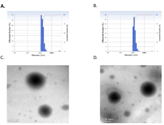
Furthermore, we expected that LPL would self-assemble to form polymeric nanoparticles because of amphipathic property of LPL. Indeed, the spherical nanoparticles with particle size of 300-400 nm was observed by TEM as shown in Fig. 3. The mean hydrodynamic diameter of PL and LPL nanoparticles as measured by DLS was 370 nm and 380 nm, respectively
(Fig. 3).
Stability of LPL and PL nanoparticles
The release of active drug from polymer-drug conjugate (prodrug) is important to be used as drug delivery in cancer (Duncan, 2006). However, our aim was to develop PEG-LCA as a permanent polymer-drug conjugate through amide bond. The amide bond is often the bond of choice when linking the polymer to the drug to make permanent polymer drug conjugates because amide bonds are very stable to aqueous hydrolysis at physiological pH, and to enzymatic hydrolysis (D'Souza and Topp, 2004). Therefore, we tested the stability of PL and LPL nanoparticles at physiological condition by incubating the nanoparticles in water at 37°C for 3 days. We did not observe any change in the morphology of the nanoparticles by TEM analysis (Supplementary Fig. 2). The results demonstrated that the nanoformulations of PL and LPL were highly stable. Therefore, we concluded PL and LPL as permanent drugs with no release of LCA from them.
Toxicity of LPL and PL nanoparticles
We tested the toxicity of PL and LPL in LO2 and HepG2 to compare anti- tumor effect between normal and cancer cells. Fig. 4 shows the cell viability of LO2 and HepG2 treated with LCA, PL and LPL at two different
concentrations and incubation periods. The cell viability of LO2 cells at 50 μM concentration of LCA and PL was almost 100% whereas cell viability was about 75% with LPL treatment. On the other hand, the cell viability of HepG2 cells treated with LCA, PL and LPL at 50 μM concentration was 60%, 65% and 20%, respectively.
The results indicated that PL at a concentration of 50 μM is not cytotoxic to normal cells whereas PL killed HepG2 cells, indicating that PL selectively killed cancer cells, while sparing normal cells. Similar effect was observed in our experiment when treated with LCA, as reported before (Goldberg et al., 2011).
Significantly, the efficiency of LPL at 50 μM concentration is 3-fold higher than LCA in killing the cancer cells. Above all, the anti-tumor effect of LPL in HepG2 cells was stronger than that of PL due to the specific interaction between galactose ligands in the LPL and asialoglycoprotein receptors in the HepG2 cells. Treatment of LCA and PL at a concentration of 100 μM in normal cells showed only slight cytotoxicity, whereas the cell viability of HepG2 cells was ≤10%. These results provoked that the nanoparticle formulation of LPL has comparable efficacy with LCA small molecules. It seemed that the mechanism of drug action of LCA is similar with LPL nanoparticles. On the other hand, the cell viability of LO2 cells was ~50%
after the treatment of LPL at a concentration of 100 μM, while the cell
viability of HepG2 cells was less than 5%. These results also indicated the efficacy of the nanoparticle formulation of LPL is higher in cancer cells than normal cells.
Ligand specificity of LPL with liver cells in vivo
To evaluate the ligand specificity of LPL in mouse liver cells in vivo, C6- loaded PL and C6-loaded LPL nanoparticles were injected into individual mouse through IV administration. Fig. 5 shows fluorescence images of several organs of mouse after injection of C6-loaded nanoparticles. It was observed that the distribution of LPL nanoparticles in the liver was higher than the distribution of PL. The result clearly depicted the ligand specificity of LPL with the mouse liver in vivo. On the other hand, we did not find any differences between LPL and PL distribution in lung and kidney, probably due to non-specific distribution of both nanoparticles in those organs.
Mechanism of apoptosis induced by LPL nanoparticles
To understand the cellular signaling processes leading to apoptosis induced by LCA nanoparticles, western blot was performed to trace the related enzymes in the signaling processes. During the early phases of apoptosis, caspase-3 is activated by proteolytic cleavage (Thornberry and Lazebnik, 1998). Caspase-3, in turn, cleaves poly (ADP-ribose) polymerase-1 (PARP) into 89-kDa fragments, thereby inactivating it (Krishnakumar and Kraus,
2010). PARP is a 116 kDa enzyme involved in DNA repair and maintenance of genomic integrity (Virág and Szabó, 2002). Therefore, detection of cleaved caspase-3 and/or PARP by western blot is routinely used for the study of apoptosis. Fig. 6 shows the detection of PARP and caspase 3 in LO2 and HepG2 cells after treatment of LCA and LPL nanoparticles as described in materials and methods.
Western blot studies showed that the expression levels of proteins related to apoptosis vary in normal and cancer cells. In contrast to LO2 cells, the blot showed the down-regulation of PARP protein levels and up-regulation of caspase 3 in HepG2 cells. Similarly, the level of Bad protein (pro-apoptotic member of the Bcl-2 family)(Kitada et al., 1998) and even actin protein expressed higher in LO2 cells in compare to HepG2 cells. While the level of cleaved PARP after treatment with LCA, LP and LPL was similar in LO2 cells, the cleaved PARP was distinctly visible in the blot only with the treatment of LPL in HepG2 cells. The cleaved PARP was not clearly visible after the treatment of LCA in HepG2 cells, probably due to very low expression of PARP. However, the expression levels of Bad with the treatment of LPL and LCA were higher in HepG2 cells compared to control.
Similarly, in compare to LO2, the expression levels of cleaved caspase-3 in HepG2 cells with the treatment of LCA and LPL were higher.
Although the results elucidated the occurrence of apoptosis with the treatment of nanoformulation of LCA in cancer cells, it seemed that a distinct mechanism of apoptosis occurs in cancer cells in contrast to normal cells. The notion is further supported by a previous report that stated doxorubicin induces apoptosis in normal and tumor cells via distinctly different mechanisms (Wang et al., 2004). The mechanism of LCA-induced apoptosis has been recently explored in several studies. The mechanism of CD95 death receptor activation by bile acids has been studied in hepatocytes and is thought to proceed through the generation of oxidative stress by these steroids (Sokol et al., 2006). Similarly, LCA is able to induce immediate oxidative stress with subsequent generation of reactive oxygen species (ROS) in colon adenocarcinoma cells (Katona et al., 2009).
The study further suggested that LCA-induced apoptosis is proceeded through a mitochondrial/caspase-9-dependent pathway initiated by caspase- 8 (Katona et al., 2009).
Most recent study suggested that LCA can directly induce syncytiotrophoblastic apoptosis in vitro, possibly via the TNF-α pathway, with caspase-3 activation (Du et al., 2014).
In this study, with the treatment of LPL, the catalytic activity of PARP and caspase-3 in HepG2 was higher than in LO2, indicating that the mode of
anti-tumor effect of LPL is similar with the apoptosis induced by LCA via a caspase-3-dependent mechanism.
We further determined the apoptosis induction by LPL nanoparticles using Annexin V-FITC staining. As shown in Fig. 7, treatment with PL and LPL induces 1.4 and 3.1-fold increases in apoptotic cells at 50 µM concentrations compared to LCA. It was clearly seen in the figure that the cells underwent early apoptosis and late apoptosis which was comparatively higher when the cells were treated with LPL nanoparticles than LCA. Therefore, Annexin- FITC study also revealed that the nanoformulation of LCA nanoparticles induced high apoptosis resulting high cell death.
Scheme 1
HO
H H
OH O
H
H
H2N O
NH2
[ ]
nO O O HO
OH OH HO
OH
HO
OH OH OH
PEG
LCA-PEG-LBA(LPL)
N O
N
O O O
OH OH HO
OH
HO
OH OH OH
HO
H H
O
H
H
H H
[ ]
nFigure 1
A
B
1,2,3,4,5,6,7,8,9,1 011
12
O O O HO
OH OH HO
OH
HO
OH OH OH
234 5
6 1 109 8 11 7 12
C
D
Figure 1. 1H NMR spectra of LCA (A), LBA (B), PEG (C) and LPL (D).
Figure 2
Figure 2. FTIR spectrum of LPL. The characteristic IR peak of LBA, PEG and LCA are indicated by arrowheads.
Figure 3
Figu re 3. S ize a nd mo rp ho lo g y o f na no part ic le s. The mean hydrodynamic diameter of LPL (A) and PL (B) nanoparticles as measured by DLS. TEM images of LPL (C) and PL nanoparticles (D). Scale Bar = 200 nm.
A.
Figure 4
Figure 4. Cell viability test with LPL and PL nanoparticles. LO2 and HepG2 cells were treated with LPL, PL, LCA, DMSO, and untreated group as a control, for 24 h. Cell viability of the cells was analyzed by CCK-8 assay (n=3 independent experiments).
CellViability (%of Control)
100uM LO2 cell line 24hr LO2 cell line 24hr
150 100
50
0
100uM
HEPG2 cell line 24hr 50uM
HEPG2 cell line 24hr
50uM
A B
C. D
150 100
50
0
(%of Control) 150
100
50
0
(%of Control) 150
100
50
(%of Control) 0
CellViability CellViability
CellViability
Control DMSO LCA PL LPL
Control DMSO LCA PL LPL Control DMSO LCA PL LPL
Control DMSO LCA PL LPL
Figure 5
Figure 5. Fluorescent images of tissue sections to detect the cell specificity of LPL and PL nanoparticles. C6-loaded LPL or PL was administered in mice (n=4) by IV injection. After 2 h, the mice were sacrificed and several organs were taken for tissue section to observe the distribution of the nanoparticles.
Liver Lung Kidney
Control
LPL
PL
Figure 6
Figure 6. Effects of LPL and PL nanoparticles on the expression of apoptotic proteins in cells. LO2 and HepG2 cells were treated with LPL, PL, LCA, DMSO, and untreated group as a control, for 48 h. Whole-cell lysates were separated by SDS- PAGE and analyzed with specific antibodies on western blot.
Figure 7
Figure 7. Apoptosis detection by Annexin V-FITC analysis. HepG2 ce lls were treated with LCA, PL and LPL at concentration of 50 µM f or 24 h. Early and late apoptosis are indicated.
79.69
12.32 4.6
7
3.32
60.27 9.72
28.45 1.56
48.69 11.62
26.23 13.62
Conclusions
In conclusion, we have successfully prepared and evaluated the nanoformulation of LPL for efficient hepatocyte-targeted anti-cancer therapy.
Moreover, the nanoparticle formulation of LPL selectively kills liver cancer cells sparing normal liver cells. Current investigations are underway to evaluate the efficacy of anti-cancer drugs-loaded LPL nanoparticles for targeted combinatorial cancer therapy.
Chapter II
Suppression of Tobacco Carcinogen-Induced Lung Tumorigenesis by Aerosol-Delivered
Glycerol Propoxylate Triacrylate-Spermine Copolymer/Short HairpinRab25 RNA
Complexes in Female A/J Mice
(Published in 2016, Journal of Aerosol Medicine and Pulmonary Drug Delivery)
Abstract
Rab25 belongs to the Rab family of small GTPases and is implicated in the development of various types of human cancer including lung cancer that is the leading cause of cancer deaths worldwide. In this study, I report the gene therapeutic effect of small hairpin Rab25 (shRab25) on 4- (Methylnitrosamino)-1-(3-pyridyl)-1-butanone (NNK)-induced lung tumorgenesis in female A/J mice. Initially, six-week-old mice were treated with single dose of NNK (2 mg/0.1 ml saline/mouse) by intraperitonal injection to induce the tumor. eight weeks later, shRab25 was delivered with GPT-SPE complex into tobacco-induced lung cancer models through aerosol delivery. Remarkably, aerosol-delivered shRab25 significantly decreased the expression level of Rab25 and other prominent apoptosis related proteins in female A/J mice. The apoptosis in these mice were determined by detecting the expression level of PCNA, Bcl-2 and Bax, and further confirmed by TUNEL assay. These results strongly support the tumorigenic role of Rab25 in tobacco induced-lung cancer and hence demonstrate aerosol delivery of shRab25 as a promising approach for lung cancer treatment.
Key words: lung cancer, gene therapy, aerosol- deliverered shRab25, NNK, A/J mice
Introduction
Lung cancer is the main cause of cancer-related deaths in the world (1.6 million, 19.4%), and the development of more effective lung cancer therapy still remains challenging (Ferlay et al., 2015). In fact, 80~90% of lung cancers are strongly associated with tobacco smoking. Among the thousands of carcinogenic compounds found in tobacco products, 4- (Methylnitrosamino)-1-(3-pyridyl)-1-butanone (NNK), is the main player to lung carcinogenesis (Akopyan and Bonavida, 2006; Stepanov et al., 2008).
Rab25 belongs to the Rab-related small guanosine triphosphatase (GTPases) proteins; these proteins are considered key regulators of intracellular membrane trafficking involved in various diseases, including cancer (Subramani and Alahari, 2010; Tanos and Rodriguez-Boulan, 2008).
Several Rab GTPases, including Rab25, have been examined for being potential drivers in carcinogenesis (Luen Tang, 2010). Studies have shown that Rab25 contributes to the aggressiveness of breast and ovarian cancers (Cheng et al., 2004). Notably, knockdown of Rab25 expression inhibits the growth of ovarian cancer cells (Fan et al., 2006), indicating the essential role for Rab25 in tumor progression and aggressiveness (Agarwal et al., 2009).
Specifically, knockdown of Rab25 by siRNA suppressed cell migration and
invasion in non-small-cell lung carcinoma (NSCLC) cells (Ma et al., 2015).
While Rab25 expression is positively correlated with E-cadherin, it is negatively associated with ZEB1 in NSCLC cell lines (Roche et al., 2013).
Particularly, the effects of Rab25 knockdown on tumor proliferation and apoptosis were confirmed in mice xenografted with gefitinib-sensitive lung cancer cells (Jo et al., 2014).
Many scientists have therefore largely focused on RNA interference (RNAi)-based knock down for cancer therapy. Usually, two kind of RNAi- based therapeutics are well-known: a vector-based short hairpin RNA (shRNA) based on a vector or a chemically synthesized double-stranded small interfering RNA (siRNA)(Rao et al., 2009). A number of studies have been conducted with several RNAi-based therapies, such as shSCRN1 in lung cancer cell lines (Kim et al., 2016), siDDR1in cancer cell lines (Ali- Rahmani et al., 2016), shCul4A in lung cancer cells (Hung et al., 2016) shRab25 in colon cancer cell lines (Krishnan et al., 2013). Comparison with other methods, shRNA has demonstrated great potential as a promising tool for cancer treatment.
Aerosol-mediated gene delivery performs a noninvasive alternative for the targeting of genes to the lungs (Merlin JL, 2001). In this regard, we have previously used several non-viral or viral vectors to transport DNA or siRNA or shRNA through aerosol delivery for lung cancer treatment (Hong
et al., 2015). For example, we delivered shAkt1 by glycerol triacrylate- spermine (GT-SPE) to suppress lung cancer progression (Hong et al., 2012).
Similarly, we used polyspermine to deliver Pdcd4 gene that greatly reduced tumor size in K-rasLA1 lung cancer model mice (Kim et al., 2014). Also, we have successfully used aerosol delivery of RNAi-based therapeutics by various polymer complexes including spermine-based poly(amino ester)/Akt1shRNA complexes (Jiang et al., 2011), folate–chitosan-graft polyethylenimine/Akt1 shRNA complexes (Jiang et al., 2009), and poly(ester amine)/Akt1 siRNA complexes (Xu et al., 2008). Using lentivirus as a viral vector, several shRNAs, such as AIMP2-DX2 shRNA (Hwang et al., 2013), OPN shRNA (Minai-Tehrani et al., 2013), and PDCD4 shRNA (Hwang et al., 2010) were used for lung cancer treatements through aerosol delivery.
In this study, we delivered short hairpin Rab25 RNA (shRab25) in NNK-induced lung tumorigenesis female A/J mice through aerosol delivery to evaluate the preventive effect of shRab25 in the suppression of lung tumorigenesis.
Materials and methods
Materials
NNK was purchased from Toronto Research Chemicals Inc. (Toronto, Ontario, Canada). Rab25 antibody was purchased from Abcam(Cambridge, UK). a-tubulin Abfrontier (Seoul, Korea), Bax, Bcl-2, proliferating cell nuclear antigen (PCNA) antibodies were purchased from Santa Cruz Biotechnology (Santa Cruz, CA). DeadENDTMColorimetric TUNEL assay kit was obtained from Promega Corporation (Promega, Madison, WI).
Rab25 shRNA (shRab25) and the scrambled control shRNA (shScr) were purchased from Koram Biogen Corp (Korea).
Mouse experiment
Female A/J mice, 5 weeks old, weighing 15-19 g, were received from Central Laboratory Animal Inc. (Seoul, Korea). Mice were housed under standard light/dark cycle at a stable temperature (23°C ± 2°C) and relative humidity (50% ± 10%) in the laboratory animal facility. The mice, 6 weeks of age, were divided into 5 groups, each group containing 8 mice. Four group of mice were injected with single dose of NNK (2 mg/0.1 ml saline/mouse) through intraperitoneal (IP) route. For gene delivery, three groups (except NNK control and saline control group) mice were exposed
with the Rab25 complexes in the nose-only inhalation chamber as described previously (Kim et al., 2004; Tehrani et al., 2007). The polymer complex with DNA (shScr or shRab25) or aerosol containing polymer only were delivered 16 times (twice a week for 8 consequent weeks). Mice were sacrificed one week after exposure and tumor on lung surface were carefully counted, as mentioned previously (Singh et al., 2006). Finally, collected lungs were fixed in 10 % formalin for histopathological examination and remaining lung stored at -70°C for further experiment. All animal experiments were approved by Animal Care and Use Committee at Seoul National University (SNU-140814-3).
Construction of Rab25 shRNA and preparation of GPT–SPE/shRNA complex
The shRNA sequence targeting mouse Rab25 mRNA and scrambled control were purchased. The glycerol propoxylate triacrylate (GPT) - spermine (SPE) copolymer (GPT–SPE) was synthesized as described previously (Jiang et al., 2011). GPT-SPE/shRNA complexes were self-assembled in distilled water by slow adding of 0.8 mg of DNA to 8 mg of polymer solution under gentle mixing. The complexes (final volume 30 ml) were incubated for 30 minutes at room temperature (RT) before delivery.
Western blot analysis
Lung tissues were lysed with IKA®T10 homogenizer (IKA®, Works GmbH
& Co, Staufen, Germany) to extract the proteins. Total protein concentration was confirmed by using the PierceÒBCA reagent (Thermo scientific, USA).
Equal amounts (25 µg/well) of protein were loaded into 10-12 % SDS- polyacrylamide gels and transferred to nitrocellulose membranes using iBlot system (Life Technologies, Grand Island, NY, USA). Membranes were blocked with Tris-buffered saline with tween 20 (TTBS) containing 5%
skim milk for 1 h at RT and immunoblotting was performed by incubation overnight at 4°C with the corresponding primary antibodies. Following day, Antibody labeled with secondary horseradish peroxidase for 4 h at RT.
After washing, bands were captured using a model EZ-capture MG luminescent image detector (ATTO, Tokyo, Japan) and analyzed by using ATTO CS Analyzer windows version 3.0 (ATTO, Tokyo, Japan).
Histopathologic and immunohistochemistry analysis
Paraffin-embedded and sectioned lung tissues (4 μm) were stained by hematoxylin and eosin. For immunohistochemistry (IHC) analysis, the lung sections were then deparaffinized in xylene (each for 5 min ´ 2times) and rehydrated by alcohol gradients (2 min/each), then washed with distilled water and 3% hydrogen peroxide used for 10 min incubation. To block the unspecific binding sites, sections were incubated with 3% bovine serum albumin in TTBS for 1 hour at RT after washing with a TTBS solution.
Concerned primary antibodies (1:100) were used overnight at 4°C. Next day, the samples were rinsed by TTBS, and then incubated in secondary antibodies conjugated to horseradish peroxidase (1:200) for 3 hours at RT.
Then, 3,3′-diaminobenzidine (DAB) reagent (Life Technologies) was reacted with the tissue sections, those were analyzed by light microscopy.
The tissue sections were counterstained with a Mayer’s hematoxylin (DAKO, Carpinteria, CA, USA) and mounted using cover slide and the Permount (Fisher Scientific, Waltham, MA). The slides were observed using a Nikon eclipse fluorescence microscopy (Tokyo, Japan).
Analysis of apoptotic cells by TUNEL assay
Determination of apoptotic cells using by DeadEndTM Colometric tunel assay kit and all steps according to kit’s instructions. Briefly, for paraffin- embedded lung tissues sections were deparaffinized and rehydrated by xylene or graded ethanol (100%, 85%, 70% and 50%) at RT, respectively.
rTdT reaction mix is added on equilibrated areas and incubated at 37°C for 60 min. After several washes in PBS and 2X SSC, the slides were incubated Streptavidin HRP solution for 30 minutes at RT. For counterstaining procedure, DAB substrate was added to each slide and developed until the appearance of light brown background. Slides were observed by a light microscope to select ´1000 magnification fields (Olympus BX51, Tokyo, Japan).
Statistical analysis
Statistical analysis was performed using analysis of Student’s t-test with Microcal Origin (Microcal Software, Northampton, USA). Results were expressed as mean values ± standard error of three independent experiments (n=3). All data were compared with corresponding values (P < 0.001 was more significant, P < 0.01 was highly significant and P< 0.05 was considered statistically significant).
Results
In vivo aerosol delivery of shRab25
To determine the carcinogenic function of NNK in vivo, mouse experiment was design as illustrated in Figure 1A. The mice, 6 weeks of age, were divided into 5 groups, each group containing 8 mice. Four group of mice were injected with single dose of NNK (2 mg/0.1 ml saline/mouse) through intraperitoneal (IP) (Takeuchi et al., 2006). Single IP injection of NNK for lung tumorigenesis in A/J mice is a simple procedure that takes 16 weeks for a reasonable yield of lung tumors to develop (Yokohira et al., 2008). The experiment was carried out with one group mice that were treated with an equal volume ofsaline (vehicle control of NNK). Eight weeks later, the NNK-treated mice were exposed with GPT-SPE, GPT-SPE/shScr, GPT- SPE/shRab25 for16 times (twice a week for 8 consequent weeks). Body weights of individual mouse were recorded weekly for 8 weeks during the aerosol delivery (Figure 1B).
Aerosol delivery of Rab25 shRNA suppresses tumorigenesis in lung
As shown in Fig. 2A, the injection of NNK induced number of tumors in lungs while there was no such tumor growth in lungs of control group mice (saline). After aerosol delivery of GPT-SPE, GPT-SPE/shScr, GPT- SPE/shRab25 in NNK-induced A/J mice for 8 weeks, we found that GPT- SPE/shRab25 largely decreased the tobacco-induced tumor numbers in the lungs in compared to GPT-SPE or GPT-SPE/shScr delivered groups. The observable antitumor effects of GPT-SPE/shRab25 were illustrated with graph (Figure 2 B, C and Table 1). The results showed that aerosol- delivered GPT-SPE/shRab25 complex significantly decreased lung tumor number (n=5) (Figure 2B) and tumor volume (n=5) (Figure 2C). For further confirmation of anti-tumor effect of aerosol-delivered GPT-SPE/shRab25 complex, we performed the histopathologic analysis of lung tissues of mice 8 weeks after the aerosol delivery (Figure 2D). The results demonstrated the higher tumor suppression effect of GPT-SPE/shRab25 than other GPT-SPE or GPT-SPE/shScr treated groups.
Cell proliferation was suppressed in Aerosol delivery of shRab25
To evaluate cell proliferation in tumor tissue, IHC and western blot studies were performed. Western blot results confirmed that aerosol-delivered GPT- SPE/shRab25 group had very low level of PCNA expression than GPT-SPE or GPT-SPE/shScr groups (Figure 3A). Further densitometry analyses revealed that the expression level of PCNA in GPT-SPE/shRab25 group was significantly lower than other groups (Figure 3B). We also investigated the expression level of PCNA by IHC. The experimental results demonstrated the clear evidence of lower expression of PCNA in GPT- SPE/shRab25 group (Figure 3C). In addition, PCNA counting analysis showed that delivery of GPT-SPE/shRab25 significantly decreased PCNA positive cells compared to the NNK control, NNK+GPT-SPE only, and NNK+GPT-SPE/shScr group (Figure 3D).
Down-regulation of Rab25 induced apoptotic cell death in lung cancer
To determine the expression level of Rab25 in theA/J mice lungs of NNK treated A/J mice after inhalation of shRab25, western blot analysis was accomplished. Western blot result clearly demonstrated that the expression
level of Rab25 after inhalation of GPT-SPE/shRab25 in the NNK induced A/J mice largely decreased when compared to only NNK-treated A/J mice (Figure 4A). Quantification of the western blot further confirmed the low expression of Rab25 in GPT-SPE/shRab25 delivered group (n=3, P<0.05 compared with NNK control) (Figure 4B). To corroborate the induction of apoptosis by aerosol delivery of GPT-SPE/shRab25, we investigated the expressions of Bax (a pro-apoptotic protein) and Bcl-2 (an anti-apoptotic protein) proteins in lung tissues by western blots. The results showed that the level of Bcl-2 decreased significantly in the GPT-SPE/shRab25 delivered group (n=3) than other controls. In contrast, the level of Bax increased in the GPT-SPE/shRab25 delivered group (n=3) than other groups (Figure 4C and 4E). Further, detection of apoptosis by TUNEL assay also indicated the higher amount of TUNEL-positive cells in the the NNK+GPT- SPE/shRab25 group (Figure 4 F).
Figure 1
Figure 1. (A) Experimental design of tumor induction and treatment. Six- week-old mice (eight mice/group) were injected intraperitoneally with NNK(100mg/kg). At 22 weeks, mice were sacrificed and lungs were harvested for evaluation of lung tumors. (B) Body weight of mice from 14 to 21 weeks during aerosol delivery (n = 5, *p < 0.05 saline compared to
NNK+GPT-SPE only, NNK+GPT-SPE/shScr, and NNK+GPT- SPE/shRab25). GPT-SPE glycerol propoxylate triacrylate-spermine;
shRab25, short hairpin Rab25 RNA.
Figure 2
Figure 2.Therapeutic efficiency of shRab25 through aerosol delivery in A/J mice. Therapeutic efficiency of shRab25 through aerosol delivery in A/J mice. (A) Lung tumor lesions. (B) Total tumor numbers (n = 5, ***p < 0.001
compared to NNK control, **p < 0.01 compared to NNK+GPT-SPE only and NNK+GPT-SPE/shScr). (C) Total tumor volume (**p < 0.01 compared to NNK control and NNK+GPT-SPE/shScr). (D) Histopathological features (magnification, 100´and 200´).
Figure 3
Figure 3.Inhibition of cell proliferation by aerosol delivery of shRab25. (A) Western blot analysis of PCNA. (B) Quantification of PCNA expression.
Bands were analyzed by densitometer (n = 3, **p < 0.01 compared to NNK control, NNK+GPT-SPE only, and NNK+GPT-SPE/shScr. #p < 0.05 compared to saline control).
(C) PCNA immunohistochemistry analysis (magnification, 400´). (D) PCNA counting (n = 3, **p < 0.01compared to NNK control, ***p < 0.001 compared GPT-SPE only and GPT-SPE/shScr, ###p < 0.001 compared to saline control, ##p < 0.01 compared to saline control). PCNA, proliferating cell nuclear antigen.
Figure 4
Figure 4. Determination of apoptosis mechanism by aerosol delivery of shRab25. (A, C) Western blot analysis. Statistical analysis of western blot (B) Rab25 (n = 3, **p < 0.01 compared to NNK control). (D) Bcl-2 (n = 3,
*p < 0.05 compared to NNK control, NNK+GPT-SPE only, NNK+GPT- SPE/shScr, #p<0.05 compared to saline control). (E) Bax (n = 3, ## p<0.01 compared to saline control). (F) Colorimetric TUNEL assay.
Table 1
Table 1. Summary of tumor incidence in NNK treated A/J mice
**p < 0.01 compared to NNK group, #p < 0.05 compared to GPT-SPE only,
$$$ p < 0.001 compared to GPT-SPE/shScr. GPT-SPE, glycerol propoxylate triacrylate-spermine; NNK only, mice induced with NNK as a positive control; NNK+GTP-SPE only, mice induced with NNK and treated with GPT-SPE copolymer; NNK+GTP-SPE/shScr, mice induced with NNK and treated with GPTSPE/shScr complex; NNK+GTP-SPE/shRab25, mice induced with NNK and treated with GPT-SPE/shRab25 complex; shRab25, short hairpin Rab25 RNA; STD, standard deviation