서 론
세포의 증식과 암의 발생 측면에서 세포주기 조절의 교란에 의해 비정상적인 세포주기 반복을 통한 증식이 계속되는 것을 암세포이다. 따라서 세포주기와 연관된 특정 유전자의 발현 조절 을 통한 apoptosis 유발 기전의 해석은 암을 포함한 특정 약제의 개발에 매우 중요한 의의를 가질 수 있다. 특히 apoptosis는 개체 의 발생단계나 DNA 손상, 바이러스 감염 등에 의한 유전적 조 절 하에서 일어나는 정교한 인체방어기전이라는 점에서 necrosis 와 구별된다 1,2) . 또한 apoptosis는 개체보존수준에서 손상된 세포 들의 제거를 위한 중요한 수단이며, 정상적인 세포주기의 이탈이 나 특정 세포주기 조절인자 활성의 변화가 apoptosis의 주원인이
될 수 있다 3) . Apoptosis의 유발에는 세포증식 억제에 중요한 역 할을 하는 종양적제 유전자인 p53이나 cyclin-dependent kinase (Cdk) inhibitor인 p21(CIP1/WAF1) 뿐만 아니라, apoptosis를 직 접적으로 조절하는 유전자가 관여한다는 사실이 알려지면서 apoptosis와 연관된 분자적 기전이 최근 많이 밝혀지고 있다 4,5) . 그 중 잘 알려진 유전자는 Bcl-2/Bax family 단백질로서 Bcl-2는 apoptosis를 억제하면서 세포의 증식을 유도하는 반면, Bax는 과 발현되었을 때 apoptosis를 유도한다 4) . 이들 두 단백질은 서로 dimer를 형성하면서 mitochodria에서 세포질 내로 cytochrome c 의 유리 같은 apoptosis 유발에 관여하는 인자들을 조절한다. 특 히 DNA 손상에 의한 p53의 발현 증가는 p21과 연관되어 세포주 기의 억제뿐만 아니라 Bax 유전자의 활성을 유도하고 Bcl-2 유전 자의 발현을 억제함으로서 apoptosis에 관여하는 것으로 알려져 있다 4,5) . 또한 capase라고 이름 붙여진 ICE/CED-like protease family 역시 apoptosis 유발에 중요한 역할을 하는데, 이들은
六鬱湯에 의한 인체자궁경부암세포의 증식억제에 관한 연구
최영현*․최병태2․이용태1
동의대학교 한의과대학 생화학교실․대학원 바이오물질제어학과, 1 : 한의과대학 생리학교실, 2 : 부산대학교 한의학전문대학원 해부학교실
Induction of Apoptosis by Yukwool-tang in Human Cervical Carcinoma HeLa Cells
Yung Hyun Choi*, Byung Tae Choi2, Yong Tae Lee1
Department of Biochemistry, College of Oriental Medicine and Department of Biomaterials Control, Graduate School, 1 : Department of Physiology, College of Oriental Medicine, Dongeui University,
2 : Department of Anatomy, Graduate School of Oriental Medicine, Pusan National University
Yukwool-tang (YWT) is a traditional Chinese medicine, which has been used for patients suffering from a uterine disease in Oriental medicine. In the present study, it was examined the biochemical mechanisms of apoptosis by YWT in human cervical carcinoma HeLa cells. It was found that YWT could inhibit the cell growth of HeLa cells in a dose-dependent manner, which was associated with apoptotic cell death such as formation of apoptotic bodies and DNA fragmentation. Flow cytometry analysis confirmed that YWT treatment increased populations of apoptotic-sub-G1 phase of the cell cycle. We observed the p53-independent induction of p21 proteins, down-regulation of anti apoptotic Bcl-2 expression and proteolytic activation of caspase-3 in YWT-treated HeLa cells. YWT treatment also concomitant degradation and/or inhibition of poly (ADP-ribose) polymerase (PARP), phospholipase C-1 (PLCγ1), β-catenin and DNA fragmentation factor 45/inhibitor of caspase-activated DNase (DFF45/ICAD). Taken together, these findings partially provide novel insights into the possible molecular mechanism of the anti-cancer activity of YWT.
Key words : Yukwool-tang(六鬱湯), cervical carcinoma HeLa cells, apoptosis
* 교신저자 : 최영현, 부산시 진구 양정동 산 45-1 동의대학교 한의과대학
․E-mail : [email protected], ․Tel : 051-850-7413
․접수 : 2007/08/08 ․채택 : 2007/11/13
proenzyme 형태로 존재하다가 apoptosis 유도를 활성화시키는 신호에 의하여 활성화된 cystein-related proteases로 되어 직접 또는 간접적으로 세포 내에 존재하는 많은 표적 단백질들의 분 해에 관여한다 2,4) .
본 연구진은 최근 울병에 사용하는 대표적인 처방인 육울탕
6,7) 의 생리․생화학적 기전 연구의 일환으로 구속 스트레스 부여 에 다른 육울탕의 영향을 병리조직화학 및 면역조직화학적 수준 에서 조사한 바 있다 8) . 선행 연구의 결과에 의하면 육울탕은 면 역강화 및 항염증 효능이 매우 우수할 것으로 평가되어, 부가적 인 다양한 효능의 검색 필요성을 제시하여 주었다. 따라서 본 연 구에서는 육울탕의 효능에 관한 연속적 연구의 하나로서 인체자 궁경부암 HeLa 세포 모델을 이용하여 암세포의 증식에 미치는 육울탕의 영향을 조사하였고, 이와 연관된 apoptosis의 유발에 관여하는 중요한 유전인자들의 발현 변화를 조사하였다.
재료 및 방법
1. 시료준비, 암세포주 및 배양조건
실험에 사용한 육울탕(Table 1) 수용액 추출물을 얻기 위하 여 약재 1 g당 증류수 1 ㎖을 가하여 환류 냉각장치가 장착된 가 열기에서 180~200℃의 온도로 2시간 동안 끓였다. 이를 3,000 rpm에서 20분간 원심분리 시킨 후, 그 상층액을 Whatman 필터 (No. 2)로 걸러내고 감압 농축과 가열을 통해 고형성분을 얻어내 어 잘게 마쇄하고 밀봉시켜 초저온 냉동고에 보관하였다. 실험에 사용한 HeLa 인체자궁경부암세포는 생명공학연구소(KRIBB, Korea)에서 분주받아 90%의 DMEM 배지(Gibco BRL, Grand Island, NY)와 10%의 우태아혈청 (fetal bovine serum, Gibco BRL)과 1%의 penicillin 및 streptomycin (Gibco BRL) 등이 포함 된 배지를 사용하여 37℃, 5% CO 2 조건 하에서 배양하였다.
Table 1. Prescription of Yukwool-tang used in the present study
韓藥名 生 藥 名 重 量(g)
향부자
CYPERI RHIZOMA
7.5창출
ATRACTYLODIS RHIZOMA
6.0천궁
LIGUSTICI RHIZOMA
6.0진피
CITRI RETICULATAE PERICARPIUM
4.0생강
ZINGIBERIS RHIZOMA
3.0반하(제)
PINELLIAE RHIZOMA
2.0적복령
PORIA
2.8치자
GARDENIAE FRUCTUS
2.8사인
AMOMI FRUCTUS
2.0감초
GLYCYRRHIZAE RADIX
2.0總重量 38.1
2. MTT assay에 의한 세포 증식억제 조사
세포 배양용 6 well plate에 HeLa 세포를 3 X 10 4 개/ml로 분주하고 24시간 동안 안정화시킨 다음 육울탕을 배지에 희석하 여 처리하였다. 72시간 후 배지를 제거하고 tetrazolium bromide salt (MTT, Sigma, St. Louis, MO)를 첨가하고 반응시킨 후, dimethylsulfoxide (DMSO)으로 well에 생성된 formazin을 녹인 후 ELISA reader (Molecular Devices, Sunnyvale, CA)로 540 nm에
서 흡광도를 측정하였다. 측정은 모두 세 번을 하였으며, 그에 대 한 평균값과 표준 오차를 Microsoft Excel 프로그램으로 구하였다.
3. 세포 형태 변화 관찰
세포 전체의 모양 변화 관찰은 육울탕 처리 후, 도립 현미경 (inverted microscope, Carl Zeiss, Germany)을 이용하여 200배의 배율로 관찰하였다. 육울탕 처리에 의한 apoptosis 유발 여부 확 인을 위한 핵의 형태적 변화 관찰은 준비된 세포를 3.7%
formaldehyde 용액으로 고정 후 4',6-diamidino-2-phenylindole (DAPI, Sigma) 용액으로 염색하고, 형광 현미경(Carl Zeiss)을 이 용하여 400배의 배율로 실시하였다.
4. DNA flow cytometry에 의한 분석
준비된 세포를 차가운 ethanol로 4℃에서 고정시킨 후, 1%
bovine serum albumin (BSA, Sigma)이 함유된 PBS로 2 ~ 3회 수세하였다. 여기에 DNA intercalating dye인 50 μg/ml의 propidium iodide (PI, Sigma) 및 0.1 mg/ml의 RNase (Sigma, St. Louis, MO, USA)를 처리하여 암실, 4℃에서 1시간 동안 염색 시켰다. PBS로 수세 과정을 거쳐 단일 세포로 분리시킨 후 DNA flow cytometry (Becton Dickinson, San Jose, CA)에 적용시켜 형 광반응에 따른 histogram을 ModiFit LT program을 사용하여 분 석하였다.
5. DNA fragmentation의 분석
정상 및 육울탕이 처리된 세포를 모아 lysis buffer [5 mM Tris-HCl (pH 7.5), 5 mM EDTA, 0.5% Triton X-100]를 4℃에서 30분간 처리 후, 14,000 rpm에서 20분간 원심분리하여 얻어진 상 층액에 0.5 mg/ml의 proteinase K (Sigma)를 처리한 다음 60℃
에서 3시간 동안 반응시켰다. 그 후 phenol : chloroform : isoamyl alcohol 혼합 용액(25 : 24 : 1, Sigma)을 첨가하고 30분 간 rotate시킨 다음 14,000 rpm에서 10분간 원심 분리하였다. 여 기서 얻어진 상층액에 적정량의 isopropanol과 5 M NaCl를 첨 가하였다. 24시간 정도 냉장 보관한 후, 14,000 rpm, 4℃에서 30 분간 원심분리 시킨 후 상층액을 버리고, RNase A가 적당량 들 어있는 TE buffer를 이용하여 pellet을 녹이고 gel loading dye (Bioneer, Korea)를 섞었다. 이를 1.5% agarose gel을 이용하여 분 리 후 ethidium bromide (EtBr, Sigma)로 염색하고 ultra violet (UV) 하에서 관찰하였다.
6. RT-PCR에 의한 mRNA 발현의 분석
준비된 세포에 TRIzol reagent (Invitrogen Co., Carlsbad,
CA)를 4℃에서 1시간 동안 처리하여 total RNA를 분리 및 정량
한 후, 각각의 primer (Table 2), DEPC water 그리고 ONE-STEP
RT-PCR PreMix Kit (Intron, Korea)를 넣고 Mastercycler
gradient (Eppendorf, Hamburg, Germany)를 이용하여 증폭하였
다. 각 PCR 산물들을 양적 차이를 확인하기 위하여 1x TAE
buffer로 1% agarose gel을 만들고 well 당 각각의 primer에 해
당하는 PCR 산물에 DNA gel loading solution을 섞어서
loading 한 후 100 V에서 전기영동을 하였다. 전기영동으로 DNA 분리가 끝난 gel을 EtBr을 이용하여 염색한 후 UV 하에서 확인하였다. RT-PCT 분석과정에서 glyceraldehyde-3-phosphate dehydrogenase (GAPDH)를 internal control로 사용하였다.
Table 2. Sequence of primers used for RT-PCR
Gene name Sequence
GAPDH Sense Antisense
5'-CGG AGT CAA CGG ATT TGG TCG TAT-3' 5'-AGC CTT CTC CAT GGT GGT GAA GAC-3'
p53 Sense
Antisense
5'-GCT CTG ACT GTA CCA CCA TCC-3' 5'-CTC TCG GAA CAT CTC GAA GCG-3'
p21 Sense
Antisense
5'-CTC AGA GGA GGC GCC ATG-3' 5'-GGG CGG ATT AGG GCT TCC-3'
Fas Sense
Antisense
5'-TCT AAC TTG GGG TGG CTT TGT CTT C-3' 5'-GTG TCA TAC GCT TTC TTT CCA T-3' FasL Sense
Antisense
5'-GGA TTG GGC CTG GGG ATG TTT CA-3' 5'-AGC CCA GTT TCA TTG ATC ACA AGG-3'
Bax Sense
Antisense
5'-ATG GAC GGG TCC GGG GAG-3' 5'-TCA GCC CAT CTT CTT CCA-3' Bcl-2 Sense
Antisense
5'-CAG CTG CAC CTG ACG-3' 5'-GCT GGG TAG GTG CAT-3' Bcl-XL Sense
Antisense
5'-CAG CTG CAC CTG ACG-3' 5'-GCT GGG TAG GTG CAT-3' XIAP Sense
Antisense
5'-GAA GAC CCT TGG GAA CAA CA-3' 5'-CGC CTT AGC TGC TCT CTT CAG T-3'
7. Western blot analysis에 의한 단백질 발현의 분석
정상 및 육울탕이 처리된 배지에서 자란 세포들을 PBS로 씻 어 내고 적당량의 lysis buffer [25 mM Tris-Cl (pH 7.5), 250 mM NaCl, 5 mM EDTA, 1% NP-40, 1 mM phenymethylsulfonyl fluoride (PMSF), 5 mM dithiothreitol (DTT)]를 첨가하여 4℃에 서 30분간 반응시킨 후, 14,000 rpm으로 30분간 원심분리하여 그 상층액을 취하였다. 상층액의 단백질 농도를 Bio-Rad 단백질 정 량 시약(Bio-Rad, Hercules, CA) 사용방법에 따라 정량 한 다음 동량의 Laemmli sample buffer (Bio-Rad)를 섞어서 sample을 만 들었다. 이렇게 만든 동량의 단백질을 SDS-polyacrylamide gel을 이용하여 전기영동으로 분리하고, 분리된 단백질을 함유한 gel을 nitrocellulose membrane (Schleicher and Schuell, Keene, NH)으 로 electroblotting에 의해 전이시켰다. 준비된 membrane에 해당 단백질의 1차 항체를 적정시간 처리하고 2차 항체를 사용하여 반응시킨 후, enhanced chemiluminoesence (ECL) 용액 (Amersham Life Science Corp., Arlington Heights, IL)을 적용시 킨 다음 암실에서 X-ray film에 감광시켜 특정단백질의 양을 분 석하였다. 본 실험에 사용된 항체들은 Santa Cruz Biotechnology Inc. (Santa Cruz, CA) 및 Calbiochem (Cambridge, MA)에서 구 입하였으며, 2차 항체로 사용된 peroxidase-labeled donkey anti-rabbit 및 peroxidase-labeled sheep anti-mouse immunoglobulin은 Amersham에서 구입하였다.
8. in vitro caspase-3, -8 및 -9의 activity 측정
Caspases의 in vitro 활성 측정을 위한 colorimetric assay kits는 R&D Systems (Minneapolis, MN)에서 구입하였으며, 제 시된 방법에 준하여 활성의 증감 여부를 조사하였다. 이를 위하 여 준비된 세포의 단백질을 추출하고 정량하여 각각 적정 단백 질을 fluorogenic peptide 기질이 함유된 extraction buffer [40
mM HEPES (pH 7.4), 20% glycerol (v/v), 1 mM EDTA, 0.2%
NP-40 and 10 mM DL-DTT]에 혼합하였다. 실험에 사용된 기질 은 caspase-3의 경우에는 Asp-Glu-Val-Asp (DEVD)-p-nitroaniline (pNA), caspase-8은 Ile-Glu-Thr-Asp (IETD)-p-nitroaniline (pNA), caspase-9는 Leu-Glu-His-Asp (LEHD)-pNA였다. 준비된 plate를 37℃에서 반응시킨 후 VERSAmax tunable microplate reader를 이용하여 405 nm의 흡 광도에서 반응의 정도를 측정하였다.
결과 및 고찰
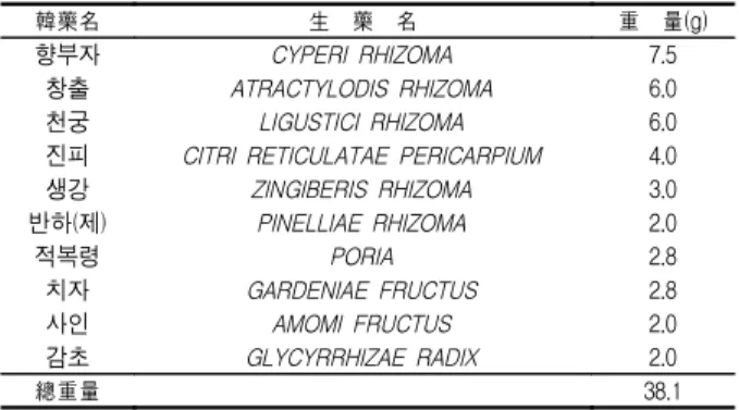
1. 육울탕에 의한 HeLa 세포의 증식억제 및 apoptosis 유발 인체자궁경부암 세포주인 HeLa 세포의 증식에 미치는 육울 탕의 영향을 알아보기 위하여 이를 72시간동안 농도별로 처리한 후 MTT assay를 이용하여 조사한 결과는 Fig. 1A에서 나타낸 바 와 같다. 결과에서 알 수 있듯이 처리 농도가 증가함에 HeLa 세 포의 증식이 억제되었으며 다양한 세포의 형태적 변형이 동반되 었다. 특히 농도가 증가할수록 세포의 밀도가 감소하면서 길고 분지를 형성하는 듯한 dendrite-like한 구조로 바뀌었으며, 부착력 을 상실하고 배지 위로 부유되는 경향성이 증가되었다(Fig. 1B).
Fig. 1. Inhibition on the cell viability and morphological changes by Yukwool-tang (YWT) treatment in HeLa human cervical carcinoma cells.
(A) Cells were seeds as described in materials and methods, and treated with various concentrations of YWT. After 72 h incubation, MTT assay was performed. The data shown are mean +/- SD of three independent experiments. (B and C) Exponentially growing cells were incubated with either vehicle alone various concentrations of YWT for 72 h. Cell morphology was visualized by inverted microscopy (Magnification, X200, B). The cells were stained with DAPI solution and then photographed by fluorescent microscope using blue filter. (Magnification, X400, C).다음은 육울탕의 처리에 의한 생존율 감소 및 형태변화가
apoptosis의 유발과 연관성이 있을 것으로 기대되어 육울탕 처리
후 HeLa 세포 핵의 형태변화를 조사하였다. Fig. 1C에 제시된 바 와 같이 정상 배지에서 자란 대조군에서는 핵의 형태에 아무런 변화가 보이지 않았지만 육울탕을 처리했을 경우는 처리 농도가 증가할수록 apoptosis 유발 특이적인 핵 내 DNA 단편화에 의한 염색질의 응축된 형태인 apoptotic body의 형성 정도가 증가되 었다. 또한 apoptotic body 형성과 함께 apoptosis 유발의 직접적 인 증거에 해당하는 DNA 단편화 여부를 조사한 결과, Fig. 2B의 결과에서처럼 육울탕의 처리에 따라 단편화의 정도 역시 증가되 었다. 따라서 육울탕 처리에 따른 apoptosis 유발의 정도를 정량적 으로 비교 평가하기 위하여 동일한 조건으로 배양된 세포들을 대 상으로 세포주기 sub-G1기에 해당하는 세포의 빈도 변화를 조사 한 결과, 육울탕의 처리 농도의 증가에 따라 sub-G1기에 해당하는 세포의 빈도가 증가하여 3 mg/ml 처리군에서는 대조군에 비하여 약 4배 증가되었음을 알 수 있었다(Fig. 2C). 이상의 결과는 육울 탕 처리에 따른 HeLa 세포의 생존율의 감소 및 형태 변화가 apoptosis 유발과 밀접한 연관성이 있음을 보여주는 결과이다.
Fig. 2. Induction of DNA fragmentation and increase of sub-G1 cell population by YWT treatment in HeLa cells.
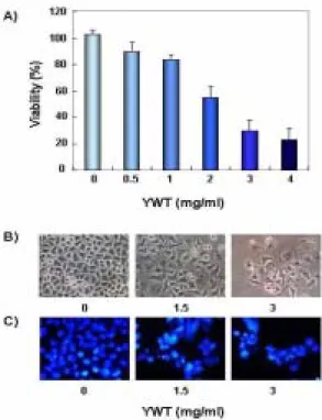
(A and C) The cells were exposed for 72 h to an increasing concentration of YWT. Then the cells were collected and stained with PI for flow cytometry analysis. The percentage of cells with hypodiploid DNA contents represent the fractions undergoing apoptotic DNA degradation. Each point represents the average of three independent experiments. (B) After YWT treatment for 72 h, the cells were collected and DNA was extracted. The fragmented DNA was separated on 1.5% agarose gel electrophoresis and visualized under UV light after stained with EtBr.2. p53 및 p21의 발현에 미치는 육울탕의 영향
다음은 육울탕 처리에 의한 HeLa 자궁경부암세포의 증식억 제 현상이 종양억제 유전자 또는 세포주기 조절 억제인자들의 발현 변화와 상관성이 있는지의 여부를 조사하기 위하여 현재까 지 알려진 종양억제 유전자 중 가장 중요한 p53 및 전체적인 세 포주기에 중요한 조절 역할을 하는 Cdk inhibitor p21의 발현에 미치는 육울탕의 영향을 조사하였다. Fig. 3의 결과에서 볼 수 있 듯이 p53 유전자의 경우는 전사 및 번역 수준에서 변화가 관찰 되지 않았지만 p21 유전자의 발현은 육울탕의 처리에 따라 전사 수준에서는 변화가 없었지만 번역 수준에서 증가하였음을 알 수 있었다. 또한 번역 수준에서 p53과 p21의 상대적인 발현 비율을
살펴 본 결과 p21은 p53에 비해 2.5배 정도 증가하는 것으로 확 인되었다. Cdk inhibitor인 p21은 p53의 발현 증가에 의하여 조 절될 수 있고 또한 p53 비의존적으로 발현이 조절될 수도 있으
며 9,10) , 암세포의 증식 억제, apoptosis 및 분화 유도에 중요한 역
할을 하는 세포주기 전반에 걸친 가장 중요한 조절인자란 점에
서 11-13) 육울탕의 처리에 의하여 p53의 발현과 상관없이 p21 유전
자가 증가되었다는 점은 매우 흥미로운 결과라고 사료된다. 그러 나 본 연구의 결과만으로 육울탕에 의한 인체자궁경부암세포의 세포주기 교란에 관한 명확한 증거는 제시할 수 없으며, 세포주 기 분석과 관련 유전자들의 kinase 활성도 검사 등을 통한 후속 적인 연구가 진행되어야 할 것이다.
Fig. 3. Induction of cyclin-dependent kinase p21(CIP1/WAF1) in YWT-treated HeLa cells.
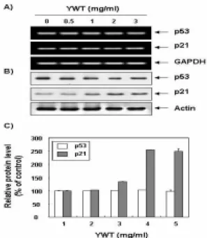
(A) After 72 h incubation with YWT, total RNAs were isolated and reverse-transcribed. The resulting cDNAs were subjected to PCR with p53 and p21 primers and the reaction products were subjected to electrophoresis in a 1% agarose gel and visualized by EtBr staining. GAPDH was used as an internal control. (B) The cellular proteins were separated by SDS-polyacrylamide gels and transferred onto nitrocellulose membranes. The membranes were probed with anti-p53 and anti-p21 antibodies. Proteins were visualized using an ECL detection system. Actin was used as an internal control. (C) Density of protein expression was determined by image analysis software. Each value indicates the mean of two separate experiments.3. Caspase의 발현 및 활성에 미치는 육울탕의 영향
Caspase는 세포의 apoptosis 유발에 핵심적인 역할을 하는
인자로서 세포 내의 핵과 mitochondria의 외막에 불활성 상태인
proenzyme 형태로 존재하다가 apoptosis를 유도하는 자극에 의
하여 활성화된 protease로 전환되어 직접 또는 간접적으로 세포
내에 존재하는 많은 표적 단백질의 분해에 관여한다 14) . 따라서
caspase의 활성화는 apoptosis의 유발에 대한 또 다른 증거가 될
수 있으며 많은 선행연구 등에서 검증되어 왔다 15) . 지금까지 알
려진 caspase 중에서 apoptosis가 유발에 상대적으로 중요한
caspase-3, -8 및 -9의 활성에 미치는 육울탕의 영향을 Western
bolt analysis를 통하여 조사하였다. Fig. 4A의 결과에서 볼 수 있
듯이 육울탕의 처리에 의한 caspase-8 단백질 발현의 변화가 없
었지만 caspase-3 및 -9의 경우는 육울탕 처리 농도 의존적으로 활성형 단백질의 발현이 증가하였다. 이러한 단백질 수준에서의 결과를 재확인하기 위하여 in vitro caspase activity assay를 통 하여 caspases의 활성 정도를 직접 분석한 결과, Fig. 4B에서 나 타난 바와 같이 육울탕 처리 농도 의존적으로 caspase-3의 활성 이 증가하는 것을 확인할 수 있었다. 하지만 caspase-8과 caspase-9의 활성 정도는 큰 변화가 관찰되지 않았으므로 육울탕 에 의한 HeLa 세포의 apoptosis 유발에 caspase-3의 활성화가 중 요한 역할을 함을 알 수 있었다.
Fig. 4. Activation of caspase-3 by YWT treatment in HeLa cells.
(A) The cells were treated with the indicated concentrations of YWT for 72 h and collected.Western blots were detected with indicated antibodies and ECL detection. (B) After 72 h incubation with YWT, aliquots (150 g protein) were incubated with substrates, DEVD-pNA, IETD-pNA and LEHD-pNA, for in vitro caspase-3, -8 and -9 activity, respectively, at 37℃ for 3 h. The released fluorescent products were measured. Data are means average of two separate experiments.
4. PARP, β-catenin 및 PLC-γ1의 발현에 미치는 육울탕의 영향 한편 Poly(ADP-ribose) polymerase (PARP) 단백질은 DNA repair나 genomic stability의 유지에 매우 중요한 역할을 하지만, apoptosis 유발과정 중 활성화된 caspase에 의하여 분해가 일어 나면 PARP의 효소적 기능의 상실로 인하여 정상적인 DNA repair 과정이 억제되어진다. 정상적인 세포의 경우 PARP 단백 질은 116 kDa의 분자량을 가지지만 apoptosis가 일어난 경우 85 kDa 크기의 단편이 관찰되거나 주 band의 발현이 감소된다 16,17) . 또한 catenin family에 속하는 단백질(α, β 및 γ)은 모든 세포에 서 발현되는 세포 연접기능에 중요한 역할을 하는 것으로 알려 져 있는데, 특히 β-catenin은 세포내 골격의 유지와 다양한 부착 성 세포의 전사 조절에 중요하며 세포 유착과 관계된 apoptosis 조절과 연관성을 가지고 있다. 정상 세포의 경우 β-catenin은 92 kDa의 분자량을 가지나 세포 유착성 apoptosis가 일어나면 활성 화된 caspase에 의하여 62-72 kDa로 단편화가 일어난다 18,19) . 그 리고 phospholipase C-γ1 (PLC-γ1)은 phosphatidylinositol 4,5-bisphosphate를 hydrolyze시켜 protein kinase C activator인
diacylglycerol 및 세포 내 Ca 2+ 조절에 중요한 역할을 하는 inositol 1,4,5-trisphosphate (IP3)를 생산한다 20) . 따라서 phosphatidylinositol 3-kinase나 Ras와 같은 세포성장 신호분자 와 같이 PLC-γ1 역시 세포의 증식에 중심적인 역할을 하는 것으 로 알려져 있으나, apoptosis가 유발될 경우 활성화된 caspase 효 소에 의하여 PLC-γ1 단백질은 분해될 수 있기 때문에 상기 두 종류의 단백질과 함께 apoptosis 유발의 생화학적 표식자로 사용 이 가능하다 21) . 따라서 육울탕에 의한 HeLa 세포의 apoptosis 유 발 시 이러한 단백질들의 단편화가 동반되는지의 여부를 조사한 결과, Fig. 5A에서 나타낸 바와 같이 PARP의 경우는 정상 배지 에서 배양된 세포들은 116 kDa 위치에 강한 주 band를 볼 수 있 었고 육울탕의 처리 농도 의존적으로 85 kDa의 단편이 증가되는 것을 관찰할 수 있었다. β-catenin의 경우는 육울탕의 처리에 의한 62-72 kDa의 단편은 관찰되지 않았으나 주 band의 발현정도가 현 저히 감소하는 것으로 관찰되었다. 이는 육울탕에 의한 HeLa 세 포의 apoptosis가 세포 내 골격 및 부착 능력의 상실과 연관이 있 음을 알 수 있었다. 또한 육울탕의 처리 농도가 증가될수록 PLC-γ 1 단백질의 발현도 매우 빨리 감소되었음을 알 수 있었으며, 처리 농도 증가에 따른 발현의 감소 정도는 β-catenin과 매우 유사하였 다. 이상의 결과로 육울탕에 의한 인체자궁경부암세포의 apoptosis 유발에는 caspase의 활성화에 따른 이들 표적 단백질 의 분해 및 발현 감소가 중요한 역할을 하는 것으로 생각된다.
Fig. 5. Effects of YWT treatment on the expression of poly(ADP-ribose) polymerase, β-catenin, phospholipase C-γ1 and DNA fragmentation factor family proteins in HeLa cells. (
A) The cells were treated with the indicated concentrations of YWT for 72 h and collected. Western blots were detected with indicated antibodies and ECL detection. (B) Density of protein expression was determined by image analysis software. Each value indicates the mean of two separate experiments.5. DFF40/CAD 및 DFF45/IACD의 발현에 미치는 육울탕의 영향
Apoptosis의 가장 중요한 현상 중 하나가 DNA 단편화이며
여기에 관여하는 중요한 인자가 DNA fragmentation factor
(DFF)이다. DFF family는 caspase-activated DNase인 DFF40/CAD와 inhibitor of caspase-activated DNase인 DFF45/ICAD로 구성되어 있다. DFF40/CAD와 DFF45/ICAD는 서로 complex를 형성하고 있으며, apoptosis가 유발되면 caspases에 의하여 ICAD의 두 군데 Asp 잔기에서 단편화가 일 어나는 것으로 알려져 있다 22,23) . 따라서 DFF40/CAD 및 DFF45/ICAD가 육울탕의 처리에 따른 apoptosis와 연관이 있는 지를 조사한 결과, Fig. 5A에서 보는 바와 같이 DFF40/CAD의
경우는 고농도 처리군에서 약간 증가되었다. 그러나
DFF45/ICAD는 육울탕 처리농도 의존적으로 발현양의 감소가 관찰되었으며, DFF40/CAD와 DFF45/ICAD의 상대적인 발현 정 도를 조사해 본 결과 최고농도에서 약 4.8배 정도의 차이가 나타 났다(Fig. 5B). 즉 육울탕의 처리에 의한 HeLa 세포의 apoptosis 유발은 caspase의 활성화와 연관된 DFF45/ICAD의 상대적인 발 현양의 차이가 중요한 역할을 할 것으로 생각된다.
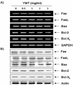
6. Fas/FasL system 및 Bcl-2 family의 발현에 미치는 육울탕의 영향
Apoptosis를 유발하는 경로는 크게 mitochondrial pathway 와 death receptor pathway의 두가지로 구분할 수 있는데, death receptor는 세포사멸의 기능을 갖는 tumor necrosis factor (TNF) 수용체군을 의미한다 24) . 현재 가장 잘 알려진 다섯 종류 중 하나 가 APO-1 또는 CD95라고도 알려진 Fas에 의한 신호전달계이다.
그리고 TNF 수용체군에 작용하는 대표적인 ligand 중의 하나가 FasL이다 25) . FasL는 type II transmembrane protein으로서 TNF 수용체 super-family인 Fas에 결합하여 apoptosis를 유발한다. 따 라서 육울탕에 의한 apoptosis 유발에 death receptor가 관여하 는지의 여부를 RT-PCR 및 Western blotting으로 조사한 결과는 Fig. 6에 나타낸 바와 같다. 결과에서 볼 수 있듯이 Fas 및 FasL 모두에서 전사 및 번역수준에서의 아무런 변화가 관찰되지 않았 다. 이는 HeLa 세포에 육울탕을 처리하였을 경우 death receptor pathway 중 Fas 및 FasL를 통한 apoptosis는 일어나지 않았음을 보여주는 결과라고 생각된다.
Bcl-2 family에 속하는 몇 가지 중요한 인자들은 apoptosis 유발 조절에 가장 대표적인 유전자인데 그 중 Bcl-2는 anti-apoptotic 분자로서 apoptosis의 유발을 억제하는 기능을 가 지며, Bax는 pro-apoptotic 분자로 Bax 단백질 발현의 증가는 apoptosis의 유발과 관계가 있는 것으로 알려져 있다 26) . 이들 두 유전자는 세포 내 소기관 중 mitochondria로부터의 cytochrome c를 유리시켜 cysteine-related protease인 caspase, 종양억제 유 전자인 p53, DNA의 단편화와 연관된 endonuclease 등의 활성을 조절한다 27) . 이들은 서로 dimer의 형태로 존재하며 그들의 발현 수준에 변화가 초래되면 apoptosis가 유발되는 것으로 알려져 있 다. 따라서 HeLa 세포에서 육울탕에 의한 apoptosis 유발에 이들 유전자가 관련되어 있는지의 여부를 RT-PCR 및 Western blotting으로 조사한 결과는 Fig. 6에 나타낸 바와 같이, Bax 및 Bcl-XL 유전자의 발현은 전사 및 번역수준에서는 거의 변화가 관찰되지 않은 반면, Bcl-2의 발현은 번역수준에서만 처리 농도
의존적으로 감소되었다. 이는 육울탕 처리에 의한 apoptosis이 유발에는 최소한 Bcl-2 family가 중요한 역할을 하고 있음을 의 미하는 것이며, Bcl-2의 발현 감소로 인한 apoptosis 유발 관련 효소들의 활성화가 이루어지고 있음을 시사하여 주는 것이다.
Fig. 6. Effects of YWT treatment on the expression of Fas/FasL system and Bcl-2 family members in HeLa cells.
(A) After 72 h incubation with YWT, total RNAs were isolated and reverse-transcribed. The resulting cDNAs were subjected to PCR with indicated primers and the reaction products were subjected to electrophoresis in a 1% agarose gel and visualized by EtBr staining. GAPDH was used as an internal control. (B) The cellular proteins were separated by SDS-polyacrylamide gels and transferred onto nitrocellulose membranes. The membranes were probed with indicated antibodies. Proteins were visualized using an ECL detection system. Actin was used as an internal control.결 론
본 연구에서는 인체자궁경부암 HeLa 세포를 대상으로 육울 탕에 의하여 항암효능의 기전을 조사하였다. 육울탕 처리에 의한 HeLa 세포의 증식억제는 p53 비의존적인 p21 단백질 발현의 증 가와 연관성이 있었으며, 육울탕의 처리에 의한 HeLa 세포의 증 식 억제는 apoptosis 유발과 밀접한 연관성이 있음을 핵의 응측 화, DNA의 단편화 및 sub-G1기 세포 빈도의 증가 등으로 확인 하였다. 육울탕의 처리는 HeLa 세포에서 특히 caspase-3의 활성 화를 유의적으로 유도하였으며, 이에 따른 caspase-3의 표적 기 질 단백질인 PARP, β-catenin 및 PLC-γ1의 단편화/분해 촉진이 관찰되었다. 육울탕 처리에 의한 apoptosis 유발은 또한 DFF 및 Bcl-2 family에 속하는 유전자들의 변화를 초래하였으나, Fas/FasL system에는 큰 변화가 없었다.
감사의 글
본 논문은 부산테크노파크 공모과제로 지원되는 연구비(과
제번호 2005QB015)로 조성되었습니다.
참고문헌