PREVENTION RESEARCH □ORIGINAL ARTICLE □
273 책임저자:황원덕, 614-052, 부산광역시 진구 양정동 산 45번지
동의대학교 한의과대학 내과학교실 Tel: 051-850-8625, Fax: 051-853-4306 E-mail: [email protected]
접수일:2007년 11월 7일, 게재승인일:2007년 12월 7일
Correspondence to:Won Deok Hwang
Department of Internal Medicine, Dongeui University College of Oriental Medicine, San 45, Yangjeong-dong, Jin-gu, Busan 614-052, Korea Tel: +82-51-850-8625, Fax: +82-51-853-4306
E-mail: [email protected]
동충하초 추출물에 의한 A549 인체폐암세포의 G2/M Arrest 유발
동의대학교 한의과대학 내과학교실, 1생화학교실 및 대학원 바이오물질제어학과
조원녕ㆍ최영현1ㆍ황원덕
Induction of G2/M Arrest of the Cell Cycle by Aqueous Extract of Cordyceps militaris in A549 Human Non-small-cell Lung Cancer Cells
Won Nyeong Cho, Yung Hyun Choi1 and Won Deok Hwang Department of Internal Medicine, Dongeui University College of Oriental Medicine,
1Department of Biochemistry, Dongeui University College of Oriental Medicine and Department of Biomaterial Control, Dongeui University Graduate School, Busan 614-052, Korea
Cordyceps militaris is a medicinal fungus, which has been used for patient suffering from cancer in Oriental medicine. It was previously reported that C. militaris extracts are capable of inhibiting tumor growth, however, the anti-poliferative effects of human cancer cells have not been poorly understood.
In this study, to elucidate the growth inhibitory mechanisms of human cancer cells by treatment of aqueous extract of C. militaris (AECM) we investigated the anti-proliferative effects of AECM in A549 human non-small-cell lung cancer cells. Exposure of A549 cells to AECM resulted in the growth inhibition in a dose-dependent manner as measured by MTT assay. The antiproliferative effect by AECM treatment in A549 cells was associated with morphological changes such as membrane shrinking and cell rounding up. AECM treatment induced G2/M arrest of the cell cycle and the levels of tumor suppressor p53 as well as cyclin-dependent kinase inhibitor p21(WAF1/CIP1) in a concentration-dependent manner. Taken together, these findings suggest that AECM-induced inhibition of A549 cell proliferation is associated with the induction of cell cycle arrest at G2/M phase via induction of p53 and p21, and C. militaris may have therapeutic potential in human lung cancer. (Cancer Prev Res 12, 273-280, 2007)
Key Words: Cordyceps militaris, A549, G2/M arrest, p53, p21
서 론
동충하초(冬蟲夏草)는 자낭균강(Ascomycetes), 맥각균목 (Clavicitales), 맥각균과(Clavicipitaceae)에 속하며 현재까지 전 세계적으로 약 800여종, 국내에는 78종 정도가 알려 져 있다.1) 동충하초는 포자가 곤충의 유충, 번데기 또는 성충 내로 침입하여 기주 안에서 내생균핵을 만든 후 밖 으로 자실체를 형성하고, 성숙된 자실체는 다시 포자를 형성함으로서 다른 곤충에 기생하는 특이한 생활주기를
가진다. 따라서 식물체를 기주로 하여 발생하는 영지버 섯, 표고버섯, 느타리버섯 등과는 전혀 다른 동물성과 식 물성 유효성분들이 같이 존재하는 버섯이다. 자실체를 형성하는 동충하초속균으로는 Cordycepes 속이 대표적이 며, 고대로부터 중국에서 결핵, 천식, 해독, 자양강장제 등의 한방약재로 사용되어온 대표적인 동충하초는 C.
militaris와 C. sinensis이다.2) 최근까지 본 연구와 관련 있는 결과들을 살펴보면, C. sinensis의 추출물 또는 유효성분들 은 수지상 세포의 성숙,3) 백혈병세포 증식억제효과,4,5) 폐암세포의 전이 억제 효과,6) 항염증효과,7) 암세포 세포
주기 교란 및 apoptosis 유도 효과,5,8∼12) 암세포의 혈관신 생억제작용13) 등이 있는 것으로 보고된 바 있다. 그리고 C. militaris 역시 세포독성 및 유전독성 억제효과,14) 항산 화성 및 항돌연변이 효과,15) 혈관신생 및 종양생성 억제 효과13) 및 고형암 성장 억제 및 면역활성 작용,16) apoptosis 유발에 의한 암세포증식억제 효과17,18) 등이 있는 것으로 알려져 있다. 그러나 현재까지 암세포의 세포주기 조절 측면에서의 연구는 거의 이루어진 바 없다.
한편 세포주기 조절은 1950년대에 Swift 및 Howard에 의해 G1, S, G2 및 M기에 대한 정의가 내려지면서 성립 되었다.19) 그 후 생화학 및 분자생물학적 실험방법들의 성장과 더불어 1980년대에 세포주기조절 인자들이 동정 되었으며, 많은 선행 연구자들은 이들 인자들의 합성, 인 산화 및 분해 과정 등의 연구를 통해서 이들이 전 세포 주기에 걸쳐 매우 다양하게 존재한다는 것이 알려지게 되었다.20) 이런 세포주기 조절 인자와 그 외의 분자들의 상호 작용에 의해 특정 세포의 증식이 조절되어지며, 또 한 그 인자들의 변화에 의해 억제 또는 증폭 되는 것이 다. 세포주기 조절 측면에서 암은 세포주기의 조절이 되 지 않는 세포의 집단으로 정의 내릴 수 있으므로, 암세포 의 세포주기 진행을 적절히 억제할 수 있는 물질은 항암 제로서의 탐색이 가능하다. 따라서 최근 천연물에 함유 되어 있는 암세포주기 억제 물질의 탐색은 암예방 및 항 암제 개발을 위한 기본 단계로 인식되어지고 있다.
본 연구에서는 인체 폐암세포의 세포주기 진행에서 동충하초 추출물이 미치는 영향을 조사하였으며, 몇 가 지 유의적인 결과를 얻었기에 이를 보고하고자 한다. 이 를 위하여 A549 세포주가 사용되었으며, 동충하초 추출 물 처리에 의한 A549 세포의 증식억제 효과, 형태변화, 세포주기 변화 및 세포주기 조절 주요 유전자들의 발현 에 미치는 동충하초 추출물의 영향을 비교하였다.
재료 및 방법
1. 암세포배양 및 동충하초 수용액의 추출 및 처리
A549 인체 폐암세포(A549 human non-small-cell lung cancer cell line)는 American Type Culture Collection (Rockville, MD, USA)에서 분주 받아 사용하였으며, 암세포의 배양을 위 해 90%의 RPMI-1640 배지(Gibco BRL, Grand Island, NY, USA), 10%의 우태아혈청(fetal bovine serum, FBS) 및 1%의 penicillin 및 streptomycin (Biofluids, Rockville, MD, USA)이 포함된 배지를 사용하여 37oC, 5% CO2 조건 하에서 배양 하였다. 본 연구에 사용된 동충하초(C. militaris)는 대전대 학교 한의과대학 부속 한방병원에서 공급받았으며 100
g을 1,000 ml의 증류수에 3시간 이상 끓인 후, 3,000 rpm 으로 20분간 원심 분리시켜 침전물을 제거하였다. 이를 다시 0.45μm의 여과지를 이용하여 부유 성분을 걸러낸 후 수용성분을 동결 건조하여 사용하였다. 동충하초 수 용액 추출물[aqueous extract of C. militaris (AECM)]의 처리 를 위하여 세포를 0.05% trypsin-EDTA를 이용하여 세포 배양용 페트리 접시로부터 부유시킨 다음 새로운 세포 배양용 페트리 접시에 6×10⁵ 개/ml 정도로 분주하여 24 시간 동안 안정화 시켰다. AECM을 세포에 처리하기 직 전 적정 농도로 성장배지에 첨가하여 녹인 다음, 0.22 μ m의 pore size를 가진 주사기용 필터유닛을 사용하거나 1회용 펌프 필터 유닛을 사용하여 미생물 및 불순물을 걸러낸 다음, 배지를 갈아주면서 직접 처리하였다.
3. MTT assay를 이용한 세포 증식률의 측정
세포 배양용 6 well plate에 A549 세포를 3×10⁴ 개/ml의 개수로 well 당 2 ml씩 분주하고 24시간 동안 안정화시킨 다음 AECM을 배지에 적정 농도로 처리한 후 48시간동 안 배양하였다. 48시간 후 배지를 제거하고 tetrazolium bromide salt (MTT, Amresco, Solon, Ohio, USA)를 0.5 mg/ml 농도가 되게 배지에 희석하여 2 ml씩 처리 후, 3시간동안 CO2 incubator에서 반응시킨 다음 MTT 시약을 제거하고 dimethylsulfoxide (DMSO)를 1 ml씩 처리하여 well에 생성 된 formazan을 모두 녹인 후 96 well에 200μl씩 옮겨서 ELISA reader (Molecular Devices, Sunnyvale, CA, USA)로 540 nm에서 흡광도를 측정하였다. 3번의 측정값을 평균값과 표준 오차를 Microsoft EXCEL program을 이용하여 분석하 였다.
4. DNA flow cytometry에 의한 세포주기 분석
준비된 세포들을 PBS로 두세 번 씻어내고, 고정액(70%
ethyl alcohol, 0.5% Tween 20)을 첨가하여 4oC에서 고정시 킨 후, propidium iodide (PI, concentration, 50μg/ml; Sigma) 와 10 kunit의 RNase (Sigma)를 처리하여 4oC에서 1시간동 안 염색하였다. 이를 다시 PBS로 두 번 씻어낸 후, nylon mesh로 세포를 하나씩 분리시킨 후 DNA flow cytometry (Becton Dickinson, San Jose, CA, USA)에 적용시켜 형광반 응에 따른 histogram을 ModiFit LT (Becton Dickinson) 프로 그램으로 분석하였다.
5. SDS-polyacrylamide gel 전기영동 및 Western blot analysis
세포배양용 패트리 접시에 6×10⁵ 개/ml 정도로 A549 세포를 분주하여 24시간 동안 안정화시킨 다음 AECM을
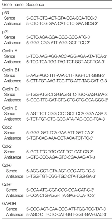
Table 1. Gene-specific primers for RT-PCR Gene name Sequence
p53 Sence Antisence
5'-GCT-CTG-ACT-GTA-CCA-CCA-TCC-3' 5'-CTC-TCG-GAA-CAT-CTC-GAA-GCG-3' p21
Sence Antisence
5'-CTC-AGA-GGA-GGC-GCC-ATG-3' 5'-GGG-CGG-ATT-AGG-GCT-TCC-3' Cyclin A
Sence Antisence
5'-TCC-AAG-AGG-ACC-AGG-AGA-ATA-TCA-3' 5'-TCC-TCA-TGG-TAG-TCT-GGT-ACT-TCA-3' Cyclin B1
Sence Antisence
5'-AAG-AGC-TTT-AAA-CTT-TGG-TCT-GGG-3' 5'-CTT-TGT-AAG-TCC-TTG-ATT-TAC-CAT G-3' Cyclin D1
Sence Antisence
5'-TGG-ATG-CTG-GAG-GTC-TGC-GAG-GAA-3' 5'-GGC-TTC-GAT-CTG-CTC-CTG-GCA-GGC-3' Cyclin E
Sence Antisence
5'-AGT-TCT-CGG-CTC-GCT-CCA-GGA-AGA-3' 5'-TCT-TGT-GTC-GCC-ATA-TAC-CGG-TCA-3' Cdc2
Sence Antisence
5'-GGG-GAT-TCA-GAA-ATT-GAT-CA-3' 5'-TGT-CAG-AAA-GCT-ACA-TCT-TC-3' Cdk2
Sence Antisence
5'-GCT-TTC-TGC-CAT-TCT-CAT-CG-3' 5'-GTC-CCC-AGA-GTC-CGA-AAG-AT-3' Cdk6
Sence Antisence
5'-ACG-GGT-GTA-AGT-GCC-ATC-TG-3' 5'-TGG-TGT-CGG-TGC-CTA-TGG-GA-3' Cdk6
Sence Antisence
5'-CGA-ATG-CGT-GGC-GGA-GAT-C-3' 5'-CCA-CTG-AGG-TTA-GAG-CCA-TC-3' GAPDH
Sence Antisence
5'-CGG-AGT-CAA-CGG-ATT-TGG-TCG-TAT-3' 5'-AGC-CTT-CTC-CAT-GGT-GGT-GAA-GAC-3' 처리하였다. 48시간까지 배양한 후, 세포를 PBS로 씻어
내고 다음 원심분리를 하여 세포를 모았다. 이렇게 모아 진 세포에 적당량의 lysis buffer [250 mM NaCl, 25 mM Tris-HCl (pH 7.5), 5 mM EDTA pH 8.0, 1% NP-40, 0.1 M phenymethylsulfonyl fluoride; PMSF, 1 M 1,4-dithio-DL- threitol; DTT, protease inhibitor cocktail]를 첨가하여 4oC에 서 30분간 반응시킨 후, 13,500 rpm으로 30분간 원심분리 하여 그 상층액을 취하였다. 상층액의 단백질 농도는 Bio-Rad 단백질 정량 시약(Bio-Rad, Hercules, CA, USA)의 사용법에 따라 동량으로 맞춘 다음 동량의 Laemmli sample buffer (β-melcaptomethanol 5%, Laemmli sample buffer 95%, Bio-Rad)를 섞어서 sample을 만들었다. 이렇게 만든 sample 동량을 sodium dodesyl sulfate (SDS) polyacrylamide gel 전기 영동으로 분리하였다.
단백질발현 분석을 위한 Western blot analysis를 위하여 분리된 단백질을 함유한 acrylamide gel을 nitrocellulose membrane (Schleicher and Schuell, Keene, NH, USA)으로 electroblotting에 의해 전이시킨 후, 10% skim milk를 함유 한 PBS-T (0.1% Tween 20 in PBS)에 담구어 상온에서 2시 간 정도 incubation하여 비특이적인 단백질들에 대한 blocking을 실시하고 PBS-T로 15분(5분간 3번씩) 정도 세 척하였다. 세척 후 1차 항체(PBS-T에 1:500 또는 1:
1,000으로 희석하여 사용)를 처리하여 상온에서 1시간 이상 또는 4oC에서 over night시킨 다음 PBS-T로 세척하 고 처리된 1차 항체에 적절한 2차 항체(PBS-T로 1:1500 으로 희석하여 사용)를 사용하여 상온에서 1시간 정도 반응시켰다. 다시 PBS-T로 세척하고 enhanced chemilu- minoesence (ECL) 용액(Amersham Life Science Corp., Arlington Heights, IL, USA)을 적용시킨 다음 암실에서 X-ray film에 감광시켜 특정단백질의 발현 양상을 비교 분석하였다.
본 실험에 사용된 항체들은 Santa Cruz Biotechnology Inc.
(Santa Cruz, CA, USA) 및 Calbiochem (Cambridge, MA, USA) 에서 구입하였으며, 2차 항체로 사용된 peroxidase-labeled donkey anti-rabbit immunoglobulin 및 peroxidase-labeled sheep anti-mouse immunoglobulin은 Amersham Corp.에서 구입하 였다.
6. Reverse transcription-polymerase chain re- action 분석
동일한 조건에서 배양된 A549 세포를 대상으로 RNAzol B (TEL-TEST, Inc., Texas, USA)를 이용하여 total RNA를 분리하였다. 분리된 RNA를 정량한 후, Choi 등21)의 방법 에 준하여 oligo dT primer와 AMV reverse transcriptase를
이용하여 2μg의 RNA에서 ss cDNA를 합성하였다. 이 cDNA를 template로 사용하여 관찰 대상 유전자를 poly- merase chain reaction (PCR) 방법으로 증폭하였다(Table 1).
이때 housekeeping 유전자인 glyceraldehyde-3-phosphate dehydrogenase (GAPDH)를 internal control로 사용하였다.
각 PCR 산물들을 1% agarose gel을 이용하여 전기영동하 고 ethidium bromide (EtBr, Sigma)로 염색한 후 UV 하에서 확인하였다.
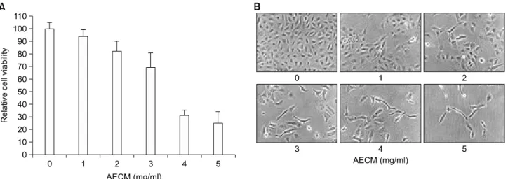
Fig. 1. Inhibition of proliferation and morphological changes of A549 human non-small-cell lung cancer cells following incubation with aqueous extract of C. militaris (AECM). (A) Cells were seeded as described in materials and methods, and treated with various concentrations of AECM. After 48 h incubation with AECM, the growth inhibition was measured by the metabolic-dye-based MTT assay. Results are expressed as the means±S.E. of three independent experiments. (B) Exponentially growing cells were incubated with AECM for 48 h. Cell morphology was visualized by inverted microscopy. Magnification, ×200.
Fig. 2. DNA-fluorescence histogram after AECM treatment in A549 human non-small-cell lung cancer cells. Ex- ponentially growing cells were incu- bated with various concentrations of AECM for 48 h. Cells were trpy- sinized and pellets were collected.
The cells were fixed and digested with RNase, and then cellular DNA was stained with PI. DNA flow cyto- metric cell cycle analysis was per- formed comparing untreated controls with cells treated with AECM. The data is a representative example for duplicate tests.
결과 및 고찰
1. AECM이 A549 세포의 증식에 미치는 영향
먼저 A549 세포의 증식에 미치는 AECM의 영향을 알아 보기 위하여 실험재료 및 방법에서 서술한 것처럼 인체 A549 세포를 적정 시간 안정화 시킨 후 48시간 동안 배 지에 AECM을 적정 농도로 희석하여 처리한 후, MTT assay를 실시하여 Fig. 1A에 나타내었다. Fig. 1A에 나타낸
바와 같이 48시간 동안 정상 배지에서 자란 A549 세포에 비하여 AECM이 함유된 배지에서는 AECM의 첨가 농도 의존적으로 세포의 증식이 감소하였음을 알 수 있었다.
즉 3.0 mg/ml 처리군의 경우 대조군에 비하여 약 30% 이 상 세포증식이 억제되었으며, 5.0 mg/ml 처리군에서는 약 75% 정도의 세포증식 억제현상을 관찰할 수 있었다.
그러나 동일 조건에서 수행된 선행연구인 U937 인체백 혈병세포의 결과와 비교해 볼 때, A549 세포는 다소 감 수성이 낮은 것으로 나타났다.10)
Fig. 3. Effects of AECM treatment on the levels of cyclins in A549 human non-small-cell lung cancer cells. (A) After 48 h incubation with AECM, total RNAs were isolated and reverse-transcribed. The resulting cDNAs were subjected to PCR with indicated primers, and the reaction products were subjected to electrophoresis in a 1% agarose gel and visualized by EtBr staining. GAPDH was used as an internal control. (B) The cells were lysed and then cellular proteins were separated by SDS-polyacrylamide gels and transferred onto nitrocellulose membranes. The membranes were probed with indicated antibodies. Proteins were visualized using an ECL detection system. Actin was used as an internal control.
Fig. 4. Effects of AECM treatment on the levels of Cdks in A549 human non-small-cell lung cancer cells. (A) After 48 h incubation with AECM, total RNAs were isolated and reverse-transcribed. The resulting cDNAs were subjected to PCR with indicated primers, and the reaction products were subjected to electrophoresis in a 1% agarose gel and visualized by EtBr staining. GAPDH was used as an internal control. (B) The cells were lysed and then cellular proteins were separated by SDS-polyacrylamide gels and transferred onto nitrocellulose membranes. The membranes were probed with indicated antibodies. Proteins were visualized using an ECL detection system. Actin was used as an internal control.
2. AECM이 A549 세포의 형태에 미치는 영향
AECM의 처리에 따른 상기 증식억제와 연관된 A549 세포의 형태변화를 조사하였다. 이를 위하여 AECM을 각 농도별로 처리하여 48시간 동안 배양한 후 위상차 현 미경을 이용하여 관찰하였다. Fig. 1B에서 나타난 바와 같이 AECM의 처리농도가 증가하면 할수록 A549 세포의 membrane shrinking 현상을 관찰할 수 있었으며, 밀도 감 소와 함께 세포 내 과립 형성과 같은 apoptosis가 유발되 었을 경우 관찰되는 세포변형 현상을 관찰할 수 있었다.
또한 정상 조건에서 배양된 세포에 비하여 AECM이 처 리된 경우 세포의 길이가 길어지고 denrite와 같은 분지 의 형성이 많아졌다. 따라서 인체 A549 세포의 AECM 처 리 농도 의존적인 형태적 변화와 밀도의 감소는 AECM
처리에 따른 세포 증식 억제와 부합되는 결과임을 알 수 있었다.
3. A549 세포의 세포주기 분포에 미치는 AECM의 영향
다음은 AECM의 처리에 의한 A549 세포의 증식억제가 세포주기 특정 시기의 교란 현상과 연관성이 있는지의 여부를 조사하였다. 이를 위하여 AECM이 함유된 배지 에서 48시간 동안 배양된 세포를 대상으로 DNA flow cytometry 분석을 실시하였으며, 그 결과는 Fig. 2에 나타 낸 바와 같다. 제시된 결과에서 알 수 있듯이 정상 배지 에서 자란 A549 세포의 경우 G1기가 전체의 약 64.16%
를 차지하고 있었으며, S기와 G2/M기에 속하는 세포가 각각 17.96% 및 17.02% 정도였다. 그리고 2.0 mg/ml의 AECM 농도에서 48시간 배양된 세포의 경우 G1기가
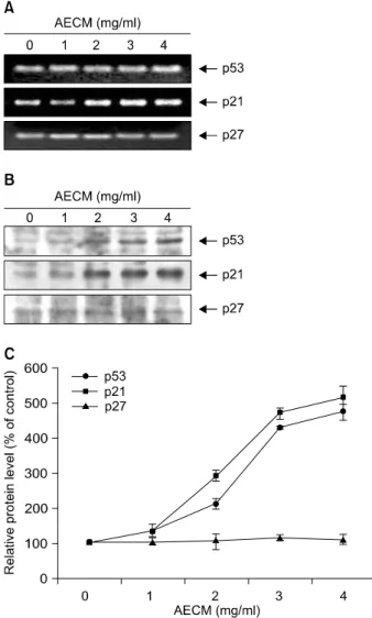
Fig. 5. Induction of tumor suppressor p53 and Cdk inhibitor p21 expression by AECM treatment in A549 cells. (A) After 48 h incubation with AECM, total RNAs were isolated and reverse-transcribed. The resulting cDNAs were subjected to PCR with p53 and p21 primers, and the reaction products were subjected to electrophoresis in a 1% agarose gel and visualized by EtBr staining. GAPDH was used as an internal control. (B) The cells were lysed and then cellular proteins were separated by SDS-polyacrylamide gels and transferred onto nitrocellulose membranes. The membranes were probed with anti-p53 and anti-p21 antibodies. Proteins were visualized using an ECL detection system. Actin was used as an internal control and protein levels were normalized against actin (C).
70.55%로 다소 증가되었으며, S기와 G2/M기에 속하는 세포의 빈도는 약 4.86% 및 22.29%로 나타났다. 그러나 3.0 mg/ml 및 4.0 mg/ml 처리군의 경우 G2/M기에 속하는 세포의 빈도는 각각 38.65% 및 39.49%로서 대조군에 비 하여 각각 약 2.27배 및 2.32배 증가되었으며, G1기 및 S기에 해당되는 세포의 빈도는 상대적으로 감소되는 경 향성을 보여 주었다. 즉 AECM의 처리에 의한 A549 세포 의 증식 억제는 세포주기 G2/M arrest와 직접적인 연관성 이 있음을 알 수 있었다. 또한 apoptosis가 유발된 세포의 빈도로 추정될 수 있는 sub-G1기에 속하는 세포의 빈도 는 대조군에서 약 0.86%였으나, AECM의 처리 농도가 증 가할수록 매우 증가되어 3.0 mg/ml 및 4.0 mg/ml 처리군 에서 3.37% 및 11.03%로 각각 약 3.79배 및 12.83배 증가 되어 AECM의 처리에 의한 A549 세포의 증식억제는 G2/M arrest와 연관된 apoptosis 유발의 결과임을 알 수 있 었다.
4. Cylins 및 Cdks의 발현에 미치는 AECM의 영향
일반적으로 진핵세포에서 세포주기의 진행을 위해서 는 세포주기 특이적인 cyclin의 발현 증가가 우선적으로 이루어져야한다. 즉 mid G1에서 D-type cyclin (cyclin D1, D2 및 D3)의 발현을 필요로 하며, late G1에서 S기로의 진입을 위해서는 cyclin E의 발현이 증가되어야한다.22) 그 리고 S기에서 G2기로의 진입 및 G2기와 M기 진행 동안 에는 각각 cyclin A 및 B-type cyclin (cyclin B1 및 B2)의 발 현이 증가되어야한다. 그리고 이들 cyclin들은 특정 Cdks 와 특이적인 결합에 의하여 cyclin/Cdk complex를 형성하 여 세포주기진행을 조절한다.23) 특히 Cdk2는 cyclin A와 결합하여 S기와 G2기 동안 역할을 하는24,25) 반면 Cdc2는 cyclin B1과 결합하면 핵막의 histone H1과 lamin이 인산화 에 의해 kinase 활성이 증가하여 핵막의 붕괴 및 염색체 의 재배열이 일어나서 M기로 진행이 된다.26,27) 따라서 AECM의 처리에 의한 A549 세포의 G2/M arrest와 연관된 cyclins 및 cyclin-dependent kinases (Cdks)의 발현 변화를 조 사하기 위하여 RT-PCR 및 Western blot 분석을 실시하였 다. Fig. 3은 cyclins에 대한 결과로서 mRNA 수준의 경우 cyclin B1의 발현이 AECM의 처리에 따라 점차 감소되었 으며, 단백질 수준에서는 조사된 4가지 cyclin 중 cyclin E 를 제외하고 모두 AECM의 처리 농도 의존적으로 발현 이 감소되었음을 알 수 있었다. 그리고 Cdks의 경우 Cdk6 이 AECM 처리에 따라 단백질 수준에서 발현이 매우 감 소되었다(Fig. 4).
5. 종양억제유전자 p53 및 Cdk inhibitor p21의 발현 에 미치는 AECM의 영향
Cdk inhibitor는 cyclin/Cdk complex와 결합하여 그 활성을 억제하는 것으로 알려져 있는데,28) 특히 CIP/KIP 군에 속 하는 p21 및 p27은 종양 억제유전자인 p53에 의하여 활 성화되어 세포의 전반적인 증식을 억제할 수 있는 것으
로 알려져 있으나,29,30) 세포의 종류 및 환경의 조건에 따라 p53 비의존적인 경로를 통하여 활성화될 수도 있다.31∼34) 따라서 본 실험에서는 AECM의 처리에 의한 인체 폐암 세포의 증식억제 효과가 이들 두 유전자의 발현과 연관 성이 있는지를 RT-PCR 및 Western blot analysis를 통하여 조사하였다. Fig. 5의 결과에서 알 수 있듯이 p53은 AECM의 처리에 따라 전사수준에서의 변화는 거의 없었 으나, 단백질 수준에서의 발현은 AECM 처리에 따라 매 우 증가되었다. 그리고 p21의 경우 전사 및 번역 수준 모두에서 AECM의 처리에 따라 발현이 증가되었으며, 증가되는 경향성은 p53 단백질의 발현 양상과 유사하였 다. 그러나 동일 조건에서 p27의 발현은 유의적인 변화 가 관찰되지 않았다. 이상의 결과에서 AECM에 의한 세 포주기 G2/M arrest 유발에는 Cdk inhibitor p21의 발현 증 가와 연관성이 있을 것으로 추정되며, AECM에 의한 p21 의 발현이 p53 의존적인지의 여부는 정확하지 않다. 그 러나 p21 뿐만 아니라 p53 단백질의 발현 증가는 세포주 기 진행 억제뿐만 아니라 apoptosis 유발에서도 중요한 역 할을 할 수 있으므로 AECM에 의한 p53의 발현 증가는 A549 세포의 증식억제에 중요한 작용을 할 것으로 생각 된다.
결 론
본 연구에서는 동충하초 열수 추출물의 처리에 의한 인체폐암세포의 증식 억제에 관한 기전 해석을 시도하 였다. 이를 위하여 A549 세포주가 사용되었으며 다음과 같은 결과를 얻었다. AECM의 처리 농도 의존적으로 A549 세포의 증식율이 현저하게 감소되었으며, 이러한 암세포 증식억제 효과는 암세포의 심한 형태적 손상과 연관이 있었다. AECM 처리에 의한 A549 세포의 증식억 제에는 세포주기 G2/M arrest 및 apoptosis 유발에 의한 것 이었다. AECM 처리에 의한 세포주기 G2/M arrest는 전사 수준에서의 cyclin B1 및 Cdk6의 발현 감소와 연관성이 있었다. 또한 AECM의 처리 농도 의존적으로 A549 세포 에서 종양억제 유전자 p53 및 Cdk inhibitor p21의 발현이 매우 증가되었다.
참 고 문 헌
1) Sung JM. Lee HK, Choi YS, Kim YO, Kim SH, Sung GH.
Distribution and taxonomy of entomopathogenic fungal species from Korea. Kor J Mycol 25, 231-252, 1997.
2) Buenz EJ, Bauer BA, Osmundson TW, Motley TJ. The
traditional Chinese medicine Cordyceps sinensis and its effects on apoptotic homeostasis. J Ethnopharmacol 96, 19-29, 2005.
3) Kim GY, Ko WS, Lee JY, Lee JO, Ryu CH, Choi BT, Park YM, Jeong YK, Lee KJ, Choi KS, Heo MS, Choi YH. Water extract of Cordyceps militaris enhances maturation of murine bone marrow-derived dendritic cells in vitro. Biol Pharm Bull 29, 354-360, 2006.
4) Chen JL, Greider CW. Telomerase RNA structure and function: implications for dyskeratosis congenita. Trends Bio- chem Sci 29, 183-192, 2004.
5) Buenz EJ, Weaver JG, Bauer BA, Chalpin SD, Badley AD.
Cordyceps sinensis extracts do not prevent Fas-receptor and hydrogen peroxide-induced T-cell apoptosis. J Ethnopharmacol 90, 57-62, 2004.
6) Nakamura K, Yamaguchi Y, Kagota S, Kwon YM, Shinozuka K, Kunitomo M. Inhibitory effect of Cordyceps sinensis on spontaneous liver metastasis of Lewis lung carcinoma and B16 melanoma cells in syngeneic mice. Jpn J Pharmacol 79, 335- 341, 1999.
7) Shahed AR, Kim SI, Shoskes DA. Down-regulation of apo- ptotic and inflammatory genes by Cordyceps sinensis extract in rat kidney following ischemia/reperfusion. Transplant Proc 33, 2986-2987, 2001.
8) Kuo YC, Weng SC, Chou CJ, Chang TT, Tsai WJ. Activation and proliferation signals in primary human T lymphocytes inhibited by ergosterol peroxide isolated from Cordyceps cica- dae. Br J Pharmacol 140, 895-906, 2003.
9) Zhang QX, Wu JY. Cordyceps sinensis mycelium extract induces human premyelocytic leukemia cell apoptosis through mitochondrion pathway. Exp Biol Med (Maywood) 232, 52-57, 2007.
10) Lee H, Kim YJ, Kim HW, Lee DH, Sung MK, Park T.
Induction of apoptosis by Cordyceps militaris through activation of caspase-3 in leukemia HL-60 cells. Biol Pharm Bull 29, 670-674, 2006.
11) Zhang Q, Wu J, Hu Z, Li D. Induction of HL-60 apoptosis by ethyl acetate extract of Cordyceps sinensis fungal mycelium.
Life Sci 75, 2911-2919, 2004.
12) Yang LY, Huang WJ, Hsieh HG, Lin CY. H1-A extracted from Cordyceps sinensis suppresses the proliferation of human mesangial cells and promotes apoptosis, probably by inhibiting the tyrosine phosphorylation of Bcl-2 and Bcl-xL. J Lab Clin Med 141, 74-83, 2003.
13) Yoo HS, Shin JW, Cho JH, Son CG, Lee YW, Park SY, Cho CK. Effects of Cordyceps militaris extract on angiogenesis and tumor growth. Acta Pharmacol Sin 25, 657-665, 2004.
14) Kim MN, Cui CB, Lee DS, Ham SS. Cytotoxicity and antigenotoxic effect of Cordycepes militaris extracts. J Kor Soc Food Sci Nutr 30, 921-927, 2001.
15) Kim MN, Oh SW, Lee DS, Ham SS. Antioxidative and antimutagenic effects of the ethanol extract from Cordycepes militaris. Kor J Posthavest Sci Technol 8, 109-117, 2001.
16) Lee H, Lee Y, Park T. Tumor growth inhibitory and
immunomodulatory activities of Cordycepes militaris water extracts in ICR mice bearing Sarcoma-180 solid tumor. J Kor Soc Food Sci Nutr 33, 59-65, 2004.
17) Hong SH, Kam CW, Choi YH, Park DI. Induction of apoptotic cell death by an aqueous extract of Cordyceps militaris in A549 human lung carcinoma cells. Kor J Oriental Physiol Pathol 18, 1102-1106, 2004.
18) Park C, Hong SH, Lee JY, Kim GY, Choi BT, Lee YT, Park DI, Park YM, Jeong YK, Choi YH. Growth inhibition of U937 leukemia cells by aqueous extract of Cordyceps militaris through induction of apoptosis. Oncol Rep 13, 1211-1216, 2005.
19) Howard A, Pelc SR. Synthesis of deoxyribonucleic acid in normal and irradiated cells and its relation to chromosome breakage. Heredity 6, 261, 1953.
20) Minshull J, Pines J, Golsteyn R, Standart N, Mackie S, Colman A, Blow J, Ruderman JV, Wu M, Hunt T. The role of cyclin synthesis, modification and destruction in the control of cell division. J Cell Sci 12, S77-S97, 1989.
21) Choi YH, Kong KR, Kim YA, Jung KO, Kil JH, Rhee SH, Park KY. Induction of Bax and activation of caspases during β-sitosterol-mediated apoptosis in human colon cancer cells.
Int J Oncol 23, 1657-1661, 2001
22) Weinberg RA. The retinoblastoma protein and cell cycle control. Cell 81, 323-330, 1995.
23) Elledge SJ, Harper JW. Cdk inhibitors: on the threshold of checkpoints and development. Curr Opin Cell Biol 6, 847-852, 1994.
24) Girard F, Strausfeld U, Fernandez A, Lamb NJ. Cyclin A is required for the onset of DNA replication in mammalian fibroblasts. Cell 67, 1169-1179, 1991.
25) Guadagno TM, Ohtsubo M, Roberts JM, Assoian RK. A link between cyclin A expression and adhesion-dependent cell cycle progression. Science 262, 1572-1575, 1993.
26) Krek W, Nigg EA. Differential phosphorylation of vertebrate p34cdc2 kinase at the G1/S and G2/M transitions of the cell cycle: identification of major phosphorylation sites. EMBO J 10, 305-316, 1991.
27) Ohsumi K, Katagiri C, Kishimoto T. Chromosome condensa- tion in Xenopus mitotic extracts without histone H1. Science 262, 2033-2035, 1993.
28) Harper JW. Cyclin dependent kinase inhibitors. Cancer Surv 29, 91-107, 1997.
29) Li Y, Jenkins CW, Nichols MA, Xiong Y. Cell cycle expression and p53 regulation of the cyclin-dependent kinase inhibitor p21. Oncogene 9, 2261-2268, 1994.
30) Taylor WR, Stark GR. Regulation of the G2/M transition by p53. Oncogene 20, 1803-1815, 2001.
31) Xiong Y, Hannon GJ, Zhang H, Casso D, Kobayashi R, Beach D. p21 is a universal inhibitor of cyclin kinases. Nature 366, 701-704, 1993.
32) Jiang H, Lin J, Su ZZ, Collart FR, Huberman E, Fisher PB.
Induction of differentiation in human promyelocytic HL-60 leukemia cells activates p21, WAF1/CIP1, expression in the absence of p53. Oncogene 9, 3397-3406, 1994.
33) Zeng YX, El-Deiry WS. Regulation of p21WAF1/CIP1 expression by p53-independent pathways. Oncogene 12, 1557- 1564, 1996.
34) Choi YH, Lee WH, Park KY, Zhang L. p53-independent induction of p21 (WAF1/CIP1), reduction of cyclin B1 and G2/M arrest by the isoflavone genistein in human prostate carcinoma cells. Jpn J Cancer Res 91, 164-173, 2000.