• 검색 결과가 없습니다.

Inhibitory Effect of Bee Venom Toxin on Lung Cancer NCI H460 Cells Growth Through Induction of Apoptosis via Death Receptor Expressions

N/A
N/A
Protected

Academic year: 2021

Share "Inhibitory Effect of Bee Venom Toxin on Lung Cancer NCI H460 Cells Growth Through Induction of Apoptosis via Death Receptor Expressions"

Copied!
10
0
0

로드 중.... (전체 텍스트 보기)

전체 글

(1)

Inhibitory Effect of Bee Venom Toxin on Lung Cancer NCI H460 Cells Growth Through Induction of

Apoptosis via Death Receptor Expressions

Keun Young Hur and Ho Sueb Song*

Department of Acupuncture & Moxibustion Medicine, College of Oriental Medicine, Gachon University

[Abstract]

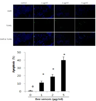
Objectives : I investigated whether bee venom inhibit cell growth through enhancement of death receptor expressions in the human lung cancer cells, NCI-H460.

Methods : Bee venom(1-5 μg/ml) inhibited the growth of NCI-H460 lung cancer cells by the induction of apoptotic cell death in a dose dependent manner.

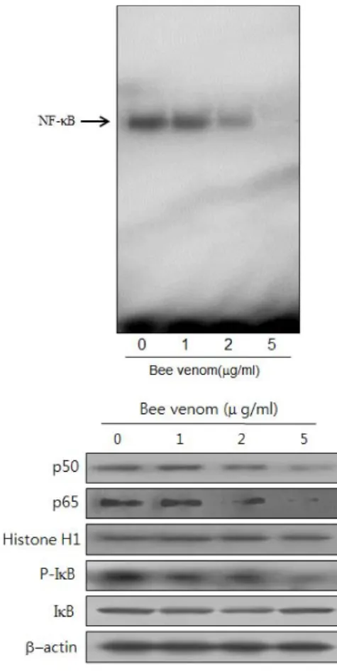
Results : Consistent with apoptotic cell death, expression of TNF-R1, TNF-R2, FAS, death receptors(DR) 3, 4, 5 and 6 was increased in the cells. Expression of DR downstream pro-apoptotic proteins including Caspase-8, -3, -9 was upregulated and Bax was concomitantly overwhelmed the expression of Bcl-2. NF-kB were inhibited by treatment with bee venom in NCI-H460 cells through TNF response change led by TNF-R1 and TNF-R2.

Conclusions : These results suggest that bee venom should exert anti-tumor effect through induction of apoptotic cell death in NCI-H460 human lung cancer cells via enhancement of death receptor expression, and that bee venom could be a promising agent for preventing and treating lung cancer.

Key words :

Bee venom;

Lung cancer;

NCI-H460;

Death receptor;

Apoptosis

Received : 2014. 02. 15.

Revised : 2014. 02. 24.

Accepted : 2014. 02. 24.

On-line : 2014. 03. 20.

※ This reserch was supported by the Gachon University Research Fund in 2013(GCU-2013-R421)

✱ Corresponding author : Department of Acupuncture & Moxibustion Medicine, Gil Oriental Medicine Hospital of Gachon Univercity, 1200-1, Guwol-dong, Namdong-gu, Incheon, 405-760, Republic of Korea

Tel : +82-70-7120-5012 Email : [email protected] ail.net

This is an Open-Access article distributed under the terms of the Creative Commons Attribution Non-Commercial License (http://creativecommons.org/licenses/by-nc/3.0) which permits unrestricted non-commercial use, distribution, and reproduction in any medium, provided the original work is properly cited.

The Acupuncture is the Journal of Korean Acupuncture & Moxibustion Medicine Society. (http://www.TheAcupuncture.or.kr)

Copyright © 2014 KAMMS. Korean Acupuncture & Moxibustion Medicine Society. All rights reserved.

(2)

Ⅰ. Introduction

The more aging population is increased, the higher lung cancer incidence rate becomes, which reaches about 30 % in patients over 70 years 1,2) . Small-cell lung cancer and non – small-cell lung cancer(NSCLC) is the most common type and it represents more than 80 % of the lung cancer 3) .

This is regarded to be the major cause of cancer mortality in the whole world, because of its difficulty of early diagnosis and high potential to invade locally and metastasize to distant organs 4,5) .

The incidence of lung cancer in Republic of Korea accounted for 11 %, ranking 4th, and the mortality due to it continued to increase to rank 1st position in cancer mortality, 2008 6) .

Although there are many therapeutic strategies including multimodality therapy, concurrent chemo- radiotherapy superior to radiotherapy or chemotherapy alone, high systemic toxicity and drug resistance prevent the successful outcomes and reveal their limitation in most cases 7-9) .

Therefore, developing novel alternatives for this malignancy is of great importance. Apoptosis, the programmed cell death plays critical role in anti-cancer effects of chemotherapeutics, which can be induced by various intracellular and extracellular stimuli via either an intrinsic or extrinsic pathway in different cells 10,11) .

In the extrinsic pathway, apoptosis is generally activated by death receptor(DR)s of tumor necrosis factor(TNF) family such as Fas, tumor necrosis factor receptor(TNFR)-1, TNFR-2, DR 3, DR 4, DR 5 and DR 6, which are implicated in various kinds of cancers 12-19) .

According to the several previous reports 20-23) , it is true that DR mediated apoptosis has emerged as an effective strategy for cancer therapy. However, certain types of cancer cells are intrinsically resistant to DR-mediated apoptosis. In an effort to identify agents that can sensitize NCI-H460 lung cancer cells to DR-induced apoptosis, I confirmed whether Bee Venom Toxin(BV) exerts anti-cancer

activity as an effective sensitizer of DR-mediated apoptosis.

Ⅱ. Materials and method

A. Materials

Bee venom was purchased from You-Miel Bee Venom Ltd(Hwasoon, Jeonnam, Korea). The composition of the bee venom was as follows: 45~50 % melittin, 2.5~3 % mast cell degranulating peptide, 12 % phospholipase A2, 1 % lysophospholipase A, 1~1.5 % histidine, 4~5 % 6-pentyl a-pyrone lipids, 0.5 % secarpin, 0.1 % tertiapin, 0.1 % procamine, 1.5~2 % hyaluronidase, 2~3 % amine, 4~5 % carbohydrate, and 19~27 % of others, including protease inhibitor, glucosidase, invertase, acid phosphomonoesterase, dopa- mine, norepinephrine, and unknown amino acids, with 99.5 % purity. all of the secondary antibodies such as Bax, Bcl-2, Caspase-3, -8, -9, Cleaved Caspase-3, -8, -9, TNF-R1, TNF-R2, FAS, DR 3, DR 4, DR 5 and DR 6 used in western blot analysis were purchased from Santa Cruz Biotechnology(Santa Cruz, CA). T4 polynucleotide kinase was obtained from Promega (Madison, WI). Poly(dI-dC), horseradish peroxidase- labeled donkey anti-rabbit secondary antibody, and ECL detection reagent were obtained from Amersham Pharmacia Biotech(Piscataway, NJ). Reagents for sodium dodecyl sulfate(SDS) – polyacrylamide gel elec- trophoresis were purchased from Bio-Rad(Hercules, CA).

B. Cell culture

The NCI-H460 lung cancer cell lines were purchased from the American Type Culture Collection(Manassas, VA). NCI-H460 cancer cells were cultured in RPMI- 1640 medium supplemented with 10 % fetal bovine serum(FBS) and penicillin/streptomycin(100 U/ml).

Cell cultures were then maintained at 37 ℃ in a

humidified atmosphere with 5 % CO 2 .

(3)

C. Cell viability assay

To determine the cell number, NCI-H460 lung cancer cells were plated in 24-well plates(5×10 4 cells/well), and subconfluent cells were subsequently treated with bee venom(1, 2 and 5 μ g/ml) for 24 hr.

After treatment, cells were trypsinized and pelleted by centrifugation for 5 min at 1,500 rpm, resuspended in 5 ml of phosphate-buffered saline(PBS), and 0.1 ml of 0.2 % trypan blue was added to the cancer cell suspension in each of the solutions(0.9 ml each). Subsequently, a drop of suspension was placed into a Neubauer chamber and the living cancer cells were counted. Cells that showed signs of staining were considered to be dead, whereas those that excluded trypan blue were considered viable. Each assay was carried out in triplicate.

D. Western blot analysis

Cells were homogenized with lysis buffer(50 mM Tris, pH 8.0, 150 mM NaCl, 0.02 % NaN3, 0.2 % SDS, 1 mM phenylmethylsulfonyl fluoride, 10 μ g/ml aprotinin, 1 % igapel 630 [Sigma], 10 mM NaF, 0.5 mM EDTA, 0.1 mM EGTA, and 0.5 % sodium deoxycholate) and centrifuged at 23,000 g for 1 hour. Equal amounts of proteins(80 μ g) were separated on SDS/12 %- polyacrylamide gels and then transferred to a nitro- cellulose membrane(Hybond ECL; Amersham Pharmacia Biotech). Blots were blocked for 2 hours at room temperature with 5 %(w/v) nonfat dried milk in Tris buffered saline(10 mM Tris, pH 8.0, 150 mM NaCl) containing 0.05 % Tween 20. The membrane was incubated for 5 hours at room temperature with specific antibodies: rabbit polyclonal for caspase-3, cleaved caspase-3, caspase-8, cleaved caspase-8, caspase-9, cleaved caspase-9, TNF-R1, TNF-R2, fas, DR 3, DR 4, DR 5, DR 6(1 : 1,000 dilution, Cell Signaling Technology, Inc, Beverly, MA), Bcl-2, Bax, goat polyclonal antibody to p50, p65(1 : 500 dilution, Santa Cruz Biotechnology, Inc) and phospho-IkBa(1 : 200, Santa Cruz Biotechnology, Inc). The blot was

then incubated with the corresponding conjugated anti-rabbit and anti-mouse immunoglobulin G-horse- radish peroxidase(1 : 2,000 dilutions, Santa Cruz Biotechnology, Inc). Immunoreactive proteins were detected with the ECL western blotting detection system.

E. Apoptosis evaluation

NCI-H460 lung cancer cells(2.5 × 10 5 cells/well) were cultured on 8-chamber slides. The cells were treated with bee venom(1, 2 and 5 μ g/ml). The cells were washed twice with PBS and fixed by incubation in 4 % paraformaldehyde in PBS for 1 hr at room temperature. Membrane was permeabilized by exposure to 0.1 % Triton X-100 in phosphate-buffered saline for 5 min at room temperature. TdT-mediated dUTP nick and labeling(TUNEL) assays were performed by using the in situ Cell Death Detection Kit(Roche Diagnostics GmbH, Mannheim, Germany) according to the manufacturer’s instructions. For 4'-6-Diamidino- 2-phenyl indole(DAPI) staining, slides were incubated for 15 min at room temperature in the dark with mounting medium for fluorescence containing DAPI (Vector Laboratories, Inc, Burlingame, CA). The cells were then observed through a fluorescence micro- scope(Leica Microsystems AG, Wetzlar, Germany).

F. Preparation of nuclear extracts and electromobility shift assays

It was performed according to the manufacturer’s

recommendations(Promega, Madison, WI). Briefly, 1 ×

10 6 cells/ml was washed twice with 1 × PBS, followed

by the addition of 1 ml of PBS, and the cells were

scraped into a cold Eppendorf tube. Cells were spun

down at 15,000 g for 1 min, and the resulting

supernatant was removed. Solution A(50 mM HEPES,

pH 7.4, 10 mM KCl, 1 mM EDTA, 1 mM EGTA, 1 mM

dithiothreitol, 0.1 μ g/ml phenylmethyl- sulfonyl

fluoride, 1 μ g/ml pepstatin A, 1 μ g/ml leupeptin,

10 μ g/ml soybean trypsin inhibitor, 10 μ g/ml aprotinin,

(4)

and 0.5 % Nonidet P-40) was added to the pellet in a 2 : 1 ratio(v/v) and allowed to incubate on ice for 10 min. Solution C(solution A + 10 % glycerol and 400 mM KCl) was added to the pellet in a 2 : 1 ratio(v/v) and vortexed on ice for 20 min. The cells were centrifuged at 15,000 g for 7 min, and the resulting nuclear extract supernatant was collected in a chilled Eppendorf tube. Consensus oligonucleotides were end-labeled using T4 polynucleotide kinase and [g -32 P] ATP for 10 min at 37 ℃ . Gel shift reactions were assembled and allowed to incubate at room temperature for 10 min followed by the addition of 1 μ l(50,000~200,000 cpm) of 32 P-labeled oligonu- cleotide and another 20 min of incubation at room temperature. For supershift assays, nuclear extracts from cells treated with bee venom(1~5 μ g/ml) were incubated with specific antibodies against the p50, p65, IkB and Rel-A NF- κ B isoformsp-IkB for 1 hr before EMSA. For competition assays, nuclear extracts from cells treated with bee venom(1~5 μ g/ml) were incubated with unlabelled NF- κ B oligonuclaotide (50×, 100× and 200×) or labeled SP-1(100×) and AP-1(100×) for 30 min before EMSA. Subsequently 1 μ l of gel loading buffer was added to each reaction and loaded onto a 6 % nondenaturing gel and electrophoresed until the dye was three-fourths of the way down the gel. The gel was dried at 80 ℃ for 1hr and exposed to film overnight at 70 ℃ . The relative density of the DNA-protein binding bands was scanned by densitometry using MyImage(SLB, Seoul, Korea), and quantified by Labworks 4.0 software(UVP Inc, Upland, California).

G. Colony formation assay

Anchorage-independent growth was determined by colony formation using the soft-agar method which investigate every cell in a given population for its ability to undergo unlimited division and form colonies.

Briefly, NCI-H460 cells(8×10 3 cells/ml) were suspended in 2 ml of 0.3 % agar containing basal medium Eagle’s agar containing 10 % FBS and colonies were then fed with growth media and bee venom(1, 2 or 5 μ g/ml).

Thereafter, the cultures were maintained at 37 ℃ in a 5 % CO 2 atmosphere for 2 weeks, and cell colonies >80 μ m in diameter were scored.

H. Data analysis

The data were analyzed using the GraphPad Prism 4 ver. 4.03 software(GraphPad Software, La Jolla, CA). Data are presented as mean±SD. The differences in all data were assessed by one-way analysis of variance(ANOVA). When the P value in the ANOVA test indicated statistical significance, the differences were assessed by the Dunnett’s test. A value of p<0.05 was considered to be statistically significant.

Ⅲ. Results

A. Effect of bee venom on cell growth in NCI-H460 lung cancer cells

To assess the inhibitory effect of bee venom on cell growth of lung cancer NCI-H460 cells, we analyzed cell viability by direct cell counting. The cells were treated with several concentrations of bee venom(1, 2, and 5 μ g/ml) for 24 hr. As shown in Fig. 1, bee venom inhibited cell proliferation of lung cancer cells in a concentration-dependent manner. Twenty-four hour treatment of bee venom inhibited NCI-H460 cell growth with IC 50 value of 3.14 μ g/ml(Fig. 1).

Morphologic observation showed that the cells were gradually reduced in size and changed into a small round single cell shape by the treatment of 5 μ g/ml of bee venom in NCI-H460 cells(Fig. 1).

B. Apoptotic cell death by bee venom in NCI-H460 lung cancer cells

To determine the inhibition of cell growth by bee

venom was due to the induction of apoptotic cell

death, we evaluated the changes in the chromatin

(5)

Fig. 1 Effect of bee venom on cell viability in NCI-H460 lung cancer cells

Concentration-dependent effect of bee venom was shown on the cell viability assay in lung cancer NCI-H460 cells. After treatment of bee venom(1, 2 and 5 μg/ml) for 24 hr, the cells were harvested by trypsinization and stained with 0.2 % trypan blue. Relative cell survival rate was determined by counting live and dead cells. The results were expressed as a percentage of viable cells. Morphologic observation with the treatment of bee venom in NCI-H460 cells. Columns, means of three experiments, with triplicates of each experiment; bars, SD.

* : p <0.05, significantly different from untreated control cells.

morphology of cells by using DAPI staining followed by TUNEL staining assays, and then the double labeled cells were analyzed by fluorescence microscope. Conversely well with cell growth inhibition, DAPI-stained TUNEL-positive cells were significantly increased in bee venom treated cells concentration dependently. The treatment of bee venom(5 μ g/ml) resulted in about 50 % induction of apoptotic cell death in NCI-H460 lung cancer cells(Fig. 2).

C. Expression of death

receptors(DRs) in NCI-H460 lung cancer cells by bee venom

Apoptosis can be induced by stimulation of DRs expression. Therefore, to investigate expression of DRs in cancer cells undergoing apoptotic cell death, we performed western blot analysis. The Results

Fig. 2 Effect of bee venom on apoptotic cell death

The lung cancer cells, NCI-H460 were treated with bee venom(1,2 and 5 μg/ml) for 24 hr, and then labeled with DAPI and TUNEL solution. Total number of cells in a given area was determined by using DAPI nuclear staining (fluorescent microscope). The green color in the fixed cells marks TUNEL- labeled cells. The apoptotic index was determined as the DAPI-stained TUNEL-positive cell number/total DAPI stained cell number(magnification, 200x). Columns, means of three experiments, with triplicates of each experiment; bars, SD.

* : p <0.05, significantly different from bee venom-untreated control cells.

showed that bee venom treatment increased TNF-R1, TNF-R2, FAS, DR 3, DR 4, DR 5 and DR 6 expres- sions in NCI-H460 cells(Fig. 3).

D. Effect of bee venom on the expression of apoptotic

regulatory proteins in NCI-H460 lung cancer cells

To investigate the relationship between the induc-

tion of apoptotic cell death and increase of DR expres-

sion, and the expression of their regulatory proteins

by bee venom, expression of apoptotic cell death

related proteins was investigated by western blots. The

expression of pro-apoptotic proteins overwhelmed anti-

apoptotic protein: Bcl-2 was decreased. However,

Bax, caspase-3, -8, -9, cleaved form of caspase-3,

(6)

Fig. 3. Effect of bee venom on death recptors expression in NCI-H460 lung cancer cells

Cells were treated with bee venom(1, 2, and 5 μg/ml) for 24 hr, and examined for expressions of TNF-R1, TNF-R2, FAS, DR 3, DR 4, DR 5, DR 6 by western blot analysis. β-actin was used as an control. Each band is representative for three experiments.

Fig. 4 Effect of bee venom on the expression of apoptosis regulatory proteins

Expression of apoptosis regulatory proteins was determined using western blot analysis. The NCI-H460 lung cancer cells were treated with different concentrations of bee venom(1, 2, and 5 μg/ml) for 24 hr. Equal amounts of total proteins(50 μg/lane) were subjected to 12 % or 8 % SDS-PAGE. Expression of Caspase-3, Caspase-8, Caspase-9, Cleaved Caspase-3,-8,-9, Bax, Bcl-2, and β-actin were detected by western blotting using specific antibodies. β -actin protein here was used as an internal control. Each band is representative for three experiments.

-8 and -9 was increased by treatment of bee venom in a concentration dependent manner(Fig. 4).

Fig. 5 Inhibition of NF-κB in lung cancer NCI-H460 cells by bee venom

Activation of NF-κB was determined by electrophoretic mobility shift assay(EMSA), as described in Materials and Methods. Nuclear extracts from lung cancer NCI-H460 cells with bee venom(1, 2, 5 μg/ml) were incubated in binding reactions of 32 P-labeled oligonucleotide containing the B sequence. NF-κB DNA binding activity was determined by EMSA. Equal amounts of total proteins(50 μg/lane) were subjected to 12 % or 8 % SDS-PAGE. Expression of p50, p65, IkB, p-IkB and β-actin were detected by western blotting using specific antibodies. β-actin protein here was used as an internal control. Each band is representative for three experiments.

E. Inhibition of NF-kB

NF- κ B is known to inhibitory transcription factor

of apoptosis. Whether to prevent anti-apoptotic

ability of NF- κ B is crucial for a agent causing

cancer cells go apoptosis. To determine the effect of

bee venom on the growth of lung cancer NCI-H460

cells through apoptosis, I assessed NF- κ B activity in

the cells treated for different concentration with for

24 hr bee venom by EMSA and observed NF- κ B

signal molecules in the cells by western blot analysis.

(7)

NF- κ B was highly activated in this cell. However, the activation of NF- κ B and its signal molecules were concentration-dependently decreased by the culture in the presence of bee venom in the cells(Fig. 5).

F. Effect of bee venom on the colony formation of lung cancer NCI-H460 cells

To determine the sensitivity of bee venom to lung cancer NCI-H460 cells, I next studied the ability of these 4 cell lines to form colonies on 6-well cell culture plates in the presence or absence of bee venom for 3 weeks. The number of colonies formed was reduced in a dose dependent manner, as shown in Fig. 6. At the highest concentration of bee venom(5 μ g/ml), colony formation was reduced over 90 % as compared to the untreated controls.

Fig. 6 Effect of bee venom on the colony formation of NCI-H460 lung cancer cells

NCI-H460 cells(8×10 3 cells/ml) were suspended in 2 ml of 0.3 % agar containing basal medium Eagle’s agar containing 10 % FBS and colonies were then fed with growth media and bee venom(1, 2 or 5μg/ml ). Thereafter, the cultures were maintained at 37 ℃ in a 5 % CO 2 atmosphere for 2 weeks, and cell colonies > 80 μm in diameter were scored.

Columns, means of three experiments, with triplicates of each experiment; bars, SD.

* : p <0.05, significantly different from untreated control cells.

Ⅳ. Discussion

Apoptosis, a programmed cell death can be induced by signaling the intrinsic mitochondrial mediated pathway or the extrinsic death receptor – related pathway, and interplay between these two

pathways 24) . Intrinsic pathway is majorly dependent upon mitochodrial membrane permeability, which is loosened by overwhelming Bax over Bcl-2, initiating subsequent intrinsic apoptosis including cytochrome c release from mitochondria, caspase-9 and -3 activation 25-29) . Meanwhile, extrinsic pathway is triggered by the binding of a DR to its ligands at the characteristic death domain(DD) including DR 1 to TNF ; DR 2 to FasL ; DR 3 to Apo3L ; DR 4 and DR5 to the tumor necrosis factor(TNF)-related apoptosis- inducing ligand(TRAIL, Apo2L), activating the initiator caspases such as caspase 8 and/or caspase 10 and affecting the intrinsic pathway as well 30,31) .

Several studies paid more attention to the extrinsic apoptotic pathway. Fucoidan induced apoptosis of HT-29 and HCT116 human colon cancer cells through enhanced expression of DRs 20) . α -tocopherol ether- linked acetic acid(a-TEA) induced apoptotic cell death via activation of Fas, DR 5 and Fas-associated protein with death domain(FADD) in human breast cancer cells 21) . Flavokawain B induced apoptosis via enhancing DR 5 expression in prostate cancer cell line 22) . Celastrol also enhanced anti- cancer effect of TNF-related apoptosis-inducing ligand(TRAIL)/Apo-2L via upregulation of DR 4 and DR 5 expression in human ovarian and colon cancer cells 23) . In addition, (E)-2,4-bis(p-hydroxyphenyl)-2-butenal diacetate exerted anticancer activity via inducing DR-mediated apoptosis in the lung cancer 32) .

From the above reports, targeting DR-mediated apoptosis has emerged as an effective strategy for cancer therapy. However, certain types of cancer cells are intrinsically resistant to DR-mediated apoptosis.

Therefor, in the present study, I found that the treatment of bee venom resulted in about 50 % induction of apoptotic cell death in NCI-H460 lung cancer cells(Fig. 2) and that bee venom subsequently increased expression of proapoptotic caspase-8 as well as caspase-9, -3(Fig. 4) via increase of Bax/Bcl-2 ratio and upregulation of the expression of FAS, DR 3, DR 4 DR 5 and DR 6(Fig. 3).

From the above, I confirmed that bee venom acts as

an effective sensitizer of DR-mediated and mito-

chondria-mediated apoptosis in NCI-H460 lung cancer

(8)

cells to DR-induced and intrinsic one as well, implying that bee venom exerts anti-proliferative influence upon in NCI-H460 human lung cancer cells via induction of intrinsic and extricsic apoptosis concomitantly.

TNF is a major mediator of carcinogenesis as well as inflammation 33) , which involves activation of an inflammatory or anti- apoptotic transcription factor NF- κ B as its strong initiator , binding to TNF-R1 and TNF-R2, members of the TNF receptor super- family with rich cysteine residues in their extra- cellular regions 34-36) . Although mostly, TNF-R1 plays major roles in regulating common TNF responses like cytotoxicity and proliferation through activation of NF- κ B 37,38) , it is also true that in some cell types, TNF-R2 can independently mediate similar cellular responses like it 39-41) . Among the participation of the two above receptors in the intracellular TNF responses, their involving molecular mechanisms underlying intracellular events of apoptosis in the various cancers brings more attention 42,43) .

In this study, both TNF-R1 and TNF-R2 of TNF receptor superfamilies were generally increased in their expressions together, compared to control(Fig. 3), inconsistent with the previous report 44) that expres- sion of TNF-R1 was selectively enhanced by bee venom with little change of TNF-R2 in A549 lung cancer cells. However, activity of NF- κ B was highly down regulated by bee venom in NCI-H460 lung cancer cells(Fig. 5) consistent with it. Taken together, it was suggested that bee venom should inhibit proliferation of NCI-H460 lung cancer cells through initiation of bilateral apoptosis via affection of TNF-R1 and TNF-R2 regulating TNF response resulting in upregulation of caspase family and concomitant downregulation of NF-kB. In the colony formation of NCI-H460 lung cancer cells to bee venom, it was additionally reconfirmed that bee venom inhibited over 90 % of colony formation of them, implying that bee venom deter the growth of them(Fig. 6).

Consequently, although further studies are needed to substantiate my investigation concretely, these present data provide that bee venom could be one of alternatives to enhance tumor growth inhibiting

ability of chemotherapeutics through overcoming the resistance in NCI-H460 human lung cancer via initiation of DR mediated apoptosis as well as mitichondrial mediated one.

Ⅴ. References

1. Gridelli C, Perrone F, Monfardini S. Lung cancer in the elderly. Eur J Cancer. 1997 ; 33(14) : 2313-4.

2. Havlik RJ, Yancik R, Long S, Ries L, Edwards B.

The National Institute on Aging and the National Cancer Institute SEER collaborative study on comorbidity and early diagnosis of cancer in the elderly. Cancer./ 1994 ; 74(7) : 2101-6.

3. Gridelli C, Massarelli E, Maione P et al. Potential role of molecularly targeted therapy in the management of advanced nonsmall cell lung carcinoma in the elderly. Cancer. 2004 ; 101(8) : 1733-44.

4. Casas F, Kepka L, Agarwal JP et al. Radiochemo- therapy in the elderly with lung cancer. Expert Rev Anticancer Ther. 2009 ; 9(10) : 1405-11.

5. De Petris L, Crino L, Scagliotti GV et al. Treat- ment of advanced non-small cell lung cancer.

Annals of Oncology. 2006 ; 17(2) : 1136 – 41.

6. Ministry for Health, Welfare and Family Affairs.

Annual report of cancer incidence(2007), cancer prevalence(2007) and survival(1993-2007) in Korea. Seoul : Ministry for Health, Welfare and Family Affairs. 2009.

7. Carney DN. Lung cancer: time to move on from chemotherapy. N Engl J Med. 2002 ; 346(2) : 126-8.

8. Herbst RS, Heymach JV, Lippman SM. Lung cancer.

N Engl J Med. 2008 ; 359(13) : 1367-80.

9. Giaccone G. Clinical perspectives on platinum re- sistance. Drugs. 2000 ; 59(4) : 9 – 17.

10. Fadeel B, Orrenius S. Apoptosis: a basic biological phenomenon with wide-ranging implications in human disease. J Int Med. 2005 ; 258(6) : 479-517.

11. Ghobrial IM, Witzig TE, Adjei AA. Targeting

apoptosis pathways in cancer therapy. CA-Cancer

(9)

J Clin. 2005 ; 55(3) : 178-94.

12. O'Donovan TR, O'Sullivan GC, McKenna S. In- duction of autophagy by drugresistant esophageal cancer cells promotes their survival and recovery following treatment with chemotherapeutics.

Autophagy. 2011 ; 7(5) : 509 – 24.

13. Kang YJ, Kim IY, Kim EH et al. Paxilline en- hances TRAIL-mediated apoptosis of glioma cells via modulation of c-FLIP, survivin and DR5. Exp Mol Med. 2011 ; 43(1) : 24 – 34.

14. Zhu DM, Shi J, Liu S, Liu Y, Zheng D. HIV infection enhances TRAIL-induced cell death in macrophage by down-regulating decoy receptor expression and generation of reactive oxygen species. PLoS One. 2011 ; 6(4) : 18291.

15. Yoshida T, Horinaka M, Sakai T. “Combination- oriented molecular -targeting prevention” of cancer: a model involving the combination of TRAIL and a DR5 inducer. Environ Health Prev Med. 2010 ; 15(4) : 203 – 10.

16. Inoue N, Matsuda F, Goto Y, Manabe N. Role of cell-death ligand-receptor system of granulosa cells in selective follicular atresia in porcine ovary. J Reprod Dev. 2011 ; 57(2) : 169 – 75.

17. Ashkenazi A. Targeting the extrinsic apoptosis pathway in cancer. Cytokine Growth Factor Rev.

2008 ; 19(3-4) : 325 – 31

18. Elrod HA, Sun SY. Modulation of death receptors by cancer therapeutic agents. Cancer Biol Ther.

2008 ; 7(2) : 163 – 73.

19. Sun SY. Understanding the role of the death receptor 5/FADD/caspase-8 death signaling in cancer metastasis. Mol Cell Pharm. 2011 ; 3(1) : 31 – 4.

20. Kim EJ, Park SY, Lee JY, Park JH. Fucoidan present in brown algae induces apoptosis of human colon cancer cells. BMC Gastroenterol.

2010 ; 10(8) : 96.

21. Li J, Yu W, Tiwary R et al. A-TEA-induced death receptor dependent apoptosis involves activation of acid sphingomyelinase and elevated ceramide- enriched cell surface membranes. Cancer Cell Int.

2010 ; 10(40) : 40-53.

22. Tang Y, Li X, Liu Z, Simoneau A, Xie J, Zi X.

Flavokawain B, a kava chalcone, induces apoptosis via up-regulation of death-receptor 5 and Bim expression in androgen-receptor negative, hormonal refractory prostate cancer cell lines and reduces tumor growth. Int J Cancer. 2010 ; 127(8) : 1758-68.

23. Zhu H, Liu XW, Ding WJ et al. Upregulation of death receptor 4 and 5 by celastrol enhances the anti-cancer activity of TRAIL/Apo-2L. Cancer Lett. 2010 ; 297(2) : 155-64.

24. Ghobrial IM, Witzig TE, Adjei AA. Targeting apoptosis pathways in cancer therapy. CA-Cancer J Clin. 2005 ; 55(3) : 178-94.

25. Green DR, Reed JC. Mitochondria and apoptosis.

Science. 1998 ; 28(8) : 1309 – 12.

26. Hengartner MO. The biochemistry of apoptosis.

Nature. 2000 ; 407(6805) : 770 – 6.

27. Reed JC. Apoptosis-regulating proteins as targets for drug discovery. Trends Mol Med. 2001 ; 7(7) : 314 – 19.

28. Li JY, Xu ZJ, Tan MY, Su WK, Gong XG. 3- (4-(Benzo[d]thiazol-2-yl)-1-phenyl-1Hpyrazol- 3-yl) phenyl acetate induced HepG2 cell apoptosis through a ROS-mediated pathway. Chem Biol Interact. 2010 ; 183(3) : 341 – 48.

29. Cain K, Brown DG, Langlais C, Cohen GM. Caspase activation involves the formation of the aposome, a large(approximately 700 kDa) caspaseactivating complex. J Biol Chem 1999 ; 274(32) : 22686 – 92.

30. Costantini P, Jacotot E, Decaudin D, Kroemer G.

Mitochondrion as a novel target of anticancer chemotherapy. J Natl Cancer Inst. 2000 ; 92(13) : 1042 – 53.

31. Wallach D, Varfolomeev EE, Malinin NL, Goltsev YV, Kovalenko AV, Boldin MP. Tumor necrosis factor receptor and Fas signaling mechanisms.

Annu Rev Immunol. 1999 ; 17(1) : 331 – 7.

32. Lee US, Ban JO, Yeon ET et al. Growth Inhibitory Effect of(E)-2,4-bis(p-hydroxyphenyl)-2-Butenal Diacetate through Induction of Apoptotic Cell Death by Increasing DR3 Expression in Human Lung Cancer Cells. Biomol Ther. 2012 : 20(6) : 538-43.

33. Old LJ. Tumor necrosis factor. Sci Am. 1988 ;

258(5) : 59-60.

(10)

34. Tartaglia LA, Weber RF, Figari IS, Reynolds C, Palladino MA Jr, Goeddel DV. The two different receptors for tumor necrosis factor mediate distinct cellular responses. Proc Natl Acad Sci.

1991 ; 88(20) : 9292-6.

35. Beutler B, Van Huffel C. Unraveling function in the TNF ligand and receptor families. Science.

1994 ; 264(5159) : 667-8.

36. Smith CA, Farrah T, Goodwin RG. The TNF receptor superfamily of cellular and viral proteins:

activation, costimulation, and death. Cell. 1994 ; 76(6) : 959-62.

37. Schutze S, Potthoff K, Machleidt T, Berkovic D, Wiegmann K, Kronke M. TNF activates NF-kappa B by phosphatidylcholine-specific phospholipase C-induced “Acidic” sphingomyelin breakdown.

Cell. 1992 ; 71(5) : 765-76.

38. Laegreid A, Medvedev A, Nonstad U et al. Tumor necrosis factor receptor p75 mediates cell -specific activation of nuclear factor kappa B and induction of human cytomegalovirus enhancer. J Biol Chem. 1994 ; 269(1) : 7785-91.

39. Mukhopadhyay A, Suttles J, Stout RD, Aggarwal BB. Genetic deletion of the tumor necrosis factor receptor p60 or p80 abrogates ligand-mediated activation of nuclear factor-kappa B and of mitogen-

activated protein kinases in macrophages. J Biol Chem. 2001 ; 276(34) : 31906-12.

40. Grell M, Becke FM, Wajant H, Mannel DN, Scheurich P. TNF receptor type 2 mediates thymocyte proliferation independently of TNF receptor type 1. Eur J Immunol. 1998 ; 28(1) : 257-63.

41. Medvedev AE, Espevik T, Ranges G, Sundan A.

Distinct roles of the two tumor necrosis factor (TNF) receptors in modulating TNF and lympho- toxin alpha effects. J Biol Chem. 1996 ; 271(16) : 9778-84.

42. Rothe M, Sarma V, Dixit VM, Goeddel DV. TRAF2- mediated activation of NF-kappa B by TNF receptor 2 and CD40. Science. 1995 ; 269(5229) : 1424-7.

43. Rothe M, Wong SC, Henzel WJ, Goeddel DV. A novel family of putative signal transducers associated with the cytoplasmic domain of the 75 kDa tumor necrosis factor receptor. Cell. 1994 ; 78(4) : 681-92.

44. Jang DM, Song HS. Inhibitory Effects of Bee Venom on Growth of A549 Lung Cancer Cells via Induction of Death Receptors. Journal of Korean Acupuncture & Moxibustion Medicine Society.

2013 ; 30(1) : 57-70.

수치

Fig.  1  Effect  of  bee  venom  on  cell  viability  in  NCI-H460 lung cancer cells
Fig.  4  Effect  of  bee  venom  on  the  expression  of apoptosis regulatory proteins
Fig.  6  Effect  of  bee  venom  on  the  colony  formation of NCI-H460 lung cancer cells

참조

관련 문서

It also suggest that cell proliferation inhibits by a novel signal transduction for adenosine effect in human FaDu hypopharynx squamous cell carcinoma cells....

Inhibitory effects of classified methanol extracts of Smilax china L on the COX-2 and iNOS expression of human colorectal cancer cell lines... Inhibitory

Glutamate excitotoxicity induced by excessive activation of NMDA receptor causes various damage to cells, which leads to cell death.. In previous studies, increased ROS

Effect of cell migration of JMJD6 transcript variants over- expressed MCF-7 cells using Transwell.. Effect of cell migration of JMJD6 transcript variants

PI3K inhibition decreased antioxidants/GD-induced apoptosis in A549 cells, and PI3K inhibitor LY294002 had inhibitory effect on antioxidants/GD-induced caspase-3

These results suggest that the bilobalide inhibits the cell proliferation and induces the apoptotic cell death in FaDu human pharyngeal squamous cell carcinoma via both

In the present study we investigated the proliferation effect of human oral cancer KB cell treated with pulsatilla koreana extract.. We analyzed the effects of this

Taken together, these results suggested that latex containing the ficin inhibited the cell growth and induced apoptosis by caspase and Bcl-2 family signaling pathway in