저작자표시-비영리-동일조건변경허락 2.0 대한민국 이용자는 아래의 조건을 따르는 경우에 한하여 자유롭게
l 이 저작물을 복제, 배포, 전송, 전시, 공연 및 방송할 수 있습니다. l 이차적 저작물을 작성할 수 있습니다.
다음과 같은 조건을 따라야 합니다:
l 귀하는, 이 저작물의 재이용이나 배포의 경우, 이 저작물에 적용된 이용허락조건 을 명확하게 나타내어야 합니다.
l 저작권자로부터 별도의 허가를 받으면 이러한 조건들은 적용되지 않습니다.
저작권법에 따른 이용자의 권리는 위의 내용에 의하여 영향을 받지 않습니다. 이것은 이용허락규약(Legal Code)을 이해하기 쉽게 요약한 것입니다.
Disclaimer
저작자표시. 귀하는 원저작자를 표시하여야 합니다.
비영리. 귀하는 이 저작물을 영리 목적으로 이용할 수 없습니다.
동일조건변경허락. 귀하가 이 저작물을 개작, 변형 또는 가공했을 경우 에는, 이 저작물과 동일한 이용허락조건하에서만 배포할 수 있습니다.
의학석사 학위논문
The reproducibility and usefulness of motor unit number index (MUNIX) using abductor digiti minimi and tibialis
anterior muscles in ALS patients
근위축성 측삭경화증 환자에서 소지외전근 및 전경골근을 이용한 MUNIX 검사의 재현성과 유용성에
관한 연구
2014 년 2 월
서울대학교 대학원 의학과 뇌신경과학 전공
신 제 영
의학석사 학위논문
The reproducibility and usefulness of motor unit number index (MUNIX) using abductor digiti minimi and tibialis
anterior muscles in ALS patients
근위축성 측삭경화증 환자에서 소지외전근 및 전경골근을 이용한 MUNIX 검사의 재현성과 유용성에
관한 연구
2014 년 2 월
서울대학교 대학원 의학과 뇌신경과학 전공
신 제 영
The reproducibility and usefulness of motor unit number index (MUNIX) using abductor digiti minimi and tibialis
anterior muscles in ALS patients
지도 교수 이 상 건
이 논문을 의학석사 학위논문으로 제출함
2013년 10월
서울대학교 대학원
의학과 뇌신경과학 전공
신 제 영
신제영의 의학석사 학위논문을 인준함
2014년 1월
위 원 장 홍 윤 호 (인) 부위원장 이 상 건 (인) 위 원 채 종 희 (인)
학위논문 원문제공 서비스에 대한 동의서
본인의 학위논문에 대하여 서울대학교가 아래와 같이 학위논문 제공하는 것에 동의합니다.
1. 동의사항
① 본인의 논문을 보존이나 인터넷 등을 통한 온라인 서비스 목적으로 복제할 경우 저작물의 내용을 변경하지 않는 범위 내에서의 복제를 허용합니다.
② 본인의 논문을 디지털화하여 인터넷 등 정보통신망을 통한 논문의 일부 또는 전부의 복제․배포 및 전송 시 무료로 제공하는 것에 동의합니다.
2. 개인(저작자)의 의무
본 논문의 저작권을 타인에게 양도하거나 또는 출판을 허락하는 등 동의 내용을 변경하고자 할 때는 소속대학(원)에 공개의 유보 또는 해지를 즉시 통보하겠습니다.
3. 서울대학교의 의무
① 서울대학교는 본 논문을 외부에 제공할 경우 저작권 보호장치(DRM)를 사용하여야 합니다.
② 서울대학교는 본 논문에 대한 공개의 유보나 해지 신청 시 즉시 처리해야 합니다.
논문 제목: The reproducibility and usefulness of motor unit number index (MUNIX) using abductor digiti minimi and tibialis anterior muscles in ALS
patients
학위구분: 석사 þ · 박사 □ 학 과: 의학과 뇌신경과학 학 번: 2009-21840 연 락 처:
저 작 자: 신 제 영 (인) 제 출 일: 2014 년 1 월 29 일
서울대학교총장 귀하
i
ABSTRACT
Introduction: Amyotrophic lateral sclerosis (ALS) is a neurodegenerative disorder characterized by progressive loss of motor neuron. The motor unit number index (MUNIX) is a novel diagnostic technique developed to quantify axonal loss in ALS patients. The objective of this study was to establish the reproducibility and usefulness of the MUNIX using the abductor digiti minimi (ADM) and tibialis anterior (TA) muscles in ALS patients and normal controls.
Methods: The MUNIX values were obtained from bilateral ADM and TA muscles in 30 ALS patients and 27 normal controls. The MUNIX, compound muscle action potential (CMAP), Medical Research Council (MRC) sum score, and ALS functional rating scale (ALSFRS) were evaluated, and their correlations were examined by using Pearson correlation analysis. All muscles were recorded twice to assess the reproducibility and coefficient of variation (COV). The mean values of all 4 (bilateral ADM and TA) muscles were obtained, and their correlations with functional status were analyzed.
Results: The mean values of CMAP amplitude and MUNIX showed no significant difference. The COVs for CMAP and MUNIX were within the acceptable range of less than 20%. There was a significant correlation between MUNIX and CMAP amplitude in both ADM (r = 0.918, P < 0.01) and TA (r = 0.850, P < 0.01) muscles in ALS patients. In normal controls, there was also a significant correlation between MUNIX and CMAP amplitude in both ADM (r = 0.629, P < 0.01) and TA (r = 0.894, P < 0.01)
ii
muscles. In ALS patients, MUNIX values significantly correlated with ALSFRS scores in the ADM (r = 0.439, P = 0.015), but not in the TA (r = 0.357, P= 0.053). There was a significant correlation between MUNIX and MRC sum score in the TA (r = 0.474, P < 0.01), but not in ADM (r = 0.349, P
= 0.058). Using the mean values of all 4 muscles, there was a more significant correlation between CMAP and MRS sum score (r = 0.480, P < 0.01), and between CMAP and ALSFRS (r = 0.517, P < 0.01). There was also a more significant correlation between MUNIX and MRS sum score (r = 0.493, P <
0.01) and between MUNIX and ALSFRS (r = 0.481, P < 0.01).
Conclusions: This study demonstrated the reproducibility of the MUNIX using ADM and TA muscles. Both ADM and TA muscles showed good correlations, but their patterns of correlations in the MUNIX were slightly different. The overall mean value of the 4 muscles was more useful for assessing functional status and disease progression. Further prospective follow-up studies with a larger sample size are needed to confirm our results.
---
Keywords: ALS, compound muscle action potential, MUNIX, abductor digiti minimi, tibialis anterior
Student number: 2009-21840
iii
CONTENTS
Abstract ... i
Contents... iii
List of tables and figures ... iv
Introduction ... 1
Methods ... 4
Results ... 7
Discussion ... 17
References ... 21
Abstract in Korean... 25
iv
LIST OF TABLES AND FIGURES
Table 1. The characteristics of ALS patients and healthy controls ... 9 Table 2. The demographics of ALS patients ... 10 Table 3. The results of CMAP and MUNIX of the ADM muscle in ALS patients ... 11 Table 4. The results of CMAP and MUNIX of the TA muscle in ALS patients ... 12 Table 5. The reproducibility of MUNIX measurements in ALS
patients and normal controls ... 13
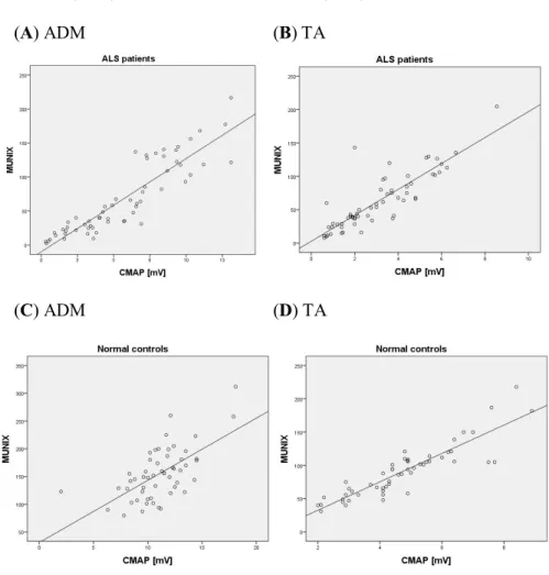
Figure 1. MUNIX values plotted against the CMAP amplitude of the
muscle tested in ALS patients and normal controls – bilateral ADM
muscles (A, C) and bilateral TA muscles (B, D). ... 14
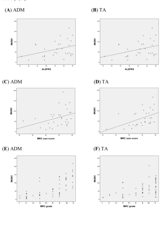
Figure 2. The mean MUNIX value of bilateral muscles plotted against
the ALSFRS, MRC sum score (C, D). MUNIX values are plotted
against the power levels of the ADM and TA muscles graded on the
MRC scale (E, F). ... 15
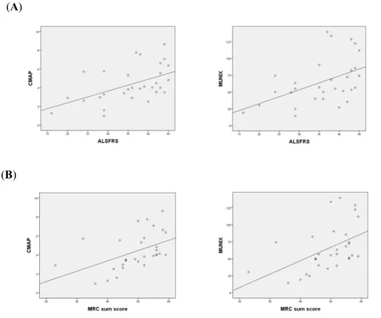
Figure 3. The mean CMAP and MUNIX values of 4 muscles plotted
against the ALSFRS (A) and MRC sum score (B) ... 16
v
LIST OF ABBREVIATIONS
abductor digiti minimi, ADM amyotrophic lateral sclerosis, ALS
amyotrophic lateral sclerosis functional rating scale, ALSFRS coefficient of variation, COV
compound muscle action potential, CMAP Medical Research Council, MRC
motor unit number estimation, MUNE motor unit number index, MUNIX
surface electromyographic interference pattern, SIP
tibialis anterior, TA
1
INTRODUCTION
Amyotrophic lateral sclerosis (ALS) is a neurodegenerative disorder characterized by progressive loss of upper and lower motor neurons, with a median survival of 2-3 years (1). The incidence of ALS is fairly uniform at about 2-6 per 100,000 person-years (2). There is no standard treatment for ALS. Prediction of prognosis and survival is important for the appropriate treatment of individual patients.
There are few diagnostic studies for evaluating disease progression or predicting progression. Although the needle electromyography (EMG) technique is a gold standard for the diagnosis of ALS, it is difficult to apply in the early stages of disease and is not efficient for the follow up of disease progression. EMG is also partially invasive and time-consuming method. For the quantification of motor unit loss of an ALS patient, the motor unit number estimation (MUNE) technique had been used. The MUNE technique has the advantage of revealing motor neuron loss in the early stages of ALS, while the compound muscle action potential (CMAP) amplitude does not change until 50% of motor units are lost. Unfortunately, the MUNE technique is difficult to perform and time consuming, allowing only a few muscles to be studied in a clinical setting (3).
The motor unit number index (MUNIX) is a novel electrophysiologic technique that can be used to study the number of motor units in a muscle.
The major advantages of the MUNIX technique are that it is noninvasive, quick to perform, and technically not very challenging and that it can be
2
applied to any muscle in which CMAPs after supramaximal electrical stimulation can be evoked(4). MUNE and MUNIX values have been shown to decline in ALS patients more rapidly than strength, and to predict disease course. Therefore, these techniques are more useful for predicting disease progression rather than for predicting the early stages of ALS (5),(6).
Recently, some studies have demonstrated the reproducibility and feasibility of the MUNIX in ALS patients and normal controls (7). Also, other studies have shown that the MUNIX can serve as a potential biomarker of disease progression (8). However, previous studies of the MUNIX in ALS patients have only been performed in the abductor digiti minimi (ADM) muscle (9).
Although there have been some MUNIX measurements in the abductor pollicis brevis, abductor digiti minimi, biceps brachii, tibialis anterior, abductor hallucis, and extensor digitorum brevis muscles of normal controls (10), there have been few studies to evaluate the MUNIX of the lower extremities or large muscles in ALS patients. ALS patients can be divided into subtypes according to symptom onset sites: bulbar, cervical, thoracic, and lumbosacral. They have various types of onset symptoms related to onset regions. Indeed, some ALS patients have good thenar muscle power in early stages, while others do not. Therefore, it is important to study the MUNIX of large muscles in ALS patients to reflect the overall function. A previous study showed the usefulness and reproducibility of the MUNIX in a large muscle, the tibialis anterior (TA) muscle in prior polio(11). However, there have been few studies on the usefulness of MUNIX of larger muscles in ALS patients.
Each ALS patient has different onset regions and the spreading pattern of
3
symptoms is slightly different. Ideally, the MUNIX of muscles in all 4 extremities in combination with other methods will be helpful in assessing disease progression more precisely.
The aim of this study was to evaluate the reproducibility of MUNIX measurements in the TA muscle compared to the ADM muscle and the usefulness of mean MUNIX values in bilateral ADM and TA muscles.
4
METHODS
Subjects
ALS patients and healthy volunteers were recruited from the ALS clinic of Seoul National University Hospital between June 2013 and September 2013.
Thirty patients (11 women and 19 men; age, 27-77 years) diagnosed with probable laboratory-supported, probable, or definite ALS according to the revised El Escorial criteria (12) were included. Patients who [1] were clinically severely affected (MRC grade ≤1), [2] were on a ventilator, or [3]
had cognitive dysfunction were excluded. In this study, 27 healthy volunteers (13 women and 14 men; age, 27-75 years) were included as controls. There was no significant age difference between the ALS patients and healthy controls. There were no other neuromuscular diseases in patients and healthy controls. The baseline characteristics of ALS patients and normal controls are listed in Table 1. All patients and normal subjects were enrolled after written informed consents were obtained. This study was approved by the Institutional Review Board of Seoul National University Hospital.
Muscle strength and functional assessment
Maximal muscle strength was measured according to the Medical Research Council (MRC) scale. Grades 3 and 4 were described in more detail, and the + and – were also added. The same investigator graded bilateral shoulder abduction, elbow flexion, wrist extension, hip flexion, knee extension, and foot dorsiflexion (range, 0-5). The MRC sum score was calculated by
5
summing all 12 MRC grades (maximums 60). The ALS functional rating scale (ALSFRS) (range, 0-48) was used to score daily life activities (13).
Electrophysiologic measurement
MUNIX measurements were made with a commercially available EMG instrument (Synergy; Oxford Instruments, Hawthorne, NY). All measurements were made by skilled EMG technicians. First, surface EMG interference patterns (SIP) were recorded during voluntary muscle contraction against resistance provided by an operator. Two series of SIP recordings were obtained each at 5 increasing force levels (minimal, 25%, 50%, submaximal, and maximal levels).
To evaluate reproducibility, we recorded CMAP and the MUNIX twice. The retest was performed by the same operator. The time between these investigations was 15 minutes or more. No marks were made for recording electrode positions.
MUNIX measurements were performed in 4 (bilateral ADM and TA) muscles of each patient and control subject. The signals were saved to a hard disk and then analyzed offline using the “MUNIX” program, which is a DOS- based share-ware developed by Nandedkar et al. (14).
Statistical analysis
The percentage variation in MUNIX values in each muscle was computed as the difference between the 2 measurements divided by their mean (3). The coefficient of variation (COV) was obtained as follows:
6
COV = 100 Ⅹ Abs{(MUNIX2 – MUNIX1)/[(MUNIX1 + MUNIX2)/2]}
where “Abs” is the absolute value, and MUNIX1 and MUNIX2 are the 2 MUNIX measurement. CMAP variability was quantified in the same way.
To evaluate the association between MUNIX and CMAP/MRC grade in each muscle, the ADM and TA muscles were grouped separately. Correlation coefficients for trial pairs (MUNIX vs. CMAP/MRC sum score/ALSFRS) were evaluated by using Pearson correlation analysis in each group. For the analysis of correlations between MUNIX and MRC sum score/ALSFRS, the mean value of bilateral muscles was used. Finally, the mean MUNIX value of the 4 muscles (bilateral ADM and TA) was used to assess the usefulness of the combinations, and correlation coefficients were also evaluated. A P value of
<0.05 was considered statistically significant. The computer software package (SPSS 21.0 for Windows, SPSS, Chicago, IL) were used for statistical analysis.
7
RESULTS
The clinical characteristics and mean values of CMAP and MUNIX in both ALS patients and normal controls are summarized in Table 1. The mean age was 57.33 years (range, 31-77 years) in ALS patients, and it was 55.04 years (range, 27-75 years) in normal controls. The mean duration of symptom of ALS patients were 15.5 months (range, 3-41 months). CMAP amplitudes and MUNIX measurements were significantly lower in ALS patients than in normal controls in all muscles. The mean MRC sum score was 49.2 points (range, 23-59 points) and the mean ALSFRS score was 35.7 points (range, 16- 45 points). The demographics of the patients are summarized in Table 2, and their MUNIX and CMAP results of the ADM and TA muscles are shown in Tables 3 and 4, respectively. According to the El Escorial criteria, 7 of the 30 patients were diagnosed with definite ALS, 13 were diagnosed with probable ALS, and 10 were diagnosed with probable laboratory-supported ALS. Onset symptoms developed from the bulbar segment in 5 patients, from the cervical segment in 19 patients, and from the lumbosacral segment in 6 patients.
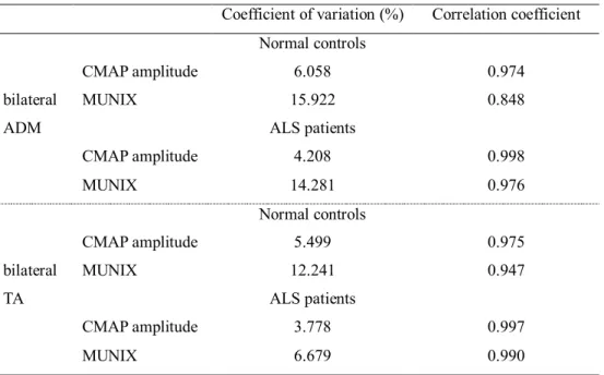
The mean values of CMAP amplitude and MUNIX showed no significant difference. The reproducibility of MUNIX and CMAP amplitude is summarized in Table 5. The COVs for CMAP and MUNIX were within the acceptable range of less than 20% in ALS patients and normal controls. In ALS patients, the COVs were higher in the ADM muscle than in the TA
8
muscle, whereas in normal controls they were higher in the TA muscle than in the ADM muscle. Correlations between MUNIX values obtained from test and retest were significant in both ADM and TA muscles. (P < 0.01)
There was a significant correlation between MUNIX and CMAP amplitude in both ADM (r = 0.918, P < 0.01) and TA (r = 0.850, P < 0.01) muscles in ALS patients (Figure 1, A and B). In normal controls, there was also a significant correlation between MUNIX and CMAP amplitude in both ADM (r = 0.629, P < 0.01) and TA (r = 0.894, P < 0.01) muscles (Figure 1, C and B).
In ALS patients, the MUNIX significantly correlated with the ALSFRS in the ADM (r = 0.439, P = 0.015), but not in the TA (r = 0.357, P= 0.053) (Figure 2, A and B). There was a significant correlation between MUNIX and MRC sum score in the TA (r = 0.474, P < 0.01) muscle, but not in the ADM (r = 0.349, P = 0.058) muscle (Figure 2, C and D). In both ADM and TA muscles of ALS patients, MUNIX values were lower in the weaker muscles, but the range was variable in muscles with mild weakness―MRC Grade IV, IV+, and V (Figure 2, E and F)
Finally, the mean values of all 4 muscles were analyzed in the same way.
There was a more significant correlation between CMAP and MRS sum score (r = 0.480, P < 0.01) and between CMAP and ALSFRS (r = 0.517, P < 0.01).
There was also a more significant correlation between MUNIX and MRS sum score (r = 0.493, P < 0.01), and between MUNIX and ALSFRS (r = 0.481, P
< 0.01).
9
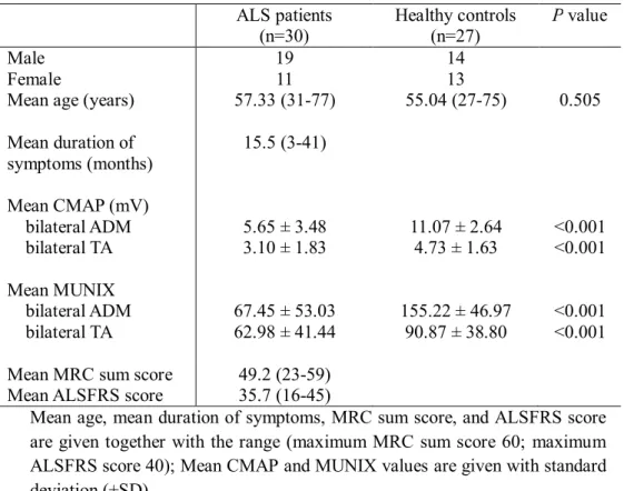
Table 1. The characteristics of ALS patients and healthy controls ALS patients
(n=30)
Healthy controls (n=27)
P value
Male 19 14
Female 11 13
Mean age (years) 57.33 (31-77) 55.04 (27-75) 0.505 Mean duration of
symptoms (months)
15.5 (3-41)
Mean CMAP (mV) bilateral ADM bilateral TA
5.65 ± 3.48 3.10 ± 1.83
11.07 ± 2.64 4.73 ± 1.63
<0.001
<0.001 Mean MUNIX
bilateral ADM bilateral TA
67.45 ± 53.03 62.98 ± 41.44
155.22 ± 46.97 90.87 ± 38.80
<0.001
<0.001 Mean MRC sum score 49.2 (23-59)
Mean ALSFRS score 35.7 (16-45)
Mean age, mean duration of symptoms, MRC sum score, and ALSFRS score are given together with the range (maximum MRC sum score 60; maximum ALSFRS score 40); Mean CMAP and MUNIX values are given with standard deviation (±SD)
10 Table 2. The demographics of ALS patients
Patient
No. Age
(years) Gender El Escorial
classification Initial
Symptom Duration
(months) ALSFRS- Revised MRC
sum score
1 54 F Probable LS 7 35 51
2 71 F Probable-lab B 16 34 46
3 63 F Probable C 9 42 49
4 70 F Definite B 12 35 52
5 67 F Probable C 13 38 55
6 73 F Probable C 10 29 43
7 77 F Probable-lab B 9 45 56
8 48 F Probable C 12 42 56
9 63 F Definite C 14 24 45
10 51 F Definite B 15 29 50
11 57 F Definite C 20 16 40
12 57 M Probable C 36 24 32
13 63 M Probable-lab LS 12 44 55
14 58 M Probable-lab B 13 44 58
15 61 M Probable C 25 29 36
16 62 M Probable-lab C 14 43 44
17 45 M Definite C 6 29 52
18 71 M Probable C 13 43 59
19 47 M Probable LS 16 37 53
20 58 M Probable-lab C 24 36 52
21 54 M Probable C 13 44 46
22 65 M Probable-lab C 11 40 42
23 63 M Probable C 8 45 59
24 61 M Probable C 12 39 57
25 43 M Probable-lab LS 19 28 45
26 31 M Definite C 3 38 50
27 37 M Probable-lab LS 41 20 23
28 53 M Probable C 5 43 58
29 47 M Probable-lab C 26 36 56
30 50 M Definite LS 31 41 56
C, LS, and B refer to the cervical, lumbosacral and bulbar region, respectively. The duration of disease is time from symptom onset to MUNIX measurement.
11
Table 3. The results of CMAP and MUNIX of the ADM muscle in ALS patients
Patient No.
Right ADM Left ADM
MRC grade
CMAP 1
CMAP 2
MUNIX 1
MUNIX 2
MRC grade
CMAP 1
CMAP 2
MUNIX 1
MUNIX 2
1 IV 3.3 3.3 25 22 IV+ 7.3 7.5 132 138
2 III 3.6 3.7 26 26 IV 6.2 6.1 66 70
3 IV- 6.4 6.6 48 46 II 3.6 3.3 10 5
4 IV 9.6 9.6 118 132 IV 1.7 1.7 17 16
5 IV 4.6 4.4 39 35 IV+ 4.4 4.5 57 62
6 III 0.5 0.5 6 6 IV- 3 3 30 31
7 V 8.5 8.3 141 108 V 7 6.9 78 77
8 III+ 3.3 3.3 35 35 III- 2.4 2.4 22 22
9 II 0.9 0.9 18 18 I 0.6 0.6 8 8
10 III 1.8 2.3 26 34 IV 6.5 6.8 137 150
11 II 1 1.2 14 20 II 0.5 0.7 5 9
12 II 3.8 3.9 18 17 III 8.7 8.6 109 123
13 IV+ 11.2 10.9 119 135 V 9.5 9.2 145 171
14 V 12.7 13 178 170 V 10.3 10.5 103 95
15 II 1.9 1.8 34 14 II 0.5 0.6 2 3
16 III 8.4 8.5 130 155 II 4.6 4.7 34 42
17 IV 9.9 9.5 93 72 II 5.7 5.8 35 34
18 IV+ 6.5 6.4 56 58 V 5.1 5 67 87
19 V 13.1 13.1 216 194 V 10.9 11 168 169
20 I 1.7 1.7 21 21 II 4.2 4.2 48 57
21 V 9.3 9.2 139 151 III 1.5 1.5 23 21
22 III 4 4.5 40 52 III 4 4 40 41
23 IV+ 13 13.7 121 144 IV- 6.8 7.5 64 68
24 III 3.4 3.6 28 43 IV- 6.6 6.8 61 72
25 II 1.6 1.5 9 9 IV 7.9 8.6 135 140
26 IV+ 9.3 9.2 122 114 IV+ 10.3 10.4 156 128
27 IV+ 7.1 7.3 85 75 IV- 3.2 3.3 16 15
28 IV+ 7.3 7.6 127 143 IV- 4.9 5.2 58 63
29 IV 2.4 2.3 40 35 IV 6.9 6.5 31 26
30 IV 5.7 6 35 30 IV+ 8.3 8 82 70
12
Table 4. The results of CMAP and MUNIX of the TA muscle in ALS patients
Patient No.
Right TA Left TA
MRC grade
CMAP 1
CMAP 2
MUNIX 1
MUNIX 2
MRC grade
CMAP 1
CMAP 2
MUNIX 1
MUNIX 2
1 IV 4 4 68 68 IV+ 6.7 6.6 136 125
2 III+ 2 2 29 26 III- 1.8 1.8 40 40
3 V 2.1 2.2 40 42 V 2.1 2.1 44 44
4 V 2.2 2.3 50 52 V 1.8 1.8 43 53
5 V 3.4 3.4 97 97 V 3.2 3.2 80 80
6 IV- 1 1 24 26 IV- 1.9 1.8 39 37
7 IV 2 1.9 38 37 IV 1.9 1.9 40 37
8 V 6 6.1 119 135 V 6.3 6.1 113 115
9 IV+ 4.4 4.7 85 86 IV 4.6 4.7 89 86
10 IV 2.3 2.7 16 20 IV 2.6 2.7 41 45
11 III 0.8 1 24 26 IV- 2.8 3 34 38
12 III 4.8 4.4 68 59 III 5.6 5.7 103 122
13 IV 3.8 3.8 41 34 IV+ 3.8 3.5 37 42
14 V 5.7 6 102 114 V 5.9 6 106 109
15 III 0.9 0.9 14 14 III 0.6 0.5 8 6
16 IV- 4.8 4.7 66 57 III 4.4 4.1 100 89
17 IV 4.2 4.2 64 68 IV 3.3 3.5 61 65
18 IV 1.7 1.6 38 37 IV 2.7 2.7 53 51
19 IV 3.3 3.3 95 95 IV 3.7 3.7 79 79
20 V 4.4 4.4 75 75 III 1.4 1.4 15 15
21 V 1.2 1.2 28 26 V 2 2 36 35
22 V 0.7 0.8 12 11 V 1.4 1.4 16 13
23 V 3.6 3.6 120 120 V 2 2 143 143
24 V 3 2.9 58 59 V 3.5 3.5 73 72
25 II 1.4 1.4 24 24 II 1 1 29 29
26 V 5.4 5.5 129 127 V 5.3 5.4 128 121
27 I 0.7 0.7 10 10 I 0.6 0.6 11 11
28 V 5.8 6.2 126 131 IV+ 8.5 8.8 204 230
29 IV+ 3 2.8 54 44 IV+ 3.7 3.5 75 70
30 III 1.4 1.4 29 29 IV+ 0.7 1 60 70
13
Table 5. The reproducibility of MUNIX measurements in ALS patients
and normal controls
Coefficient of variation (%) Correlation coefficient
bilateral ADM
CMAP amplitude MUNIX
CMAP amplitude
Normal controls 6.058 15.922 ALS patients
4.208
0.974 0.848
0.998
MUNIX 14.281 0.976
bilateral TA
CMAP amplitude MUNIX
CMAP amplitude
Normal controls 5.499 12.241 ALS patients
3.778
0.975 0.947 0.997
MUNIX 6.679 0.990
14
Figure 1. MUNIX values plotted against the CMAP amplitude of the muscle tested in ALS patients and normal controls―bilateral ADM muscles (A, C) and bilateral TA muscles (B, D)
(A) ADM (B) TA
(C) ADM (D) TA
15
Figure 2. The mean MUNIX value of bilateral muscles plotted against the ALSFRS (A, B) and MRC sum score (C, D). MUNIX values are plotted against the power levels of the ADM and TA muscles graded on the MRC scale (E, F)
(A) ADM (B) TA
(C) ADM (D) TA
(E) ADM (F) TA
16
Figure 3. The mean CMAP and MUNIX values of 4 muscles plotted against the ALSFRS (A) and MRC sum score (B).
(A)
(B)
17
DISCUSSION
In this study, we showed the reproducibility of MUNIX of the TA muscle in ALS patients and found a good correlation between MUNIX and CMAP in both ADM and TA muscles The mean and lower limit values in normal controls were almost the same as those reported by Nandedkar et al. (15).
The reproducibility test showed some interesting results. The COV of MUNIX was higher for the ADM muscle than for the TA muscle in both ALS patients and normal controls, and it was higher in normal controls than in ALS patients in both muscles. The COV of CMAP showed similar findings. These results are slightly different from those of previous study (16). COV values usually expected to be high when the MUNIX is low. However, our results showed lower COVs in lower MUNIX values. Previous studies showed relatively variable MUNIX measurements compared to ours. The COVs were generally lower in our study than in previous studies. Our MUNIX values also showed relatively uniform values. The reason for this result may be due to the different skill of EMG technicians or the small sample size. The TA muscle is relatively difficult to take clear CMAPs compared to the ADM muscle. A nerve conduction study of the TA muscle was done by stimulating the peroneal nerve below the fibula head. Strong stimuli are needed because the nerve lies deep in the muscle. For this reason, the CMAPs are relatively lower in the TA muscle than in the ADM muscle(17). This technical difficulty may have produced relatively uniform CMAP values in our study. To reduce this effect, a larger sample size will be needed.
18
Consistent with previous investigations, the MUNIX well correlated with CMAP amplitude in normal controls as well as in ALS patients. However, there have been few reports to evaluate the MUNIX of the TA muscle in ALS patients. However, a recent study of prior polio patients evaluated the MUNIX and CAMP of the TA muscle and showed a good correlation between MUNIX and CMAP(11).
In our study, MUNIX values significantly correlated with ALSFRS in the ADM muscle, but not in the TA muscle. The ALSFRS score consists of functions affected by the bulbar, cervical, thoracic, and lumbosacral segments (18). However, the majority of scores are related to respiratory function and weakness of the upper extremities. Respiratory function may be largely due to weakness of the bulbar segment, but the lumbosacral segment is located distant from the bulbar segment than from the cervical segment. The TA is a muscle of the lower extremity which is innervated by roots of the lumbosacral segment. Thus, the MUNIX of the TA mainly reflects the function of the lower extremity, not the bulbar or cervical function which is a major part of the ALSFRS. However, most ALS patients progress to advanced stages, and all segments can be affected. Therefore, if the enrolled number of patients was sufficiently large, the results may have shown a good correlation even in the TA muscle. This is a coincidence with recent studies that reported the spreading pattern of symptoms (19-21).
The correlation between MUNIX and MRC sum score was statistically significant in the TA muscle, but not in the ADM muscle. The TA is a large muscle compared to the ADM and is a proximal muscle with more abundant
19
muscle mass. Because of these characteristics of the TA, it may be more strongly related to the MRC sum score. This fact supports the hypothesis that the MUNIX in a large muscle more efficiently predict the progression of weakness. A previous study has evaluated the MRC sum scores in ALS patients by summing the muscle power levels of upper extremities (22), which was different from ours. In our study, MRS sum scores were calculated by summing the motor power levels of both upper and lower extremities. The MRC sum score has been widely used in clinical studies.
The ALSFRS and MRC sum score reflect both upper and lower motor neuron functions (23), whereas the MUNIX assesses only lower motor function. For this reason, there are some limitations to the MUNIX for accurate evaluation of muscle activity in ALS patients.
We also found more significant correlations between the mean values of the 4 muscles. Since ALS usually begins focally in the upper lower limb or bulbar muscles and then spreads to other regions, it might be beneficial to estimate the MUNIX in multiple muscles rather than in a single muscle. It is hypothesized that combined results of the 4 muscles will offset the distortion that may appear and show functional status. Our study results support this hypothesis. Therefore, this combination method can help predict disease progression regardless of onset regions.
MUNIX values were also found to be lower in the weaker muscles in ALS patients. Interestingly, the range of MUNIX values seems to be wider in moderately weak muscles, suggesting that the MUNIX has the potential to differentiate loss of motor units from compensatory reinnervation. There has
20
been some reports showing a decrease in the MUNIX in diseases other than ALS (24, 25). MUNIX values of motor neuron have been described in other diseases.
In conclusion, this study showed good correlations between MUNIX and CMAP/MRS sum score/MRC grade in the TA muscle. The pattern of their correlations in the TA muscle was not significantly different from that in the ADM muscle. The TA is a large muscle and easy to measure the MUNIX.
Therefore, the MUNIX can also be used in large muscles in ALS patients. In addition, the mean values of bilateral ADM and TA muscles reflected functional status more precisely. They can help predict disease progression and survival. This is the first study that revealed the reproducibility and usefulness of the MUNIX in muscles of the 4 extremities. Further prospective studies with sufficiently longer follow-ups are needed to confirm our results.
21
REFERENCES
1. Kiernan MC, Vucic S, Cheah BC, Turner MR, Eisen A, Hardiman O, et al. Amyotrophic lateral sclerosis. Lancet. 2011;377(9769):942-55.
2. Brooks BR. Clinical epidemiology of amyotrophic lateral sclerosis.
Neurologic clinics. 1996;14(2):399-420.
3. Bromberg MB. Updating motor unit number estimation (MUNE).
Clinical neurophysiology : official journal of the International Federation of Clinical Neurophysiology. 2007;118(1):1-8.
4. Nandedkar SD, Nandedkar DS, Barkhaus PE, Stalberg EV. Motor unit number index (MUNIX). IEEE transactions on bio-medical engineering.
2004;51(12):2209-11.
5. Shefner JM, Cudkowicz M, Brown RH, Jr. Motor unit number estimation predicts disease onset and survival in a transgenic mouse model of amyotrophic lateral sclerosis. Muscle & nerve. 2006;34(5):603-7.
6. Ahn SW, Kim SH, Oh DH, Kim SM, Park KS, Hong YH, et al.
Motor unit number estimation in evaluating disease progression in patients with amyotrophic lateral sclerosis. Journal of Korean medical science.
2010;25(9):1359-63.
7. Ahn SW, Kim SH, Kim JE, Kim SM, Kim SH, Park KS, et al.
Reproducibility of the motor unit number index (MUNIX) in normal controls and amyotrophic lateral sclerosis patients. Muscle & nerve. 2010;42(5):808- 13.
22
8. Neuwirth C, Nandedkar S, Stalberg E, Weber M. Motor unit number index (MUNIX): a novel neurophysiological technique to follow disease progression in amyotrophic lateral sclerosis. Muscle & nerve. 2010;42(3):379- 84.
9. Furtula J, Johnsen B, Christensen PB, Pugdahl K, Bisgaard C, Christensen MK, et al. MUNIX and incremental stimulation MUNE in ALS patients and control subjects. Clinical neurophysiology : official journal of the International Federation of Clinical Neurophysiology. 2013;124(3):610-8.
10. Neuwirth C, Nandedkar S, Stalberg E, Barkhaus PE, Carvalho M, Furtula J, et al. Motor Unit Number Index (MUNIX): a novel neurophysiological marker for neuromuscular disorders; test-retest reliability in healthy volunteers. Clinical neurophysiology : official journal of the International Federation of Clinical Neurophysiology. 2011;122(9):1867-72.
11. Sandberg A, Nandedkar SD, Stalberg E. Macro electromyography and motor unit number index in the tibialis anterior muscle: differences and similarities in characterizing motor unit properties in prior polio. Muscle &
nerve. 2011;43(3):335-41.
12. Brooks BR, Miller RG, Swash M, Munsat TL, World Federation of Neurology Research Group on Motor Neuron D. El Escorial revisited: revised criteria for the diagnosis of amyotrophic lateral sclerosis. Amyotrophic lateral sclerosis and other motor neuron disorders : official publication of the World Federation of Neurology, Research Group on Motor Neuron Diseases.
2000;1(5):293-9.
23
13. Cedarbaum JM, Stambler N. Performance of the Amyotrophic Lateral Sclerosis Functional Rating Scale (ALSFRS) in multicenter clinical trials. Journal of the neurological sciences. 1997;152 Suppl 1:S1-9.
14. Nandedkar SD, Barkhaus PE, Stalberg EV. Motor unit number index (MUNIX): principle, method, and findings in healthy subjects and in patients with motor neuron disease. Muscle & nerve. 2010;42(5):798-807.
15. Neuwirth C, Nandedkar S, Stalberg E, Barkhaus PE, Carvalho M, Furtula J, et al. Motor Unit Number Index (MUNIX): reference values of five different muscles in healthy subjects from a multi-centre study. Clinical neurophysiology : official journal of the International Federation of Clinical Neurophysiology. 2011;122(9):1895-8.
16. Nandedkar SD, Barkhaus PE, Stalberg EV. Reproducibility of MUNIX in patients with amyotrophic lateral sclerosis. Muscle & nerve.
2011;44(6):919-22.
17. Buschbacher RM. Reference values for peroneal nerve motor conduction to the tibialis anterior and for peroneal vs. tibial latencies.
American journal of physical medicine & rehabilitation / Association of Academic Physiatrists. 2003;82(4):296-301.
18. The Amyotrophic Lateral Sclerosis Functional Rating Scale.
Assessment of activities of daily living in patients with amyotrophic lateral sclerosis. The ALS CNTF treatment study (ACTS) phase I-II Study Group.
Archives of neurology. 1996;53(2):141-7.
24
19. Ravits J, Laurie P, Fan Y, Moore DH. Implications of ALS focality:
rostral-caudal distribution of lower motor neuron loss postmortem. Neurology.
2007;68(19):1576-82.
20. Ravits J, Paul P, Jorg C. Focality of upper and lower motor neuron degeneration at the clinical onset of ALS. Neurology. 2007;68(19):1571-5.
21. Ravits JM, La Spada AR. ALS motor phenotype heterogeneity, focality, and spread: deconstructing motor neuron degeneration. Neurology.
2009;73(10):805-11.
22. Boekestein WA, Schelhaas HJ, van Putten MJ, Stegeman DF, Zwarts MJ, van Dijk JP. Motor unit number index (MUNIX) versus motor unit number estimation (MUNE): a direct comparison in a longitudinal study of ALS patients. Clinical neurophysiology : official journal of the International Federation of Clinical Neurophysiology. 2012;123(8):1644-9.
23. Gordon PH, Miller RG, Moore DH. Alsfrs-R. Amyotrophic lateral sclerosis and other motor neuron disorders : official publication of the World Federation of Neurology, Research Group on Motor Neuron Diseases. 2004;5 Suppl 1:90-3.
24. Drey M, Grosch C, Neuwirth C, Bauer JM, Sieber CC. The Motor Unit Number Index (MUNIX) in sarcopenic patients. Experimental gerontology. 2013;48(4):381-4.
25. Li X, Jahanmiri-Nezhad F, Rymer WZ, Zhou P. An Examination of the Motor Unit Number Index (MUNIX) in Muscles Paralyzed by Spinal Cord Injury. IEEE journal of biomedical and health informatics. 2012.
25
국문 초록
서론: 근위축성 측삭경화증은 신경퇴행성 질환으로 진행성 운동신경 소실을 특징으로 한다. MUNIX(motor unit number index)는 근위 축성 측삭경화증 환자에서 신경 축삭의 손상 정도를 평가할 수 있 는 새로운 진단기술이다. 이 연구의 목적은 근위축성 측삭경화증 환 자와 정상 대조군에서 전경골근과 소지외전근을 이용한 MUNIX 검 사의 재현성과 유용성을 증명하는 것이다.
방법: 30 명의 근위축성 측삭경화증 환자와 27 명의 정상대조군에서 MUNIX 검사를 시행하였다. MUNIX, 복합운동근육활동전위, MRC 총근력지수, 근위축성 측삭경화증 임상점수(ALSFRS)를 평가하고 그들 사이의 연관성을 피어슨 상관 분석을 통하여 계산하였다. ALS 환자에서 재현성을 평가하기 위해서 모든 근육에 대하여 두번씩 기 록하여 변동계수를 분석하였다. 또 4 가지 근육 모두의 평균값을 계 산하여 임상적 상태와의 연관성을 분석하였다.
결과: 검사와 재검사간에 복합운동근육활동전위 및 MUNIX 의 평균 값은 유의미한 차이가 없었다. 복합운동근육활동전위와 MUNIX 의
26
변동계수는 20% 이하로 적절한 범위였다. 근위축성 측삭경화증 환 자에서 소지외전근(r = 0.918, P < 0.01)과 전경골근(r = 0.850, P
< 0.01)모두에서 MUNIX 값과 복합운동근육활동전위 사이에 통계 적으로 유의미한 상관관계가 있었다. 정상 대조군에서도 소지외전근 (r = 0.629, P < 0.01)과 전경골근(r = 0.894, P < 0.01) 의 MUNIX 와 복합운동근육활동전위는 유의미한 상관관계가 있었다.
근위축성 측삭경화증 환자에서 소지외전근(r = 0.439, P < 0.015) 에서는 MUNIX 와 ALSFRS 간에 유의미한 상관관계가 있었으나, 전경골근(r = 0.357, P < 0.053)에서는 유의미한 상관관계가 없었 다. 전경골근(r = 0.474, P < 0.01) 에서는 MUNIX 와 MRC 총근력 지수 사이의 유의미한 상관관계가 있었으나소지외전근(r = 0.349, P = 0.058)에서는 유의미한 상관관계가 없었다. 4 곳의 근육의 평 균값을 이용하여 분석하였을 때, 복합운동근육활동전위와 MRC 총 근력지수 및 복합운동근육활동전위와 ALSFRS 사이에 유의미한 상 관관계가 있었으며, MUNIX 와 MRC 총근력지수 및 MUNIX 와 ALSFRS 간에도 유의미한 상관관계가 있었다.
결론: 이 연구를 통해 근위축성 측삭경화증 환자에서 소지외전근과 전경골근을 이용한 MUNIX 검사가 운동신경 소실을 효과적으로 반 영해주는 것을 확인할 수 있었다. 소지외전근과 전경골근 모두 좋은 상관관계를 보였으나 MUNIX 상관관계의 패턴은 약간 달랐다. 4 곳 근육의 조합은 환자의 기능 상태와 질병의 진행을 판단하는데 좀더
27
유용하였다. 이 연구 결과를 뒷받침하기 위해 앞으로 더 많은 환자 를 포함하는 전향적 연구가 필요할 것이다.
--- 주요어 : 근위축성 측삭경화증, MUNIX, 복합운동활동전위, 소지외전 근, 전경골근
학 번 : 2009-21840