저작자표시-비영리-변경금지 2.0 대한민국 이용자는 아래의 조건을 따르는 경우에 한하여 자유롭게
l 이 저작물을 복제, 배포, 전송, 전시, 공연 및 방송할 수 있습니다. 다음과 같은 조건을 따라야 합니다:
l 귀하는, 이 저작물의 재이용이나 배포의 경우, 이 저작물에 적용된 이용허락조건 을 명확하게 나타내어야 합니다.
l 저작권자로부터 별도의 허가를 받으면 이러한 조건들은 적용되지 않습니다.
저작권법에 따른 이용자의 권리는 위의 내용에 의하여 영향을 받지 않습니다. 이것은 이용허락규약(Legal Code)을 이해하기 쉽게 요약한 것입니다.
Disclaimer
저작자표시. 귀하는 원저작자를 표시하여야 합니다.
비영리. 귀하는 이 저작물을 영리 목적으로 이용할 수 없습니다.
변경금지. 귀하는 이 저작물을 개작, 변형 또는 가공할 수 없습니다.
수의학박사학위논문
Helicobacter 감염에 의한 위염에서 위 점막 수지상세포의 역할
The roles of gastric mucosal dendritic cell in Helicobacter-induced gastritis
2019 년 8 월
서울대학교 대학원
수의학과 수의병리학 전공
고 두 민
i
ABSTRACT
The roles of gastric mucosal dendritic cell in Helicobacter-induced gastritis
(Supervisor: Dae-Yong Kim, D.V.M., Ph.D.)
Du-Min Go
Department of Veterinary Pathology, College of Veterinary Medicine, Graduate School, Seoul National University
Dendritic cells (DCs) are known to be important immune-regulator in all organs, but DCs within the stomach remain poorly characterized. DCs have been reported to play a role in regulating host immune responses to Helicobacter infection, but the precise role of gastric DCs in gastritis remains unclear. Here, gastric DCs were finely characterized and their roles in Helicobacter-induced gastritis were investigated. Gastric tissues from DC-deficient mouse models and control mice with Helicobacter infection were comparatively analyzed using histopathology, flow cytometry, immunohistochemistry or immunofluorescence, quantitative real-time PCR and luminex immunoassays. Programmed death ligand 1 (PD-L1)–deficient mouse models were also used to confirm the T cell regulation
ii
mechanism of gastric DCs. The clinical aspects were confirmed by conducting multiplexed immunohistochemistry on paraffin-embedded gastric tissue microarrays of patients with gastritis. Gastric DCs were composed of CD103+CD11b-, CD103-CD11b+, and CD103-CD11b- subsets, and all subsets expanded during Helicobacter infection. Helicobacter-infected classical DC- deficient mice showed severe gastritis and T cell accumulation compared to control mice. However, the degree of infiltration of regulatory T cells was not different between the two groups. Gastric DCs expressed considerably higher level of PD- L1 than other immune cells and co-localized with T cells in mouse and human stomach. Blockade of PD-L1 exacerbated gastritis with severe T cell accumulation during Helicobacter infection. This study indicate that gastric classical DCs attenuate Helicobacter-induced gastritis by regulating T cell recruitment through PD-L1 expression and that considering the possibility of inflammatory side-effects on the stomach would be important when treating cancer patients with PD-L1 inhibitors.
---
Keywords: dendritic cell, gastritis, Helicobacter, programmed death ligand 1 (PD- L1), programmed death 1 (PD-1), T cell
Student Number: 2012-21535
iii
CONTENTS
ABSTRACT ... i
CONTENTS ... iii
ABBREVIATIONS ... 1
LITERATURE REVIEW ... 3
Introduction ... 3
Initiation of T cell immunity ... 4
Development and function of DC subsets ... 5
DCs-mediated immune tolerance ... 8
Function of DCs in Helicobacter infection ... 9
Mouse model of Helicobacter infection ... 10
Objective ... 12
CHAPTER I. Gastric classical dendritic cells attenuate Helicobacter-induced gastric inflammation via controlling T cell immunity ... 13
Abstract ... 14
Introduction ... 15
Materials and Methods ... 18
Mice ... 18
Bone marrow transplantation ... 18
Helicobacter culture and infection ... 18
Diphtheria toxin injection ... 19
Necropsy and tissue preparation ... 19
iv
Histopathologic examinations ... 20
Single-cell preparations and flow cytometric analysis ... 20
Antibodies for flow cytometry ... 22
Immunohistochemistry and immunofluorescence ... 23
Helicobacter felis quantification ... 24
Quantitative reverse transcription-PCR ... 24
Magnetic luminex immunoassay ... 25
Statistical analysis ... 25
Results ... 27
Gastric DCs have three distinct subpopulations ... 27
Helicobacter infection increased the number of DCs in the stomach... 32
Flt3 KO mice have more severe gastric inflammation with less Helicobacter colonization ... 36
Flt3 KO mice showed more prominent CD8+ T cell accumulation than WT mice during Helicobacter infection ... 43
Enhanced gastric inflammation of Helicobacter-infected Flt3 KO mice was hematopoietic cell-intrinsic ... 52
Depletion of cDCs enhanced Helicobacter-induced gastric inflammation ... 56
Discussion ... 63
CHAPTER II. Programmed death ligand 1 expressing gastric classical dendritic cells attenuate Helicobacter-induced gastric inflammation via controlling local T cell immunity ... 67
Abstract ... 68
Introduction ... 69
Materials and Methods ... 71
Mice ... 71
v
Bone marrow transplantation ... 71
PD-L1 in vivo blockade ... 71
Helicobacter culture and infection ... 72
Necropsy and tissue preparation ... 72
Histopathologic examinations ... 73
Single-cell preparations and flow cytometric analysis ... 73
Antibodies for flow cytometry ... 74
Immunohistochemistry and immunofluorescence ... 76
Helicobacter felis quantification ... 77
Quantitative reverse transcription-PCR ... 77
Tissue-microarrayed human gastric tissue and multiplexed immunohistochemistry . 78 Statistical analysis ... 79
Results ... 80
Gastric cDCs expressed high level of PD-L1 and co-localized with T cells in Helicobacter-infected gastric mucosa and submucosa ... 80
Blockade of PD-L1 enhanced the gastric inflammation with severe T cell accumulation upon Helicobacter infection ... 88
PD-L1 deficiency in BM-derived cells enhanced gastric inflammation upon Helicobacter infection ... 92
PD-L1 expressing DCs co-localized with T cells in human Helicobacter-positive gastritis ... 96
Discussion ... 100
OVERALL CONCLUSION ... 104
REFERENCES ... 110
1
ABBREVIATIONS Ab: antibodies
ATPase: adenosine triphosphatase BM: bone marrow
cDCs: classical dendritic cells CDP: common-DC progenitors
CMoP: common-monocyte progenitors
CTLA-4: cytotoxic T lymphocyte–associated antigen 4 DCs: dendritic cells
DN: double-negative DT: diphtheria toxin
DTR: diphtheria toxin receptor FACS: flow cytometry
Flt3: fms-like tyrosine kinase 3
Flt3L: fms-like tyrosine kinase 3 ligand H&E: hematoxylin and eosin
HBSS: Hank’s balanced salt solution H. felis: Helicobacter felis
H. pylori: Helicobacter pylori IFN-γ: interferon gamma IHC: immunohistochemistry IL: interleukin
IRAEs: immune-related adverse events IRF: interferon regulatory factor
2
KO: knockout
M-CSF: macrophage-colony stimulating factor MDP: macrophage-DCs progenitors
MHC: major histocompatibility complex moDCs: monocyte-derived DCs
PD-1: programmed death 1 pDCs: plasmacytoid DCs
PD-L1: programmed death ligand 1 PD-L2: programmed death ligand 2 qRT-PCR: quantitative real-time PCR
SPEM: spasmolytic polypeptide-expressing metaplasia SS1: Sydney strain 1
TCR: T-cell antigen receptor Th: helper T cell
TLR: toll-like receptor
TGF: transforming growth factor
TIP DCs: tumor necrosis factor-inducible nitric oxide synthase-producing DCs TNF: tumor necrosis factor
Tregs: regulatory T cells WT: wild type
3
LITERATURE REVIEW Introduction
Dendritic cells (DCs) were initially discovered during the preparation of adherent mononuclear cells from mouse spleens (Liu and Nussenzweig 2010). DCs were recognized as novel cell populations with a characteristic morphology and movement, and functional differentiation from other cells, including macrophages in the mouse peripheral lymphoid organs (Steinman and Cohn 1973; Steinman and Cohn 1974). The “dendritic cell” was named by Ralph Steinman in 1973 due to the distinct morphological features (Steinman and Cohn 1973); however, DCs were
“accessory” cells with a small number in organs, and thus the biological significance of DCs was viewed skeptically (Mildner and Jung 2014). Steinman and his colleagues showed that DCs are representative antigen-presenting cells that initiate antigen-specific cellular immune responses, presenting invaluable evidence (Banchereau and Steinman 1998; Mildner and Jung 2014). Immunology had focused only on antigens and lymphocytes for some time, and DCs provided insights into immune responses that were not adequately explained by only these two factors (Banchereau and Steinman 1998).
Since this discovery, various aspects of DCs, such as functions, development, subsets, and migration, have been extensively studied. These studies have been carried out in vitro mainly by purifying DCs from mouse spleens or by differentiating mouse bone marrow (BM) cells or human monocytes into DCs using the granulocyte-macrophage colony-stimulating factor or the fms-like tyrosine kinase 3 ligand (Flt3L) (Liu and Nussenzweig 2010). However, DCs are a heterogeneous population composed of several subsets and share certain surface
4
markers with immune cells belonging to the mononuclear phagocyte system such as macrophages and monocytes, making it difficult to only identify pure DCs (Worbs, Hammerschmidt et al. 2017). In addition, several DC-deficient mouse models have been used to identify the role of DCs in infectious, autoimmune, and metabolic diseases and tumors, but DCs can exhibit various aspects depending on the type of disease or the target organs where the lesion occurs. Thus, detailed DCs characterization is important in the study of DCs.
Initiation of T cell immunity
DCs are superior to other immune cells in capturing and processing antigens and presenting treated antigen to T cells via the T-cell antigen receptor (TCR) (Nussenzweig, Steinman et al. 1980). DCs are indispensable for the stimulation of T cells that cannot directly recognize antigens (Banchereau and Steinman 1998).
DCs present endogenous and exogenous antigens to T cells through the major histocompatibility complex (MHC) I and the MHC II, respectively. In addition, DCs induce cytotoxic CD8+ T cell immunity by presenting exogenous antigens to T cells through MHC I, which is called cross-presentation. Cross-presentation is crucial for immunity against viruses and intracellular bacteria (Rock 2003). The binding of MHC-peptide complexes on DCs and T cells induces distinct outcomes depending on the context of antigens, signals provided by B7 family members on DCs and instructing cytokines (Mildner and Jung 2014). In relation to pathogens or danger-associated molecular patterns, DCs and T cells can recognize the contexts of antigens. In the crosstalk between T cells and DCs, pro-inflammatory T cell responses or T cell tolerance is induced depending on the co-stimulatory or co-
5
inhibitory signals transmitted by DCs or the maturation state of DCs (Mildner and Jung 2014). Thus, DCs play a central role in controlling T cell immunity.
Development and function of DC subsets
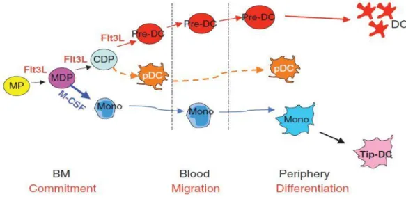
DCs are classified into classical DCs (cDCs), monocyte-derived DCs (moDCs), and plasmacytoid DCs (pDCs). They share origins initially in the BM and are divided into common-DC progenitors (CDP) and common monocyte progenitors (CMoP) according to Flt3L and the macrophage-colony stimulating factor (M-CSF) signaling in macrophage-DCs progenitors (MDP). Subsequently, cDCs and pDCs are differentiated by fms-like tyrosine kinase 3 (Flt3)-Flt3L signaling from CDP, and monocytes and their descendants are differentiated from CMoP. cDCs arise from pre-DCs differentiated from CDP in the BM, and the differentiated pre-DCs migrate through the blood and are distributed in the peripheral organs. pDCs are differentiated from CDP in BM and migrated to peripheral organs, and moDCs are differentiated from monocytes in peripheral organs (Fig. 1) (Liu and Nussenzweig 2010).
cDCs are classified into at least two subsets according to the expression of CD8α and CD103 or CD11b surface markers. First, CD8α+ cDCs are present in lymphoid tissues including the spleen, lymph node and BM. CD103+ cDCs are considered to be the same population as CD8α+ cDCs present in non-lymphoid tissues (del Rio, Rodriguez-Barbosa et al. 2007; Edelson, KC et al. 2010). CD8α+ cDCs and CD103+ cDCs are the most well-characterized cDCs subsets based on their phenotypic and gene expression characteristics (Mildner and Jung 2014).
These CD8α+ and CD103+ cDCs are referred to as the cDC1, and cDC1 is known
6
to be functionally specific for cross-presentation (den Haan, Lehar et al. 2000;
Bedoui, Whitney et al. 2009). Interferon regulatory factor (IRF) 8, Id2, BATF3, and NFIL3 have been reported as transcription factors involved in the development of cDC1 lineage (Mildner and Jung 2014). Among them, IRF8 is presumed to be the master regulator of cDC1 lineage (Seillet, Jackson et al. 2013). CD11b+ cDCs are referred to as cDC2 and are known as heterogeneous populations, unlike CD8α+ and CD103+ cDCs. Due to the complexity of this heterogeneity, the characterization of CD11b+ cDCs is relatively unclear. RelB, NOTCH2, RBP-J, IRF2, and IRF4 have been known to regulate CD11b+ cDCs development (Mildner and Jung 2014).
Of these, IRF4 has also been implicated in the function and migration of CD11b+ cDCs (Mildner and Jung 2014). Compared to CD8α+ and CD103+ cDCs, CD11b+ cDCs are ineffective for cross-presentation but have been reported to be more effective for the induction of CD4+ T cell immunity (Lewis, Caton et al. 2011).
moDCs also exist in the steady state, but are referred to as “inflammatory DCs”
because they rapidly aggregate into inflammatory sites (Goudot, Coillard et al. 2017;
Segura and Amigorena 2013). In addition, tumor necrosis factor (TNF)-inducible nitric oxide synthase-producing DCs (TIP DCs) present in pathogen-associated inflammation have been considered typical moDCs, but recent studies have suggested that TIP DCs correspond to activated effector monocytes rather than to DCs (Mildner, Yona et al. 2013). moDCs are difficult to distinguish from CD11b+ cDCs because they share surface markers such as CD11c, MHC II, and CD11b, with cDCs and can migrate to draining lymph nodes similarly to cDCs to induce adaptive immune responses (Liu and Nussenzweig 2010; Chow, Sutherland et al.
7
2017). Detailed studies that explore distinguishing between them would be helpful in understanding heterogeneous CD11b+ cDCs.
pDCs are BM-derived DCs subsets that are rarely found in peripheral blood and peripheral organs. Like cDCs, pDCs development is dependent on Flt3-Flt3L signaling, and activated pDCs are known to play a role in naive CD4+ T cell priming in lymph nodes (Kingston, Schmid et al. 2009; Sapoznikov, Fischer et al. 2007).
However, they are best known as a population that produces type I interferon in response to viral infections, and thus the role of pDCs in innate and adaptive immune responses to viral infections has been the subject of many studies (Colonna, Trinchieri et al. 2004; Yun, Lee et al. 2016). In addition, the roles of pDCs in peripheral and central immune tolerance have been reported in association with regulatory T cells (Tregs) induction (Yun, Lee et al. 2016).
Fig 1. Dendritic cell and monocyte origin and development (Liu and Nussenzweig 2010)
8
DCs-mediated immune tolerance
Although DCs induce effective immune response to antigens, they also play an important role in maintaining immune tolerance. DCs maintain immune tolerance by inducing apoptosis of autoreactive thymocytes and promoting Treg differentiation by presenting self-antigens in the thymus (central tolerance). In addition, DCs induce anergy, apoptosis of T cells in peripheral organs, and promote immune tolerance by promoting Treg response (peripheral tolerance) (Audiger, Rahman et al. 2017). Thus, DCs contribute to the balance between immunity and tolerance, which is influenced both by various DC factors and by local environmental factors that modulate the tolerogenic functions of the DCs.
Signal transduction through contact between T cells and DCs is an important process that determines immunogenic or tolerogenic pathways. The mature state of DCs has a significant influence on this signal transmission. Mature DCs, sensing pathogens by pattern recognition receptor such as toll-like receptors (TLR) (immunogenic maturation), highly express co-stimulatory molecules such as CD80, CD86, and CD40, which enhances the efficiency of TCR signaling and promotes effector T cell responses. However, immune tolerance is induced when these signals are transmitted to Tregs (Yamazaki, Iyoda et al. 2003; Salomon, Lenschow et al.
2000). On the other hand, DCs matured by self-antigens in steady states (homeostatic maturation) and immature DCs expressing low co-stimulatory molecules induce immune tolerance (Ardouin, Luche et al. 2016). In addition, DCs can induce tolerance by expressing co-inhibitory molecules, canceling stimulatory signals that are transmitted to T cells, and delivering inhibitory signals. Among these co-inhibitory molecules, programmed death ligand 1 (PD-L1) and
9
programmed death ligand 2 (PD-L2) are well known, and they transmit signals through the programmed death 1 (PD-1) expressed in T cells. In addition, the CD86 and CD80 expressed by DCs can induce T cell tolerance by binding to the cytotoxic T lymphocyte–associated antigen 4 (CTLA-4) expressed in T cells (Probst, McCoy et al. 2005).
Specific local environmental factors can also influence the immune tolerance role of DCs. DCs present at specific anatomical locations, such as the intestine which is continuously exposed to microflora and food antigens, or the skin, are known to be suitable for promoting immune tolerance (Audiger, Rahman et al.
2017). Furthermore, in inflammatory conditions, inflammation-related signals such as TLR2, TNF-α, and prostaglandin receptors, and Tregs regulate DCs to perform immunoregulatory roles (Popov, Driesen et al. 2008; Darrasse-Jèze, Deroubaix et al. 2009).
The here-described significance of DCs in maintaining immune tolerance has been demonstrated to some extent by the phenotype of DC-deficient mouse models, and tolerogenic DCs are attracting attention as a potential solution for conditions requiring immune tolerance such as autoimmune diseases (Audiger, Rahman et al.
2017). Thus, studies have been carried out on murine tolerogenic DC subsets and their human equivalents (Robbins, Walzer et al. 2008; Guilliams, Dutertre et al.
2016).
Function of DCs in Helicobacter infection
It is known that TLR2 and TLR4 play an important role for DCs to recognize Helicobacter pylori (H. pylori) and produce cytokines (Rad, Ballhorn et al. 2009).
10
Furthermore, recent studies have shown that CD11b+ gastric DC directly recognize and engulf H. pylori in the stomach (Arnold, Zhang et al. 2017).
Helicobacter-recognizing DCs express maturation markers such as CD80 and CD86, and produce pro-inflammatory cytokines such as interleukin (IL)-6, IL-12, and IL-23, and activate co-cultured autologous CD4+ T cells to secrete interferon gamma (IFN-γ) or IL-17 (Hafsi, Voland et al. 2004; Khamri, Walker et al. 2010).
Contrary to these pro-inflammatory roles, DCs also play an immune tolerance role by inducing Tregs or producing anti-inflammatory cytokines such as IL-10 and transforming growth factor (TGF)-β (Rizzuti, Ang et al. 2015; Zhang, Liu et al.
2010). These in vitro studies were performed using human moDCs or mouse BM- derived DCs. As described above, DCs have a dual role against Helicobacter.
In vivo studies to confirm the function of DCs in Helicobacter infection have been performed using the CD11c-diphtheria toxin receptor (DTR) which can ablate DCs. This study demonstrated the tolerogenic role of DCs against H. pylori (Hitzler, Oertli et al. 2011; Oertli, Sundquist et al. 2012), but other CD11c+ cells in addition to DCs could also be affected in CD11c-DTR mice (Probst, Tschannen et al. 2005;
Bennett and Clausen 2007). Therefore, in vivo studies using mouse models that ablate DCs only will help identify the role of gastric DCs in Helicobacter infection.
Mouse model of Helicobacter infection
In the 1980s, Pelayo Correa suggested chronic gastritis as a gastric cancer precursor based on clinical and epidemiological observations, proposing sequential events leading to the development of gastric adenocarcinoma (Correa 1983; Correa 1984). Chronic-active gastritis induced by unknown factors progressed to
11
multifocal atrophic gastritis with loss of parietal cells and chief cells, and intestinal metaplasia (Sakagami, Dixon et al. 1996). Afterwards, the finding that H. pylori was the cause of chronic-active gastritis provided a missing link in the Correa pathway (Sakagami, Dixon et al. 1996).
The only available animal models of H. pylori infection were germ-free piglets and non-human primates, and the limited host range of H. pylori has restricted the study of the pathogenesis of the disease (Lee, Fox et al. 1990). Although these animal models provided important information, the need for a small animal model was raised because it was unmanageable, expensive, and only available to a few investigators (Lee, O'Rourke et al. 1997). However, early attempts to colonize H.
pylori on rodents were unsuccessful (Ehlers, Warrelmann et al. 1988; Cantorna and Balish 1990). On the other hand, Helicobacter felis (H. felis) isolated from stomach of cat was more closely related to H. pylori than any other bacteria based on 16s ribosomal RNA sequencing and showed gastric trophism with stable colonization in mouse stomach (Lee, Fox et al. 1990). H. felis caused lesions similar to human gastric lesions following H. pylori infection in some mouse strains including C57BL/6 (Lee, Fox et al. 1990). In particular, C57BL/6 mice showed chronic-active gastritis, gastric atrophy, metaplasia as well as dysplasia and gastric cancer after 12-15 months of infection (Fox et al. 2002).
Meanwhile, H. pylori isolated from the stomach of patients at the gastroenterology clinic in Sydney, Australia, was subsequently colonized stably in the stomach of mice through long-term adaptation, which was named the Sydney strain 1 (SS1) (Lee, O'Rourke et al. 1997). H. pylori SS1 induced gastric pathology
12
similar to that of H. pylori-infected patients in mice, but could not induce gastric cancer (Duckworth, Burkitt et al. 2015).
Objective
DCs are the central immune cells that regulate innate and adaptive immunity in a wide range of pathophysiological conditions. DCs modulate the appropriate immune response in various organs while promoting immunity against pathogens and maintaining immune tolerance. Based on these important roles, many studies have been conducted on the overall characterization of DCs.
However, gastric DCs are relatively unknown compared to the DCs of other organs. In particular, the importance of gastric DCs has been emphasized in controlling immunity against H. pylori infection, which is closely related to chronic gastritis and gastric cancer in humans. However, in vivo studies to clarify the role of gastric DCs in Helicobacter infection are rarely reported.
In the present study, I tried to characterize gastric DCs. Gastric DCs were examined closely in vivo in both steady state and Helicobacter-induced gastritis.
Furthermore, I tried to identify the major role of gastric DCs in Helicobacter infection, and the mechanisms of the role by using genetically engineered mouse models.
13
CHAPTER I.
Gastric classical dendritic cells attenuate Helicobacter-induced
gastric inflammation via controlling T cell immunity
14
Abstract
Approximately 50% of the human population is infected by H. pylori.
However, the host’s immune system cannot eradicate colonized H. pylori in the gastric mucosa, causing chronic gastritis. H. pylori-induced chronic gastritis may progress to severe gastropathy such as peptic ulcer and gastric cancer. DCs have been reported to play an important role in regulating the host immune responses to Helicobacter infection, but the precise role of DCs in the stomach remains unclear.
To investigate the roles of gastric DCs in Helicobacter-induced gastritis, DC- deficient mice were infected with H. felis and then gastric tissues were analyzed by histopathology, flow cytometry, immunohistochemistry, quantitative real-time PCR and magnetic luminex immunoassays. It was found that gastric DCs were composed of CD103+CD11b-, CD103-CD11b+, and CD103-CD11b- subsets, and all these subsets expanded during H. felis infection. H. felis-infected Flt3 knockout (KO) and Zbtb46-DTR mice, not BDCA2-DTR mice, showed severe gastric lesions with prominent T cell infiltration compared to control group mice. This study indicate that gastric cDCs play an important role in suppressing T cell responses during Helicobacter infection.
15
Introduction
H. pylori is a gram negative bacterium that can colonize and survive in the human gastric mucosa. Approximately 50% of the human population is colonized by H. pylori, and about 15% of them develop severe lesions such as severe chronic gastritis, peptic ulcer, gastric adenocarcinoma and lymphoma (Kronsteiner, Bassaganya-Riera et al. 2014). H. pylori-induced gastropathy is influenced by various factors such as host, bacterial pathogen, and environment-related factors, and host’s immune system against H. pylori has been reported to be closely related to lesion development (Kusters, van Vliet et al. 2006).
Various components and products of H. pylori and signals from mucosal epithelial cells in contact with H. pylori cause infiltration of innate immune cells such as DCs, macrophages, monocytes and neutrophils. Among these innate immune cells, DCs have been shown to play an important role in the host T cell immune response to H. pylori (Shiu and Blanchard 2013).
DCs are representative antigen-presenting cells with an exceptional ability to modulate the overall immune response to invasive pathogens (Steinman 2012).
These properties of DCs imply the importance of DCs in the immune responses of the gastrointestinal tract, which is constantly exposed to various pathogens. To determine the role of DCs in H. pylori infection, many previous studies have focused on the direct response of DCs to H. pylori by incubating human moDCs or mouse BM-derived DCs with live H. pylori or H. pylori-related proteins in vitro.
DCs have been shown to recognize H. pylori via pattern recognition receptor such as Toll-like receptors (Rad, Ballhorn et al. 2009), and H. pylori-stimulated DCs secrete pro-inflammatory cytokines such as IL-6, IL-12 and IL-23 and activate co-
16
cultured autologous CD4+ T cells to secrete IFN-γ or IL-17 (Hafsi, Voland et al.
2004; Khamri, Walker et al. 2010). In contrast to these pro-inflammatory roles, H.
pylori-stimulated DCs also play an immune tolerance role by inducing Tregs or producing anti-inflammatory cytokines such as IL-10 and TGF-β (Zhang, Liu et al.
2010; Rizzuti, Ang et al. 2015). As described above, since DCs might have dual roles against H. pylori, in vivo studies are highly required to determine the effects of DCs on Helicobacter-infected individuals. Several previous in vivo studies used the CD11c-DTR mice in which DCs can be ablated and confirmed the tolerogenic role of DCs in H. pylori infection (Hitzler, Oertli et al. 2011; Oertli, Sundquist et al.
2012). However, as macrophages can also be ablated in CD11c-DTR mice, elucidating the function of gastric DCs in H. pylori-induced gastritis is difficult (Bennett and Clausen 2007). Meanwhile, one study demonstrated that gastric CD11b+ DCs engulf H. pylori in gastric mucosa (Arnold, Zhang et al. 2017).
However, the exact immunological roles of gastric DCs in Helicobacter-induced gastritis are largely unknown.
Many studies on human and animal models of H. pylori infection have suggested that CD4+ T cell responses such as helper T cell (Th) 1, Th17, and Tregs are predominantly controlled by gastric DCs (Bimczok, Clements et al. 2010;
Zhang, Liu et al. 2010). However, these CD4+ T cell-mediated immune responses were not effective in eradicating the colonized H. pylori in the gastric mucosa, rather causing gastritis to become chronic, which is a risk factor for the development of peptic ulcer or gastric cancer (Peek, Fiske et al. 2010). Furthermore, CD8+ T cell infiltration has been shown to be increased in patients with H. pylori infection, and IFN-γ production in response to H. pylori infection is greater in CD8+ T cells than
17
in CD4+ T cells (Bamford, Fan et al. 1998; Quiding-Järbrink, Lundin et al. 2001).
T cell-mediated IFN-γ improved Helicobacter clearance but contributed to the development of severe gastric lesions (Sayi, Kohler et al. 2009). Therefore, CD8+ T cell infiltration appear to be closely related to the severity of gastritis and Helicobacter clearance, and recent human clinical studies have emphasized the potential relevance of CD8+ T cells in the immune response to H. pylori (Kronsteiner, Bassaganya-Riera et al. 2014).
Here gastric DCs were characterized and immune regulatory effect of gastric DCs on T cells in Helicobacter-induced gastritis was identified by examining the immune responses of Helicobacter-infected DC-deficient mice, including Flt3 KO mice, BDCA2-DTR mice, and Zbtb46-DTR mice. Gastric DCs consist of three subsets: CD103+CD11b- –, CD103-CD11b+ –, and CD103-CD11b- – DCs. These DC subsets were gradually increased during Helicobacter infection. Flt3 KO mice showed more severe gastritis with profound infiltration of CD8+ T cells and lower bacterial load. Moreover, cDC-ablation increased T cell accumulation in the gastric mucosa. These findings indicate that gastric cDCs have a significant impact on the T cell immune responses to Helicobacter infection.
18
Materials and Methods
Mice
Flt3 KO, Zbtb46-DTR, CD11c-EYFP, and BDCA2-DTR mice with C57BL/6J background were individually housed in the animal facility of Seoul National University or Hanyang University and provided a normal mouse diet and water ad libitum. Male C57BL/6J wildtype (WT) mice were purchased from the Japan SLC Inc. (Hamamatsu, Shizuoka, Japan) through the Central Lab. Animal Inc. (Seoul, Korea). All mice experiments were approved by the Animal Care and Use Committees of Seoul National University or Hanyang University (certification number: SNU-140320-2-9, HY-IACUC-18-0048).
Bone marrow transplantation
BM cells from Flt3 KO, Zbtb46-DTR, or BDCA2-DTR mice were injected intravenously into lethally irradiated (500 rad twice, 3 hours interval) WT C57BL/6 mice. BM transplanted mice were fed water containing 7.6% Baytril (Bayer Korea, Seoul, Korea) for 2 weeks and monitored about, 6 weeks, until the transplanted BM is effectively reconstructed.
Helicobacter culture and Infection
H. felis (ATCC 49179) was cultured in sterile-filtered brucella broth (Becton Dickinson, Sparks, MD, USA) containing 10% FBS. Mouse-adapted H. pylori SS1 was cultured on brain heart infusion agar plates (Becton Dickinson) containing 10%
sheep blood and H. pylori Selective Supplement (Thermo Fisher Scientific,
19
Rockford, IL, USA). The brucella broth and brain heart infusion agar plates were maintained under microaerobic conditions produced using the GasPakTM EZ Campy Container System (Becton Dickinson) at 37°C. The brucella broth was maintained at 150 rpm shaking. After 24 hour fasting, 8-week-old mice were orally administered 0.2 ml suspension containing 2 × 10⁸ H. felis or 1 × 10⁹ colony- forming unit/ml H. pylori 4 times every other day.
Diphtheria toxin injection
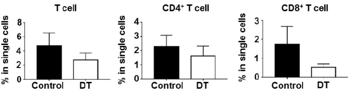
The cDCs and pDCs were depleted by intraperitoneally administering diphtheria toxin (DT; Calbiochem, San Diego, CA, USA) to Zbtb46-DTR BM transplanted mice and BDCA2-DTR BM transplanted mice, respectively. 500 ng DT was injected 2 days before H. felis infection. During the infection period, Zbtb46-DTR BM transplanted mice were treated with 100 ng DT every 2 days, and BDCA2-DTR BM transplanted mice were treated with 200 ng DT every 3 days.
The control group was treated with PBS instead of DT with the same schedule.
Necropsy and Tissue preparation
At 2, 4, and 8 weeks after H. felis infection, each group of mice was sacrificed together with the non-infected control group, and stomach, gastric lymph node, and spleen were removed. The stomach was incised along the greater curvature and spread on a filter paper. After cutting the lesser curvature, half of the stomach was used for flow cytometry (FACS) and H. felis quantification. The lesser curvature portion of the other half was fixed in 10% neutral buffered formalin for histopathologic examinations, and the remaining was stored in a -70°C deep freezer
20
for subsequent analysis. At 18 months after H. pylori infection, the H. pylori- infected groups were treated in the same manner as above.
Histopathologic Examinations
Gastric tissues fixed in 10% neutral buffered formalin were cut into two strips.
After standard tissue processing, gastric tissues were embedded in paraffin wax.
Paraffin sections were stained with haematoxylin and eosin (H&E) and Alcian Blue (pH 2.5) stain kit (Vector Laboratories, Burlingame, CA, USA) and performed immunohistochemistry (IHC) for H+/K+-adenosine triphosphatase (ATPase) to assess histopathological changes. The degree of gastritis and pathological mucosal changes were assessed on the basis of previously established updated Sydney classification (Chen, van der Hulst et al. 1999; Sayi, Kohler et al. 2009). In the sections stained with Alcian Blue, the amount of mucin in the gastric mucosa was measured using mean intensity in Image J software (National Institutes of Health, Bethesda, MD, USA). In the sections with IHC for H+/K+-ATPase, the number of positive cells was manually counted. The mucosa of two different areas was examined per slide at ×100 magnification, and the same anatomic areas were selected for each sample.
Single-Cell Preparations and Flow Cytometric Analysis
Gastric single cells were prepared to investigate the type of inflammatory cells that infiltrate into the stomach during Helicobacter infection. The gastric tissues were incubated in Hank’s balanced salt solution (HBSS) containing 2 mM EDTA (Cosmo Genetech, Seoul, Korea), 2% FBS, and 1 mM DL-Dithiothreitol (Sigma-
21
Aldrich, St. Louis, MO, USA) for 15 min at 37°C with shaking. Then, the gastric tissue was finely trimmed and grinded using gentleMACSTM Dissociator (Miltenyi Biotec, Bergisch Gladbach, Germany) and incubated in HBSS containing 100 U/ml collagenase D (Roche, Mannheim, Germany), 10% FBS, and 10 mM HEPES for 30 min at 37°C with shaking. After incubation, the gastric tissue was grinded once more using the gentleMACSTM Dissociator, filtered through a 70 μm cell strainer, and centrifuged to collect the separated single cells. Finally, the single cells were prepared by combining the cells collected from the suspension and from the gastric tissue. The gastric lymph node and spleen were incubated with 400 U/ml collagenase D at 37°C for 20 min to obtain separated single cells. The prepared single cells were primarily stained with Zombie Aqua (BioLegend, San Diego, CA, USA) to discriminate dead cells as per manufacturer’s protocol. Zombie aqua stained cells were then processed for Fc receptor block by using TruStain FcX antibody (anti-CD16/32, clone 93, BioLegend), to avoid non-specific binding. After the Fc receptors block, cells were incubated with a mixture of fluorochrome- labelled antibodies at 4°C for 30 min. To stain transcription factors (Foxp3, IRF4, IRF8 and Zbtb46), cells were fixed and permeabilized with Foxp3/Transcription Factor Staining Buffer Set (Thermo Fisher Scientific), as per manufacturer’s protocol, and then stained with antibody against each transcription factor. After washing briefly with RPMI 1640 medium with 10% FBS, cells were analyzed using BD LSRFortessa (BD Biosciences, San Jose, CA, USA) and FlowJo software (version 9.9 and 10.4; developed by FlowJo, LLC).
22
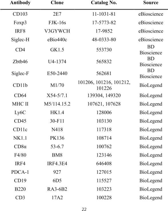
Antibodies for flow cytometry
The antibodies used for FACS are listed in Table 1.
Table 1. List of antibodies
Antibody Clone Catalog No. Source
CD103 2E7 11-1031-81 eBioscience
Foxp3 FJK-16s 17-5773-82 eBioscience
IRF8 V3GYWCH 17-9852 eBioscience
Siglec-H eBio440c 48-0333-80 eBioscience
CD4 GK1.5 553730 BD
Bioscience
Zbtb46 U4-1374 565832 BD
Bioscience
Siglec-F E50-2440 562681 BD
Bioscience CD11b M1/70 101206, 101216, 101212,
101226 BioLegend
CD64 X54-5/7.1 139304, 149320 BioLegend
MHC II M5/114.15.2 107621, 107628 BioLegend
Ly6C HK1.4 128006 BioLegend
CD45 30-F11 103130 BioLegend
CD11c N418 117318 BioLegend
NK1.1 PK136 108714 BioLegend
CD8α 53-6.7 100762 BioLegend
F4/80 BM8 123146 BioLegend
IRF4 IRF4.3E4 646408 BioLegend
PDCA-1 927 127015 BioLegend
CD19 6D5 115527 BioLegend
B220 RA3-6B2 103223 BioLegend
CD3 17A2 100228 BioLegend
23
Ly6G 1A8 127641 BioLegend
XCR1 ZET 148220 BioLegend
Rat IgG2α, κ
Isotype RTK2758 400507, 400511, 400526,
400542, 400549 BioLegend
Immunohistochemistry and Immunofluorescence
After paraffin-embedded sections were dewaxed and hydrated, antigen retrieval was performed using citrate buffer (pH 6.0; Sigma-Aldrich). Next, the sections were incubated with 0.3% hydrogen peroxide solution for 30 min to block endogenous peroxidase activity. The ImmPRESS Peroxidase Polymer kit (Vector Laboratories) was used according to manufacturer’s protocol. Briefly, paraffin- embedded sections were blocked with 2.5% normal horse serum or 2.5% normal goat serum for 1 hour and then incubated with the following primary antibodies at 4°C overnight or at 20°C for 2 hours: rabbit anti-CD3 (1:200; Dako, Glostrup, Denmark), rabbit anti-CD11c (1:400; Cell Signaling Technology, Danver, MA, USA), and mouse anti-ATPase (1:4000; MBL, Woburn, MA, USA). The paraffin- embedded sections were washed with PBS and then incubated with appropriate horseradish peroxidase-conjugated secondary antibody (Vector Laboratories) for 1 hour. ImmPACT DAB substrate (Vector Laboratories) was used for chromogenic detection. The sections were counterstained with hematoxylin. In anti-ATPase IHC, Mouse on Mouse ImmPRESS Peroxidase Polymer Kit (Vector Laboratories) was used to block endogenous mouse immunoglobulin.
For frozen sections of CD11c-EYFP mice, the sections were counterstained with DAPI and mounted using mounting medium (Vector Laboratories). The
24
images were obtained using confocal fluorescence microscope (Eclipse TE200;
Nikon, Japan).
H. felis Quantification
Total DNA was extracted from the gastric tissues (corpus and antrum) by using QIAamp DNA mini kit (Qiagen, Hilden, Germany) and used to identify H. felis DNA. Quantitative real-time PCR (qRT-PCR) was performed using specific primers for H. felis flagellar filament B (Fla-B), Rotor-Gene SYBR Green PCR kit, and Rotor-Gene Q (Qiagen). The amplified target DNA was analyzed using Rotor- Gene Q series software. The H. felis Fla-B primer–F: 5′-TTC GAT TGG TCC TAC AGG CTC AGA-3′ and R: 5′-TTC TTG TTG ATG ACA TTG ACC AAC GCA-3′–
was used (El-Zaatari, Kao et al. 2013). Next, the Warthin-Starry Stain Kit (Abcam) was used to identify the location of H. felis and to assess the extent of colonization in the stomach. Colonized H. felis were counted manually from two sections of each gastric tissue.
Quatitative Reverse Transcription-PCR
Total RNA from gastric tissues was extracted using Hybrid-R RNA purification kit (GeneAll, Seoul, Korea). The cDNA was synthesized from 0.1 μg of RNA by using QuantiTect Reverse Transcription kit (Qiagen). qRT-PCR was performed using specific primers, Rotor-Gene SYBR Green PCR kit, and Rotor- Gene Q (Qiagen). The amplified target cDNA was analyzed using Rotor-Gene Q series software against the housekeeping gene GAPDH. The specific primer pairs were F: 5′-TGC ACC ACC AAC TGC TTA G-3′ and R: 5′-GGA TGC AGG GAT
25
GAT GTT C-3′ for mouse GAPDH; F: 5′-AGC GGC TGA CTG AAC TCA GAT TGT AG-3′ and R: 5′-GTC ACA GTT TTC AGC TGT ATA GGG-3′ for mouse IFN- γ; F: 5′-GCT CCA AGA CCA AGG TGT CT-3′ and R: 5′-CTA GGT CCT GGA GTC CAG CA-3′ for mouse IL-10; and F: 5′-CTT CAA TAC GTC AGA CAT TCG GG-3′ and R: 5′-GTA ACG CCA GGA ATT GTT GCT A-3′ for mouse TGF-β.
Primers for mouse Granzyme A (cat. QT01551690) and perforin (cat. QT00282002) were purchased from Qiagen QuantiTect Primer Assay (Qiagen).
Magnetic Luminex immunoassay
For protein extraction, gastric tissues of H. felis-infected and non-infected WT and Flt3 KO mice were lysed using Cell Lysis Buffer 2 (R&D systems, Minneapolis, MN, USA). Chemokine and cytokine protein concentrations were measured using Mouse Premixed Multi-Analyte Kit (cat. LXSAMSM; R&D systems,) according to the manufacturer protocol.
Statistical Analysis
All data are shown as means ± standard errors. Statistical analysis was performed using GraphPad Prism 7.00 (GraphPad Software, San Diego, CA, USA).
Two groups were compared using Mann-Whitney U test or unpaired t-test with two- tailed distributions. When three or more groups were included, the results were analyzed using Kruskal-Wallis test or ordinary one-way ANOVA followed by Mann-Whitney U test or unpaired t-test with two-tailed distributions. Meanwhile, Pearson’s correlation coefficient was used to determine statistical dependence of H.
26
felis clearance and immune cell population. Two-tailed P values less than 0.05 were considered significant.
27
Results
Gastric DCs have three distinct subpopulations
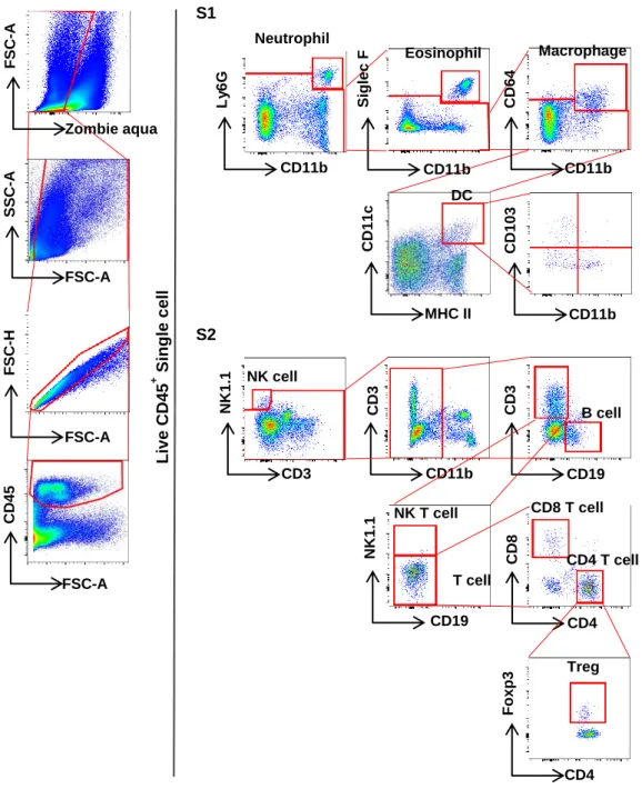
To characterize gastric DCs, CD45+ leukocytes were gated out from live cells and neutrophils (Ly6G+CD11b+), eosinophils (Siglec F+CD11b+), and macrophages (CD64+CD11b+) were sequentially excluded from the CD45+ cells. Next, gastric DCs were defined as CD64-CD11c+MHC II+ cells and their levels of CD103 and CD11b expression were further analyzed (Fig 1A). Under normal condition, the gastric CD64-CD11c+MHC II+ cells occupied about 0.24% of the total live single cells and were composed of three subsets including CD103+CD11b- (17.9%), CD103-CD11b+ (45.3%), and CD103-CD11b- (double-negative, DN; 32.1%) populations (Fig 1A and 2A).
To confirm that these subsets are DCs, the level of surface marker expression of each subset was measured by FACS analysis. The expression of Zbtb46, which is expressed specifically in human and murine cDCs (Reizis 2012; Satpathy, KC et al. 2012), was higher in all subsets, especially in the CD103+ subset, than in the isotype control. The expression of IRF4 and IRF8 was significantly higher in the CD11b+ and CD103+ subsets than in the isotype control. The DN subset also showed higher levels of IRF4 and IRF8 than those in the isotype control. The transcription factors IRF4 and IRF8 regulate CD11b+ and CD103+ cDC development, respectively (Mildner and Jung 2014). Among the known transcription factors involved in CD103+ cDC development, IRF8 appears to be the most important factor (Seillet, Jackson et al. 2013). The expression of XCR1, a potential marker for human and mouse CD103+ DCs (Bachem, Güttler et al. 2010),
28
was higher in all DC subsets than in the isotype control and was more prominent in the CD103+ subset (Fig 1B).
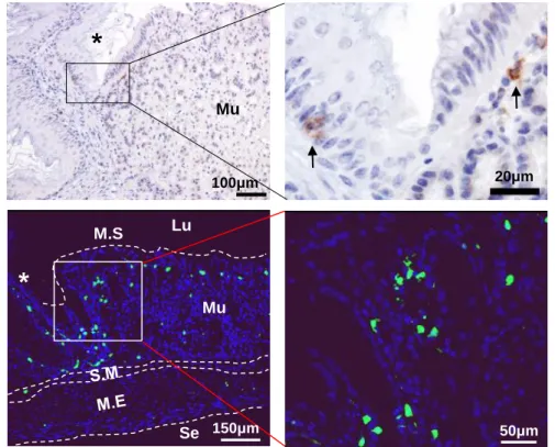
To further confirm that the CD64-CD11c+MHC II+ cells are DCs, the stomachs of Flt3 KO mice deficient in peripheral DCs were examined. Flt3 is an important regulator of DC homeostasis at the periphery, and only cDCs and pDCs are deficient in peripheral lymphoid organs of Flt3 KO mice (Waskow, Liu et al. 2008). Further, as the non-lymphoid tissues of Flt3 KO mice, the aorta have fewer cDCs and pDCs (Choi, Cheong et al. 2011; Yun, Lee et al. 2016). The number of CD64- CD11c+MHC II+ cells and CD103+, CD11b+, and DN subsets was markedly lower in the stomach of Flt3 KO mice than in that of WT mice (Fig 1C). To confirm the location of DCs in the normal mouse stomach, immunostaining of CD11c was performed and the gastric tissues of normal CD11c-EYFP mice were observed by confocal microscopy. The CD11c+ cells were very few and were mainly located in the superficial layer of the mucosal lamina propria and were well observed around the limiting ridge (Fig 1D).
29
A
CD45
FSC-A
SSC-A
FSC-A
FSC-H
FSC-A
CD64
CD11b
CD11c
MHC II CD11b
Live CD45+ cell
Ly6G
CD11b
Siglec F
CD11b Live whole cell
Single cell
CD45+ cell
Neutrophil Eosinophil Macrophage
DC CD103+ DC
CD11b+ DC DN DC
CD103
30
B
C
C
CD103+ DC
CD11b+ DC
IRF4 XCR1
Zbtb46 IRF8
DN DC
CD11c
MHC II
WT FLT3
CD45+ KO Ly6G- Siglec F- CD64-
**
**
*
DC CD103+ CD11b+
*
0 DN 0.1 0.2 0.3
% in single cells WT
Flt3 KO
31
D
Fig 1. Characterization of gastric DCs. (A) Gating strategy to identify DCs and DC subsets from isolated cells of entire glandular stomach. (B) Distinctive marker expression of DC subsets. CD103+ DCs are IRF8+XCRhigh, CD11b+ DCs are IRF4+XCRlow, and CD103-CD11b- DCs show mixed expression profile of these markers. Note that all DC subsets express Zbtb46. (C) The percentage of DCs and DC subsets in the live single cells of the stomach in the normal Flt3 KO and WT mice. Representative FACS plots (left panel). Gastric DCs and all DC subsets are deficient in Flt3 KO mice (n=9) compared to WT mice (n=11). (*P < 0.01 and **P
< 0.001) (D) Localization of gastric CD11c+ DCs (arrows) under the steady state.
Top panel: Representative images of CD11c IHC (DAB). Bottom panel:
Representative images of normal CD11c-EYFP mouse stomach. The asterisk indicates a limiting ridge that is the boundary of the glandular and nonglandular stomach. (Lu: lumen, M.S: mucosal surface, Mu: mucosa, S.M: submucosa, M.E:
muscularis externa, and Se: serosa)
Lu
Se
*
M.S
Mu 100μm 20μm
*
150μm 50μm
Mu
32
Helicobacter infection increased the number of DCs in the stomach
To investigate the role of DCs in gastritis, changes of gastric DCs in H. felis- infected WT mice were examined by sequential autopsy at 2, 4, and 8 weeks after infection. FACS analysis showed that, with an increase in the duration of H. felis infection, the infiltration of leukocytes and DCs into the stomach increased. During 2 to 8 weeks of H. felis infection, infiltration of CD45+ leukocytes and CD64- CD11c+MHC II+ DCs into the stomach was more apparent than that under steady state, approximately 1.8 to 2.8 fold and 2.1 to 4.2 fold, respectively, based on the ratio of single cells. Furthermore, the CD103+ and DN subsets increased markedly at 2, 4, and 8 weeks after H. felis infection, and the CD11b+ subset increased significantly at 4 and 8 weeks after H. felis infection (Fig 2A). To observe the changes in the distribution and number of DCs in the stomach, immunostaining of CD11c was performed. CD11c+ cells were mainly located in the inflamed submucosa and mucosal laminar propria right above the muscularis mucosa, and a few were distributed throughout the mucosal lamina propria. The number of CD11c+ cells infiltrating the stomach gradually increased during H. felis infection (Fig 2B).
33
A
CD45
FSC-A
CD11c
MHC II
CD103
CD11b CD45+ Ly6G- Siglec F- CD64-
2 weeksNormal4 weeks8 weeks
H. felis infected
17.9
32.1 45.3 28.8
40.4 14.1
49.0 34.0 16.4
44.4 35.3 15.1
26.5
38.4
42.3
28.0
2.7 3.0 2.3 2.2
34
B
2 weeks50μm Mucosa
Muscularis mucosa Submucosa
4 weeks
50μm Mucosa
Muscularis mucosa
Submucosa
35
Fig 2. Numerical increase and location of gastric DCs during H. felis infection. (A) The percentage of leukocytes, DCs, and DC subsets in the live single cells of the stomach in the WT mice with H. felis infection. Top panel: Representative FACS plots. Bottom panel: Asterisks at 2, 4, and 8 weeks after H. felis infection (n=13-14 for each group) signify significant increase compared to that in normal WT mice (n=11). (B) Localization of CD11c+ DCs (arrows) in H. felis infected WT mouse stomach. Top: CD11c IHC (DAB). Bottom: Manual cell count number of CD11c+ DCs in 2 paraffin sections (n=5 for each group) (*P < 0.01, **P < 0.001, and ***P
< 0.0001) Weeks: duration of infection
8 weeks
50μm Mucosa
Muscularis mucosa
Submucosa
36
Flt3 KO mice have more severe gastric inflammation with less Helicobacter colonization
To determine the role of DCs in Helicobacter-induced gastritis, whether the immune responses differed between H. felis-infected Flt3 KO and WT mice was investigated. First, the severity of gastritis between the two groups was compared at 2, 4, and 8 weeks after H. felis infection. Histologically, inflammation occurred mainly in the corpus. In the WT mice, infiltration of neutrophils into the submucosa was mainly observed after 2 weeks of infection, and infiltration of lymphocytes and plasma cells as well was noted at 4 weeks and the most prominently at 8 weeks after infection. These inflammatory cells mostly infiltrated into the submucosa, and, as the severity of gastritis increased, they infiltrated into the mucosal lamina propria.
In addition, lymphoid follicles were occasionally present in the submucosa or mucosa. In contrast, in Flt3 KO mice, infiltration of not only neutrophils but also many lymphocytes and plasma cells was noted at 2 weeks and continued to increase gradually until 8 weeks. The infiltration of inflammatory cells was mainly observed in the submucosa, but often diffusely expanded to the entire mucosal lamina propria.
Therefore, gastritis was more marked in Flt3 KO mice than in WT mice (Fig 3A).
In the corpus mucosa of mice in which inflammation had spread to the mucosa, varying numbers of parietal cells were lost and replaced by foveolar cell and mucous neck cell hyperplasia. In addition, some mature chief cells transdifferentiated to the metaplastic mucous-secreting cells, which appears to be a process of spasmolytic polypeptide-expressing metaplasia (SPEM) (Weis, Sousa et al. 2013). The degree of loss of parietal cells was confirmed by IHC for H+/K+ ATPase and the degree of mucous neck cell hyperplasia and transdifferentiation of
37
chief cells was confirmed by Alcian blue staining (Fig 3B). The mucosal changes were more prominent in Flt3 KO mice, which have more severe gastritis, than in WT mice (Fig 3C). Furthermore, in H. pylori infection, gastritis tended to be more severe in Flt3 KO mice than in WT mice. According to the updated Sydney classification, inflammation scores were 2 ± 1 in Flt3 KO mice and 1 or 2 in WT mice (Fig 3D).
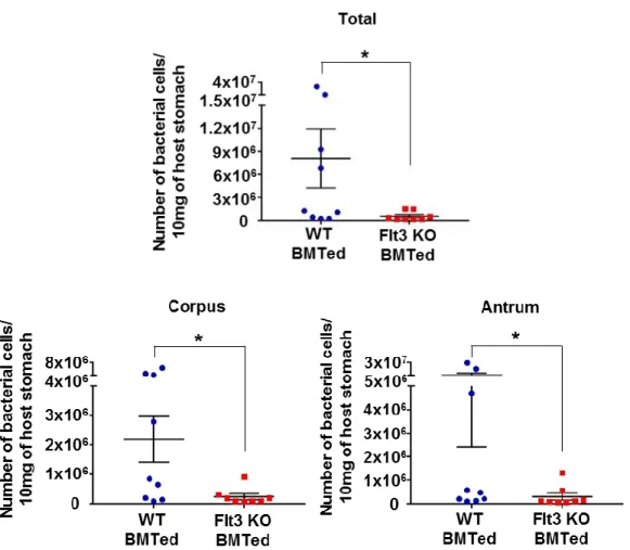
Many previous studies have shown a strong positive correlation between the degree of inflammation and clearance of infectious agents. The clearance of Helicobacter in the stomach has been reported to be dependent on CD4+ T cell- mediated IFN-γ (Sayi, Kohler et al. 2009). Therefore, the degree of H. felis colonization in each group was measured. H. felis was mainly present in the gastric pits and mucus layer throughout the glandular part (Fig 3E). The degree of H. felis colonization increased during 8 weeks of infection. Bacterial load was lower in Flt3 KO mice than in WT mice, and this difference was the most noticeable after 8 weeks of infection (Fig 3F and 3G).
38
A
C
WT Flt3 KO
2 weeks4 weeks8 weeks
66μm 66μm 66μm
66μm 66μm 66μm
Mucosa
Muscularis mucosa Submucosa
Mucosa
Muscularis mucosa Submucosa
Mucosa
Muscularis mucosa Submucosa
39
B
C
WT Flt3 KO
2 weeks 50μm 50μm
50μm 50μm
4 weeks8 weeks
50μm 50μm
50μm 50μm
50μm 50μm
50μm 50μm
*
Inflammation score
*
Mean intensity in mucosa
*
2 4 8
(weeks)
2 4 8
(weeks) WT Flt3 KO
0 1 2 3 4 5 6
0 10 20 15 25
5
*
0 400 200 600 800 1000
Parietal cell number
*
* *
2 4 8
(weeks)
*
40
D
E
WT Flt3 KO
18 months18 months
200μm 200μm
50μm 50μm
20μm 50μm 20μm
50μm 150μm
150μm WT
Flt3 KO
41
F
G
0 500 1000 1500 2000
Number of bacteria
*
WT Flt3 KO
2 4 8 (weeks)
Total
42
Fig 3. Deterioration of Helicobacter-induced gastric lesions and decrease of bacterial load in Flt3 KO mice (A) Histopathological analysis of H. felis-induced gastritis. H&E stained representative images of WT (left panel) and Flt3 KO (right panel) mice were aligned. (B) Histopathological analysis of H. felis-induced oxyntic atrophy and mucous metaplasia. Representative serial images of WT (left panel) and Flt3 KO (right panel) mice subjected to Alcian blue staining (top) and H+/K+-ATPase IHC (bottom) were aligned. (2, 4, and 8 weeks after H. felis infection). (C) Top: Inflammation score. Each mouse was scored (n=13-17 for each group). Middle and Bottom: Mean intensity of mucosal Alcian blue positive and manual cell count number of H+/K+-ATPase+ cells (parietal cells), respectively (n=10 for each group). (D) Histopathological analysis of H. pylori-induced gastritis.
H&E stained representative sections of H. pylori-infected WT (left panel) and Flt3 KO (right panel) mice (WT–n=7; Flt3 KO–n=5) (E) Localization of H. felis (white arrowheads) in WT and Flt3 KO mouse stomach (Warthin-Starry stain). (F) Manual count number of H. felis in 2 paraffin sections (n=4-7 for each group). (G) H. felis loads were evaluated using qRT-PCR for H. felis Fla-B DNA extracted from the corpus and antrum (n=7-10 for each group). (*P < 0.05 and **P < 0.01) Weeks:
duration of infection
43
Flt3 KO mice showed more prominent CD8+ T cell accumulation than WT mice during Helicobacter infection
FACS analysis was used to observe the changes in various leukocyte populations that infiltrated into H. felis-infected stomach (Fig 4). Among the leukocyte populations, T cell infiltration was the most prominent difference between WT and Flt3 KO mice. In Flt3 KO mice, infiltration of CD8+ T cell was dramatically higher than that in WT mice. In addition, infiltration of CD4+ T cell tended to be greater in Flt3 KO mice than in WT mice when the H. felis infection persisted for 8 weeks. However, no difference in the extent of Treg infiltration was noted between the two groups (Fig 5A). These phenotypes were recapitulated in the H. pylori-infected Flt3 KO mice (Fig 5B).