Surgical Outcomes of Balanced Deep Lateral and Medial Orbital Wall Decompression in Korean Population: Clinical and Computed Tomography-based Analysis
7
0
0
전체 글
(2) Korean J Ophthalmol Vol.30, No.2, 2016. ical treatments conducted to prevent disease progression, including high-dose steroids and radiation therapy, disfiguring exophthalmos eventually develops in most cases of TAO. Orbital decompression is a major method in the treatment of TAO with severe exophthalmos [5]. Bony decompression techniques, including lateral wall, inferomedial wall, balanced lateral and medial walls, or three-wall (medial, lateral, and inferior) techniques, have been introduced as principal treatments of TAO-related disfigured exophthalmos. However, conventional three-wall decompression has a relatively high incidence of postoperative diplopia [6]. Unal et al. [6] and Graham et al. [7] suggested balanced orbital decompression as a viable alternative to orbital three-wall decompression; the techniques provide comparable reduction of proptosis with low incidence of postoperative diplopia. Recently, deep lateral orbital wall decompression has been reported as a good method of decompression due to its location immediately posterior to the globe and better retroplacement with lower risk of postoperative complications of diplopia, dysesthesia, and sinusitis [8,9]. Although there are many reports on the outcomes of deep lateral orbital wall decompression, there are no reports about balanced deep lateral and medial orbital wall decompression in Korean patients. Thus, in this study, we performed balanced orbital decompressions with medial wall and deep lateral wall decompressions in Korean patients in order to determine the surgical outcomes and postoperative complications. In addition, we demonstrated the effect of each type of wall decompression by analyzing the postoperative expansion of orbital volume as measured in computed tomography (CT) images.. Materials and Methods Patients A retrospective chart review was conducted for all consecutive cases of TAO-associated exophthalmos surgically treated by a single surgeon (JKL) at Chung-Ang University Hospital between January 1, 2011 and December 31, 2013. The study protocol was approved by the institutional review board of Chung-Ang University Hospital (no. C2014116[1312]).. 86. We included 48 orbits from 24 patients that underwent balanced deep lateral and medial orbital decompression. Patients with medical history of orbital or eyelid inflammation, prior orbital surgery and/or trauma, re-operation, concomitant orbital strut removal, or prior history of orbital radiation were excluded. To ensure that the progression of TAO had ceased and to avoid active inflammatory periods, we delayed decompression surgery for more than 6 months in patients with clinical activity score less than 3 points and with prior history of medical steroid therapies within the past 6 months. Preoperative exophthalmos was measured using Hertel exophthalmometry (Oculus, Arlington, WA, USA) at less than 2 weeks prior to orbital decompression and 1 and 3 months after surgery. Orbital CT scans (Philips Brilliance 256 Slice iCT; Philips Healthcare Systems, Andover, MA, USA) was conducted routinely in all enrolled patients at less than 2 weeks prior to orbital decompression and 3 months after surgery. Postoperative complications were recorded during the follow-up periods. Surgical techniques Decompression of the deep lateral orbital wall was performed through an eyelid crease incision. The greater wing of the sphenoid bone was removed, and additional removal of the anterior department of the inferior orbital fissure was performed in most patients. Medial orbital decompression was performed via a transcaruncular incision. After exposure of the medial wall immediately posterior to the posterior lacrimal crest, the wall was fractured, and an anterior and posterior ethmoidectomy was performed using Kerrison rongeurs and Takahashi forceps. The superior limit of the medial wall removal was the ethmoidal vessels, and the inferior limit included the ethmoid-maxillary bony strut. Estimation of the decompressed volume of the orbital wall on computed tomography images CT imaging (bone window, 2.5-mm collimation) of all patients was performed on both axial and coronal planes. Orbital volume expansion after surgery was calculated using Image J software (National Institutes of Health, http://rsbweb.nih.gov/ij/) and was sorted into two groups: deep lateral orbital wall and medial orbital wall. The ex-.
(3) SU Choi, et al. Surgical Outcomes of Balanced Orbital Decompression. Data analysis Partial correlation analysis was used to compare decompression volume in the deep lateral wall and medial wall groups with exophthalmos reduction in order to determine area-adjusted surgical predictability. IBM SPSS ver. 19.0 (IBM Co., Armonk, NY, USA) was used for all statistical analyses, and a p-value less than 0.05 indicated statistical significance. The values shown represent the mean ± standard deviation.. tively). Postoperative estimated decompression volumes are shown in Table 2. The bony decompressed volume estimated by serial CT images was 0.68 ± 0.23 cm3 in the deep lateral orbital wall and 0.80 ± 0.29 cm3 in the medial orbital wall (Table 2). With regard to postoperative complications, four patients experienced change of upper eyelid fold (upper eyelid crease height asymmetry and multiple folds), four patients had facial dysesthesia, and one patient had conjunctival granuloma (Table 3). New onset strabismus was found in one patient (2 prism diopter esotropia, 7 prism diopter right hypertropia), but the issue resolved. 25. Results. Table 1. Demographics of the study population Total no. of orbits. Data 48 in 24 patients. Sex Male (n = 5). 10 (20.8). Female (n = 19). 38 (79.2). Age (yr). 15. 10. 5. Forty-eight orbits from 24 patients underwent balanced deep lateral and medial wall decompression. Nineteen patients were women and five were men. The average follow-up period was 11.46 ± 6.55 months. The mean patient age was 34.08 ± 7.03 years. Patient demographics are summarized in Table 1. The mean exophthalmos value decreased from a preoperative value of 18.91 ± 1.43 to 15.10 ± 1.53 and 14.91 ± 1.49 mm at postoperative 1 and 3 months, respectively (Fig. 1). The exophthalmic reduction was significant between the preoperative and postoperative 1 month time points, as well as the preoperative and postoperative 3 month time points ( p < 0.001, p < 0.001, respec-. Demographics. p < 0.001 ** * p < 0.001. 20 Hertel value (mm). panded area in each orbital slice was marked from the estimated preoperative wall border to the decompressed bony edge, and the marked areas were calculated with Image J software. The deep lateral wall was analyzed on axial CT planes, and medial walls were analyzed in the same manner on corneal planes. The summation of all areas (cm 2) multiplied by 0.25 cm was considered the expanded orbital volume (cm3) in each orbital wall [10].. 34.08 ± 7.03. Values are presented as number (%) or mean ± standard deviation.. 0. Preoperative. Postoperative 1 mon. Postoperative 3 mon. Fig. 1. Hertel value at preoperative and postoperative 1 and 3 months (*paired t-test, **paired t-test).. Table 2. Decompression volumes in different areas Area of decompression. Volume (cm3)*. Deep lateral orbital wall. 0.68 ± 0.23. Medial orbital wall. 0.80 ± 0.29. Values are presented as mean ± standard deviation. Estimated value according to computed tomography analysis.. *. Table 3. Postoperative complications Complication. No. of patients (%). Strabismus Postoperative 1 mon. 1 (4.17). Postoperative 3 mon. 0. Change of upper eyelid fold. 4 (16.67). Facial dysesthesia. 4 (16.67). Caruncular conjunctival granuloma. 1 (4.17). Total. 10 (41.67). 87.
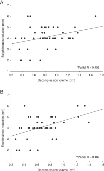
(4) Korean J Ophthalmol Vol.30, No.2, 2016. spontaneously within 3 months. Horizontal ocular deviation (exophoria) was improved after surgery in all six affected patients. In the partial correlation analysis, orbital volume expansion in deep lateral wall decompressions and medial wall decompressions were significantly correlated with exophthalmos reduction after surgery ( p = 0.002 and 0.011, respectively) (Fig. 2A and 2B). The partial correlation coefficient (partial R) of partial correlation analysis, or the correlational power between the amount of expanded decompression volume and exophthalmos reduction and consequent reflection of surgical predictability, was higher in. A. 7. Exophthalmos reduction (mm). 6 5 4 3 2 1. *Partial R = 0.402 0.2. 0.4. 0.6. 0.8. 1.0. 1.2. 1.4. 1.6. 1.8. Decompression volume (cm3). B. 7. Exophthalmos reduction (mm). 6 5 4 3 2 1. **Partial R = 0.487 0.2. 0.4. 0.6. 0.8. 1.0. 1.2. 1.4. 1.6. Decompression volume (cm3). Fig. 2. Partial correlation analysis of decompression volume in medial wall (A) and deep lateral wall (B) with postsurgical exophthalmos reduction. Partial correlation coefficient (partial R) which represent surgical predictability, was higher in deep lateral orbital wall decompression (B, partial R = 0.487) than medial orbital wall decompression (A, partial R = 0.402. *p = 0.002, **p = 0.011).. 88. deep lateral orbital wall decompression than medial orbital wall decompression (partial R = 0.487 and 0.402, respectively) (Fig. 2).. Discussion It is widely known that symmetric lateral and medial expansion of the orbit can balance shifting of the muscle cone, and balanced lateral and medial orbital wall decompression can reduce the incidence of postoperative diplopia [11]. In previous studies, however, the incidence of postoperative diplopia was reported to be as high as 30.7% to 33% after balanced lateral and medial wall orbital decompression [12,13]. These rates of postoperative diplopia are not lower than those of other conventional orbital wall decompression methods. In the case of lateral orbital wall decompression, the removed lateral orbital wall is replaced with temporalis fascia, which is soft tissue, rather than a hard structure like the lateral orbital wall. Fayers et al. [14] reported that, after lateral orbital wall decompression, 35% of the patients had noted postoperative oscillopsia. They also reported larger lateral osteotomies as a risk factor of oscillopsia. Based on these findings, after lateral orbital wall decompression, intraorbital structures become more vulnerable to mechanical insult that causes functional and positional change in the extraocular muscles and eyeball. These can affect differences in the occurrence of diplopia between previous conventional balanced orbital decompression and our study, in which we performed deep lateral rather than lateral orbital wall decompression. Therefore, it is important to determine whether balanced lateral and medial wall orbital decompression should be performed and, if so, whether the deep lateral wall is an alternative decompression target. In this study, our results showed a minimal occurrence of postoperative diplopia, which is a major advantage of the technique we used. Orbital strut was described in 1992 as an approximately 3-mm-thick strut of the orbit, which serves an important function in preventing post-decompression dystopia [15]. We maintained the orbital strut at the junction between the medial and inferior orbital walls during medial wall decompression. This was thought to reduce both inferomedial globe shifting and occurrence of diplopia. Furthermore, Ben Simon et al. [9] reported that deep lateral orbital wall decompression had no statistically.
(5) SU Choi, et al. Surgical Outcomes of Balanced Orbital Decompression. significant influence on horizontal or vertical deviation. Also, we could perform decompression surgery with orbital strut preservation because the exophthalmos of the subjects was moderate (range, 17 to 21 mm). However, the orbital strut is not always easy to preserve and might be sacrificed when further volume expansion is necessary. Wu et al. [16] reported orbital fat decompression only with relatively low occurrence of postoperative diplopia. Therefore, if additional reduction of proptosis is needed, adjunctive fatty decompression will be a good alternative instead of removal of the orbital strut in order to avoid occurrence of postoperative diplopia. Although CT-based measurement of decompressed orbital volume at the deep lateral wall (0.68 ± 0.23 cm3) produced a lower value than that of the medial wall (0.80 ± 0.29 cm3) (Table 2), volume-adjusted correlations on the partial correlation analysis (partial R) between exophthalmos reduction and expanded decompression volume were higher in deep lateral decompression than in medial decompression (partial R = 0.487 and 0.402, respectively) (Fig. 2). The differences in surgical predictability specific to the target wall of decompression are possibly due to the location of each wall. The deep lateral wall is located immediately posterior to the globe; because of this, its removal allows for better retroplacement of the globe compared to that of medial orbital wall decompression [17,18]. On the other hand, the medial orbital wall is positioned on the lateral aspect of the orbital tissue, so medial orbital wall decompression has relatively less efficiency on direct retroplacement of the eyeball compared with deep lateral orbital wall decompression. In the present study, decompression volume-adjusted exophthalmos reduction was better correlated with decompression volume in the deep lateral wall as opposed to the medial wall, which is consistent with our previous study [10]. However, the benefits of bone decompression of the deep lateral wall are limited by the naturally smaller bone volume to decompress and individual anatomical differences of the deep lateral orbital wall compared with those of the medial wall. Thus, surgeons should take into account both surgical predictability and surgical efficiency when considering balanced deep lateral wall and medial wall decompression and should balance the decompression volume of each wall relative to the preoperative exophthalmos levels. Prior volumetric studies using CT analysis have centered on defining normative data of the orbital bony structure. [18,19]. Another study using CT analysis to determine the surgical effect of orbital decompression was based not on orbital bony decompression, but on orbital fat decompression [20]. Alsuhaibani et al. [21] performed volumetric studies using CT scans with balanced orbital decompression. They showed that medial orbital wall decompression more highly affects orbital content expansion than does lateral wall decompression. This result supports our study; however, one difference was that the authors focused on orbital volume and eye position changes rather than surgical predictability and effects. Also, the difference in estimated volume of the medial wall might be caused by differences in the method used to estimate volu me. Alsuhaibani et al. [21] used computer software that presumes the volume using a 3D reconstruction method. In contrast, we analyzed collimated CT images with Image J software in order to estimate bony decompression volume. There has been no study comparing surgical effect and predictability in balanced orbital decompression based on quantitative CT analysis. Our study exclusively represents analysis of decompression volume with exophthalmic reduction and its predictability in balanced deep lateral and medial orbital decompression. Previously, Goldberg et al. [18] introduced three areas of deep bone in the lateral orbit as possible targets of lateral orbital decompression: the lacrimal keyhole, the orbital door jamb, and the basin of the inferior orbital fissure. Our surgical target of deep lateral orbital wall decompression corresponded to the orbital door jamb, with exception of the area of lateral orbital rim. The average volume of the orbital door jamb measured by Goldberg et al. [18] in a Western population was 2.9 cm3. In contrast, in our study, the average decompressed volume of the identical counterpart was 0.68 cm3. These differences can be explained in several ways. First, Goldberg measured the volume of orbital wall before decompression surgery. Such an approach was idealized for practical purposes, but it would be difficult to remove all of the bony volume of the orbital wall. Second, Goldberg included the lateral orbit rim area when measuring the volume of the orbital door jamb. However, in our study, the lateral orbital rim was preserved. Lefebvre and Yoon [22] recently reported no significant differences in volume of the sphenoid trigone between racial groups, with anaverage volume of 1.97 and 1.39 cm 3 in Asian men and women, respectively. In the present study, the proportion of female patients was 79.2%, and almost. 89.
(6) Korean J Ophthalmol Vol.30, No.2, 2016. half of the deep lateral orbital wall volume was decompressed. This is similar to the estimated volume after balanced orbital decompression reported by Alsuhaibani et al. [21]. There are some limitations to this study. When performing quantitative CT analysis, we used Image J software, which can introduce intraobserver and interobserver variation. In order to minimize this variation, all parameters were measured three times by three different ophthalmologists and the nine total measurements were averaged. Other limitations of this study are its relatively small sample size and retrospective study design. Further studies with larger sample sizes will be needed to confirm our findings on balanced deep lateral and medial orbital wall decompression. In conclusion, balanced deep lateral and medial orbital wall decompression is a safe and effective surgical method in patients with TAO and disfiguring exophthalmos. The method has beneficial results, including prevention of postoperative diplopia and significant reduction of exophthalmos levels. According to our quantitative CT analysis, the ophthalmologist can predict surgical effects of balanced deep lateral and medial orbital wall decompression. Furthermore, these results provide information that will help during preoperative counseling and surgical decision-making.. 95. 5. Lyons CJ, Rootman J. Orbital decompression for disfiguring exophthalmos in thyroid orbitopathy. Ophthalmology 1994;101:223-30. 6. Unal M, Leri F, Konuk O, Hasanreisoglu B. Balanced orbital decompression combined with fat removal in Graves ophthalmopathy: do we really need to remove the third wall? Ophthal Plast Reconstr Surg 2003;19:112-8. 7. Graham SM, Brown CL, Carter KD, et al. Medial and lateral orbital wall surgery for balanced decompression in thyroid eye disease. Laryngoscope 2003;113:1206-9. 8. Baldeschi L, MacAndie K, Hintschich C, et al. The removal of the deep lateral wall in orbital decompression: its contribution to exophthalmos reduction and influence on consecutive diplopia. Am J Ophthalmol 2005;140:642-7. 9. Ben Simon GJ, Syed HM, Lee S, et al. Strabismus after deep lateral wall orbital decompression in thyroid-related orbitopathy patients using automated hess screen. Ophthalmology 2006;113:1050-5. 10. Kim KW, Byun JS, Lee JK. Surgical effects of various orbital decompression methods in thyroid-associated orbitopathy: computed tomography-based comparative analysis. J Craniomaxillofac Surg 2014;42:1286-91. 11. European Group on Graves Orbitopathy, Mourits MP, Bijl H, et al. Outcome of orbital decompression for disfiguring proptosis in patients with Graves’ orbitopathy using various surgical procedures. Br J Ophthalmol 2009;93:1518-23.. Conflict of Interest. 12. Goldberg RA, Perry JD, Hortaleza V, Tong JT. Strabismus after balanced medial plus lateral wall versus lateral wall only orbital decompression for dysthyroid orbitopathy.. No potential conflict of interest relevant to this article was reported.. Ophthal Plast Reconstr Surg 2000;16:271-7. 13. Kacker A, Kazim M, Murphy M, et al. “Balanced” orbital decompression for severe Graves’ orbitopathy: technique. References. with treatment algorithm. Otolaryngol Head Neck Surg 2003;128:228-35. 14. Fayers T, Barker LE, Verity DH, Rose GE. Oscillopsia after. 1. Wiersinga WM, Smit T, van der Gaag R, et al. Clinical presentation of Graves’ ophthalmopathy. Ophthalmic Res 1989;21:73-82. 2. McCord CD Jr. Current trends in orbital decompression. Ophthalmology 1985;92:21-33. 3. Trokel SL, Jakobiec FA. Correlation of CT scanning and pathologic features of ophthalmic Graves’ disease. Ophthalmology 1981;88:553-64. 4. Heufelder AE. Pathogenesis of ophthalmopathy in autoimmune thyroid disease. Rev Endocr Metab Disord 2000;1:87-. 90. lateral wall orbital decompression. Ophthalmology 2013; 120:1920-3. 15. Goldberg RA, Shorr N, Cohen MS. The medical orbital strut in the prevention of postdecompression dystopia in dysthyroid ophthalmopathy. Ophthal Plast Reconstr Surg 1992;8:32-4. 16. Wu CH, Chang TC, Liao SL. Results and predictability of fat-removal orbital decompression for disfiguring graves exophthalmos in an Asian patient population. Am J Ophthalmol 2008;145:755-9..
(7) SU Choi, et al. Surgical Outcomes of Balanced Orbital Decompression. 17. Mehta P, Durrani OM. Outcome of deep lateral wall rim-sparing orbital decompression in thyroid-associated. 6. 20. Adenis JP, Robert PY, Lasudry JG, Dalloul Z. Treatment of. orbitopathy: a new technique and results of a case series.. proptosis with fat removal orbital decompression in Graves’. Orbit 2011;30:265-8.. ophthalmopathy. Eur J Ophthalmol 1998;8:246-52.. 18. Goldberg RA, Kim AJ, Kerivan KM. The lacrimal key-. 21. Alsuhaibani AH, Carter KD, Policeni B, Nerad JA. Orbital. hole, orbital door jamb, and basin of the inferior orbital fis-. volume and eye position changes after balanced orbital de-. sure: three areas of deep bone in the lateral orbit. Arch Ophthalmol 1998;116:1618-24. 19. Beden U, Edizer M, Elmali M, et al. Surgical anatomy of the deep lateral orbital wall. Eur J Ophthalmol 2007;17:281-. compression. Ophthal Plast Reconstr Surg 2011;27:158-63. 22. Lefebvre DR, Yoon MK. CT-based measurements of the sphenoid trigone in different sex and race. Ophthal Plast Reconstr Surg 2015;31:155-8.. 91.
(8)
수치
관련 문서
The thickness of the maxillary sinus lateral wall according to tooth site and measurement level was measured by using image-processing software and its histologic
유재순( 2 0 0 9 ) 은 청소년의 건강위험행위를 관리하기 위해서는 건강위험행위 전체 를 포괄하는 통합적인 접근 전략이 효과적이라고 하였고,손은성( 2 0 0 4 )
I=0 for incompressible fluid III=0 for simple shear flow.. limitations
In the efficiency review of the Trombe wall system through the experiment, if the Trombe wall was applied in the building structure, the Trombe wall had the
캐나다정부간행물목록(Weekly checklist of Canadian government publications) 에 수록된 자료 중 Folder자료, 인구센서스, 전화번호 자료 등을 제외한
In 1396, a wall with four main gates and four smaller gates was constructed to keep the city safe from invaders.. Although two of the gates are now gone, many sections of
상기 신입생 장학금 외에도 본교는 신입생장학금-재학생장학금-해외연수장학금-대학원진학장학금에 이르는 전주기 장학제도를 운영하고 있으며, 다양한 교외장학금
1 John Owen, Justification by Faith Alone, in The Works of John Owen, ed. John Bolt, trans. Scott Clark, "Do This and Live: Christ's Active Obedience as the
