MALIGNANT PERICARDIAL EFFUSION IN CARCINOMA OF THE UTERINE CERVIX
Mi Hyang Kim, MD, Tae Hwa Lee, MD, Chun June Lee, MD, Heong Yeol Kim, MD, Won Gyu Kim, MD, Sung Han Kim, MD
Department of Obstetrics and Gynecology, Kosin University College of Medicine, Busan, Korea
The occurrence of malignant pericardial effusion is rare in squamous cell carcinoma of the uterine cervix. Only six cases of antemortem diagnosis have been reported. We report a patient with squamous cell carcinoma of the cervix who received emergent pericardiocentesis. In a few reported cases, women with pericardial involvement are candidates for radiation or chemotherapy.
Early detection and prompt management of pericardial effusion is correlated with better quality of life and a decreased morbidity.
Keywords:
Pericardial effusion, Squamous cell carcinoma of uterine cervix, Pericardiocentesis
Received: 2010.12. 6. Revised: 2011. 2.21. Accepted: 2011. 3.15.
Corresponding author: Tae Hwa Lee, MD
Department of Obstetrics and Gynecology, Kosin University College of Medicine, 34 Amnam-dong, Seo-gu,
Busan 602-702, Korea
Tel: +82-51-990-6722 Fax: +82-51-244-6939 E-mail: [email protected]
Th is is an Open Access article distributed under the terms of the Creative Commons Attribution Non-Commercial License (http://creativecommons.org/licenses/
by-nc/3.0/) which permits unrestricted non-commercial use, distribution, and reproduction in any medium, provided the original work is properly cited.
Copyright © 2011. Korean Society of Obstetrics and Gynecology
Cervical cancer is the second most common cancer in women worldwide, and nearly a third of patients who present with invasive cervical cancer will die of this disease [1]. Metastasis of squamous cell carcinoma of the uterine cervix to the pericardium is an uncommonly appreciated event. Tumors often metastasize to the pericardium in patients with melanoma, lung carcinoma, lymphoma, and breast carcinoma and sarcoma [2]. The incidence of cardiac metastasis determined by autopsy data has been reported to be 0.3-3.2% in deceased patients with cervical cancer [3]. According to a review of the literature, only six previously reported cases of antemortem diagnosis of malignant pericardial effusion from carcinoma of the cervix have been presented in detail [4]. We report a patient with squamous cell carcinoma of the cervix who received emergent pericardiocentesis.
Case Report
A 52-year-old, multiparous (para 1-0-0-1) woman was referred to our hospital in June 2009 with a one-month history of dyspnea and cough. She had a negative personal and family history for other diseases. The cytology of pleural effusion was negative for malignancy.
She had a palpable mass on the left side of the neck. The examination of her left neck lymph nodes revealed squamous cell carcinoma, and her positron-emission tomography-computed tomography (PET- CT) scan showed both hydronephrosis, hypermetabolism of the uterus, and metastatic lymphadenopathies at the perigastric,
WWW.KJOG.ORG 214
peripancreatic, para-aortic, left axillary, both supraclavicular, and the left neck level II-V areas.
On pelvic examination, the cervical tumor was found to involve the vagina and the parametrium. Cervical biopsy showed squamous cell carcinoma. From June 2009 to August 2009, the patient received three cycles of 5-FU and cisplatin chemotherapy and concurrent radiation. After chemo-radiotherapy, follow-up PET-CT showed no evidence of hypermetabolism at the cervix, but metastatic lymphadenopathy was remained in the right inguinal area. However, all results showed an improved state since 22 June 2009. PET-CT showed stable disease, according to the Response Evaluation Criteria in Solid Tumors criteria.
From September 2009 to November 2009, the patient received three cycles of topotecan and cisplatin chemotherapy. Bilateral pleural effusion was slightly increased after this treatment. Follow-
CASE REPORT
Korean J Obstet Gynecol 2011;54(4):214-217 doi: 10.5468/KJOG.2011.54.4.214
pISSN 2233-5188 · eISSN 2233-5196
up pelvis magnetic resonance imaging (MRI) on 24 November 2009 showed recurrence at the right adnexa. In December 2009, a chest-tubing procedure was performed in the right side of the chest due to increased pleural effusion; the amount of drainage was 1,650 mL.
When 5-FU and cisplatin chemotherapy was completed, the patient was treated for general weakness. On 22 December 2009 she suddenly complained of dyspnea and discomfort in the left chest, and symptoms did not subsided with an oxygen mask.
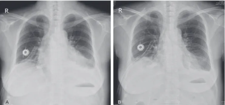
Electrocardiogram (ECG) revealed sinus tachycardia and low- voltage QRS waves. Her chest X-ray showed an increase in the size of the cardiac silhouette and small pleural effusion (Fig. 1 A). The patient’s blood pressure was 90/40 mm Hg, heart rate
was 132 and body temperature was 35.5
oC. Echocardiogram demonstrated the presence of a large pericardial effusion (Fig. 2).
A pericardiocentesis was performed under ultrasound guidance.
About 800 mL of fluid was drained, and a catheter was placed and remained for seven days. Cytological examination showed squamous cell carcinoma (Fig. 3). On 28 December 2009, the patient complained of dyspnea and discomfort of the left chest again. ECG showed no pericardial effusion, but the amount of left pleural effusion had increased. A chest-tubing procedure was performed in the left side of the chest, and the amount of drainage was 1,300 mL. After the procedure, the patient was treated with Lasix and Isoket. Sclerosis was performed in the left chest with cisplatin. Her symptoms likely would have improved,
WWW.KJOG.ORG 215
A
BFig. 1. Pericardial effusion before pericardiocentesis (A) and after pericardiocentesis (B).
A B
Fig. 2. Pericardial effusion (arrow) on parasternal long-axis view (A) and apical four-chamber view (B).
Mi Hyang Kim, et al. Malignant pericardial effusion in carcinoma of the uterine cervix
WWW.KJOG.ORG 216
but she expired one month after the pericardiocentesis.
Discussion
Malignant pericardial effusion secondary to pericardial metastasis from squamous cell carcinoma of the uterine cervix represents an infrequently seen, but potentially life-threatening problem. Mean survival periods following diagnosis of a malignant pericardial effusion in patients with solid tumors other than breast cancer are three to four months; in comparison, patients with metastatic breast cancer generally survive for more than nine months [4].
The normally compliant pericardium may slowly collect up to two liters of fluid before tamponade occurs. Early symptoms, which occur in only 5-27% of those affected prior to the hemodynamic consequences of tamponade, include dysphagia or singultus from mass effect, chest pain or pressure, cough, orthopnea, dyspnea, and/or palpitations. Later symptoms of a large effusion include exertional fatigue due to low cardiac output and systemic venous congestion [5]. Physical findings of significant effusion include jugular venous distension, quiet heart sounds, narrow pulse pressure, tachycardia, and pulsus paradoxus greater than 10 mm Hg. ECG changes include atrial arrhythmias, low voltages, atrioventricular block, repolarization changes, and more specifically, electrical alternans [6]. Radiographic studies to detect or determine the nature of effusion include serial chest X-rays showing cardiac enlargement in the absence of pulmonary vascular congestion, especially with a “water-bottle heart” configuration. Management depends on hemodynamic factors. If tamponade is present, immediatede compression by
pericardiocentesis or immediate pericardial window in centers so oriented is indicated. Subsequent steps are controversial [7].
Pericardiocentesis is best performed in an intensive-care setting with appropriate monitoring, usually under ultrasound guidance.
In this setting, major morbidity is uncommon, but mortalities have been reported from ventricular puncture [8]. Intracavitary instillation of agents such as bleomycin or tetracycline has proven both safe and effective to control malignant pleural effusion, and has experienced with their use in pericardial effusive disease and has confi rmed their utility in that setting as well [9].
Tetracycline sclerosis after pericardiocentesis is another conservative therapeutic option. This procedure is relatively simple and is 60- 80% effective in controlling symptomatic effusion with few complications in reported series [10]. Prior to 1976, various sources reported that chemotherapy for metastatic cervical squamous cell carcinoma had response rates of 0-25%. Gynecologic Oncology Group experience, reported by Thigpen et al. [11], noted a 38% response to cisplatin 50 mg/m
2each three weeks and responses of 0-19 % for other agents. Selected responses to combined agents were reported by Thigpen et al. [11] to be as high as 93% for mitomycin C and bleomycin, ranging from 6-93%
for two-to seven-drug regimens [12].
Repeated aspirations and instillations may be required for satisfactory control of malignant pericalrdial effusion. Adverse effects associated with intrapericardial instillation include severe local pain with mechlorethamine and quinacrine, bone marrow suppression with alkylating agents and rarely sepsis, arrythemias, ventricular laceration, and death [13].
The best candidates for radiotherapy are probably patients with radiosensitive tumors in whom other therapies have failed or who are chemoresistant. Studies examining various primary tumors have reported external radiotherapy success rates ranging from 50-100% for control of pericardial effusion [14].
Surgical management may therefore be reserved for patients with treatment failures, a constrictive component to the physiologic process and/or the need for histologic confi rmation. Early detection and prompt management of pericardial effusion in patients with gynecologic malignancies may decrease morbidity and prolong life [5]. Carcinoma of the uterine cervix with extrapelvic metastasis is currently an in curable disease. The occurrence of pericardial effusion in this disease is rare, and yet its early recognition is important to prevent cardiac tamponade [15].
Oncologists have previously regarded the development of a pericardial effusion as a pre-terminal event. More recent data contradict this belief. Aggressive local cardial effusion provides
Fig. 3. A few clusters of anaplastic squamous cells seen in the pericardialfl uid (H&E, ×100).
KJOG Vol. 54, No. 4, 2011
Mi Hyang Kim, et al. Malignant pericardial effusion in carcinoma of the uterine cervix
palliation of symptoms and may prolong life in women with cervical cancer [5].
Acknowledgments
This study was supported by a research grant from Kosin University College of Medicine, Korea.
References
1. Jemal A, Siegel R, Ward E, Hao Y, Xu J, Murray T, et al. Cancer statistics, 2008. CA Cancer J Clin 2008;58:71-96.
2. Lockwood WB, Broghamer WL Jr. The changing prevalence of secondary cardiac neoplasms as related to cancer therapy.
Cancer 1980;45:2659-62.
3. Hayashi Y, Iwasaka T, Hachisuga T, Kishikawa T, Ikeda N, Sugi- mori H. Malignant pericardial effusion in endometrial adeno- carcinoma. Gynecol Oncol 1988;29:234-9.
4. Nelson BE, Rose PG. Malignant pericardial effusion from squa- mous cell cancer of the cervix. J Surg Oncol 1993;52:203-6.
5. Rieke JW, Kapp DS. Successful management of malignant peri- cardial effusion in metastatic squamous cell carcinoma of the uterine cervix. Gynecol Oncol 1988;31:338-51.
6. Kralstein J, Frishman WH. Malignant pericardial diseases: diag- nosis and treatment. Cardiol Clin 1987;5:583-9.
7. Hankins JR, Satterfield JR, Aisner J, Wiernik PH, McLaughlin JS. Pericardial window for malignant pericardial effusion. Ann Thorac Surg 1980;30:465-71.
8. Celermajer DS, Boyer MJ, Bailey BP, Tattersall MH. Pericardio- centesis for symptomatic malignant pericardial effusion: a study of 36 patients. Med J Aust 1991;154:19-22.
9. Shepherd FA, Morgan C, Evans WK, Ginsberg JF, Watt D, Mur- phy K. Medical management of malignant pericardial effusion by tetracycline sclerosis. Am J Cardiol 1987;60:1161-6.
10. Shepherd FA, Ginsberg JS, Evans WK, Scott JG, Oleksiuk F. Tet- racycline sclerosis in the management of malignant pericardial effusion. J Clin Oncol 1985;3:1678-82.
11. Thigpen T, Vance RB, Balducci L, Blessing J. Chemotherapy in the management of advanced or recurrent cervical and endo- metrial carcinoma. Cancer 1981;48(2 Suppl):658-65.
12. Choo YC. Chemotherapy in advanced primary and recurrent cervical carcinoma. Int J Gynaecol Obstet 1982;20:417-23.
13. Davis S, Rambotti P, Grignani F. Intrapericardial tetracycline sclerosis in the treatment of malignant pericardial effusion: an analysis of thirty-three cases. J Clin Oncol 1984;2:631-6.
14. Cham WC, Freiman AH, Carstens PH, Chu FC. Radiation therapy of cardiac and pericardial metastases. Radiology 1975;114:701-4.
15. Jamshed A, Khafaga Y, El-Husseiny G, Gray AJ, Manji M. Peri- cardial metastasis in carcinoma of the uterine cervix. Gynecol Oncol 1996;61:451-3.
WWW.KJOG.ORG 217
악성 심낭삼출액을 보인 자궁경부암
고신대학교 의과대학 복음병원 산부인과
김미향, 이태화, 이천준, 김흥열, 김원규, 김성한
자궁경부 편평상피암 환자에서 악성 심낭삼출액의 발생은 매우 드물다. 현재까지 영어로 쓰여진 논문에서 자궁경부암 환자에서 악성 심낭 삼출액이 사전 진단된 경우는 6예에 불과하다. 본 저자는 자궁경부 편평상피암 환자에서 응급 심막천자가 필요했던 사례를 보고하고자 한 다. 다른 일부 사례연구에서, 심막으로 전이된 많은 여성에게 적극적인 방사선 또는 항암치료를 할 필요가 있다고 보고하였다. 심낭삼출액 의 조기발견 및 즉각적인 치료가 환자의 사망률을 감소시키고 보다 나은 삶의 연장과 밀접한 관계가 있다.
중심단어: 심낭삼출액, 편평상피암, 심막천자