https://doi.org/10.20307/nps.2018.24.2.132
132
Phytochemical Constituents of Capsella bursa-pastoris and Their Anti-inflammatory Activity
Joon Min Cha
1, Dong Hyun Kim
1, Tae Hyun Lee
1, Lalita Subedi
2, Sun Yeou Kim
2, and Kang Ro Lee
1,*
1
Natural Products Laboratory, School of Pharmacy, Sungkyunkwan University, 2066 Seobu-Ro, Jangan-gu, Suwon, Gyeonggi-do 440-746, Republic of Korea
2
Gachon Institute of Pharmaceutical Science, Gachon University, 191 Hambakmoero, Yeonsu-gu, Incheon 21936, Republic of Korea
Abstract − Phytochemical investigation of 80% MeOH extract of the aerial parts of Capsella bursa-pastoris yielded fourteen compounds (1 - 14). The structures of the compounds were elucidated by spectroscopic methods to be methyl-1-thio-β-D-glucopyranosyl disulfide (1), 10-methylsulphinyl-decanenitrile (2), 11-methyl-sulphinyl- undecanenitrile (3), 1-O-(lauroyl)glycerol (4), phytene-1, 2-diol (5), (3S,5R,6S,7E)-5,6-epoxy-3-hydroxy-7- megastigmen-9-one (6), loliolide (7), β-sitosterol (8), 3-hydroxy-1-(4-hydroxy-3-methoxyphenyl)-1-propanone (9), 1-feruloyl-β-D-glucopyranoside (10), pinoresinol-4'-O-β-D-glucopyranoside (11), luteolin (12), quercetin-3-O-β- D-glucopyranoside (13), and luteolin 6-C-β-glucopyranoside (14). Although compound 1 was reported as synthetic compound, 1 was first isolated from natural source. NMR spectral data assignments of 1, 2 and 3 were reported for the first time, and compounds 1 - 14 were for the first time reported from this plant source. The anti- inflammatory effects of 1 - 14 were evaluated in lipopolysaccharide (LPS)-stimulated murine microglia BV-2 cells. Compounds 12 exhibited strong inhibitory effects on nitric oxide production in LPS-activated BV-2 cells with IC
50values of 9.70 µM.
Keywords − Capsella bursa-pastoris, Cruciferae, Sulfur compound
Introduction
Capsella bursa-pastoris (L.) Medik (Cruciferae), com- monly known as Shepherd’s purse, is widely distributed throughout the world.
1The root of this plant was edible and has been used in Korean folks medicine for the treatment of edema and hypertension.
2Previous phytoche- mical investigation on this plant reported the isolation of flavonoids, terpenoids and phenolic compounds,
2-4with their biological studies, such as anti-microbial, anti- bacterial, anti-tumor, and liver catalase activities.
5-7As parts of our search for biologically active compounds from Korean natural plant sources, we investigated the constituents of the aerial parts of C. bursa-pastoris and reported the isolation of phenolic glycosides and their anti-inflammatory effects.
8In our continuing study on this plant source, we further isolated fourteen compounds, including three sulfur compounds (1 - 3). The structures
of these compounds were elucidated by physicochemical and spectroscopic methods. The isolated compounds (1 - 14) were evaluated for their potential inhibitory effects on NO production in LPS-activated microglia BV-2 cell line.
Experimental
General experimental procedures – Melting point was determined on Gallenkamp melting point apparatus.
Optical rotations were measured on a Jasco P-1020 polarimeter in MeOH. IR spectra were recorded on a Bruker IFS-66/S FT-IR spectrometer. HR-FAB-MS spectra were obtained on a JEOL JMS700 mass spectrometer.
NMR spectra were recorded on a Bruker AVANCE III 700 NMR spectrometer at 700 MHz (
1H) and 175 MHz (
13C). Preparative high performance liquid chromatography (HPLC) was conducted using a Gilson 306 pump with Shodex refractive index detector and Phenomenex-Luna- 10u-silica-(2) column (250 × 10.00 mm) or YMC J’sphere ODS-M80 column (250 × 10.00 mm). Low-pressure liquid chromatography (LPLC) was carried out on a Merck LiChroprep Lobar
®-A RP-C
18and Si 60 column (240 × 10
*Author for correspondence
Kang Ro Lee, Natural Products Laboratory, School of Pharmacy, Sungkyunkwan University, 2066Seobu-Ro, Jangan-ku, Suwon, Gyeonggi-do 440-746, Republic of Korea.
Tel: +82-31-290-7710; E-mail: [email protected]
mm) with an FMI QSY-0 pump (ISCO). Silica gel 60 (Merck, 70 – 230 and 230 - 400 mesh) and RP-C18 silica gel (Merck, 230 - 400 mesh) were used for column chromatography. The packing material for molecular sieve column chromatography was Sephadex LH-20 (Pharmacia Co.). TLC was performed using Merck precoated Silica gel F254 plates and RP-18 F254s plates. Spots were
detected by thin layer chromatography (TLC) under UV light or by heating after spraying with anisaldehyde–
sulfuric acid.
Plant materials – Whole plants of C. bursa-pastoris
(2.5 kg) were purchased from Anmyeon-Island, Chung-
cheongnam-do, Korea in March 2015. A voucher specimen
(SKKU-NPL 1410) has been deposited in the herbarium
Fig. 1. The structures of 1 - 14 isolated from C. bursa-pastoris.of the School of Pharmacy, Sungkyunkwan University, Suwon, Korea.
Extraction and isolation – The whole plants of C.
bursa-pastoris (2.5 kg) were extracted three times with 80% aqueous MeOH. The filtrate was evaporated under reduced pressure to give a MeOH extract (280 g), which was suspended in distilled water (800 mL) and solvent- partitioned to give n-hexane (33.0 g), CHCl
3(5.0 g), EtOAc (4.0 g), and n-BuOH (26.0 g). The CHCl
3layer (4.0 g) was chromatographed on a silica gel column (CHCl
3- MeOH = 40:1 → 1:1) to yielded six fractions (C1–C6).
Fraction C1 (1.1 g) was separated over a silica gel column (CHCl
3-MeOH = 40:1 → 1:1) to give eight subfractions (C11–C18). Fraction C13 (248 mg) was subjected to Sephadex LH-20 column chromatography eluted with 100% MeOH as to give seven subfractions (C131–C137).
Subfraction C137 (20 mg) was purified with a RP-C
18prep HPLC (100% MeOH) to yield 5 (20 mg). Fraction C2 (671 mg) was separated on a silica gel column (CHCl
3-MeOH = 100:1 → 1:1) to give eight subfractions (C21–C28). Subfraction C26 (207 mg) was chromato- graphed over RP-C
18silica Lobar
®-A (80% MeOH) and a RP-C
18prep HPLC (65% MeOH) to yield 2 (48 mg), 3 (20 mg) and 4 (5 mg). Subfraction C27 (116 mg) was chromatographed over RP-C
18silica Lobar
®-A (80%
MeOH) and a RP-C
18prep HPLC (40% MeOH) to yield 6 (5 mg), 7 (5 mg) and 9 (5 mg). Subfraction C28 (20 mg) was purified with a RP-C
18prep HPLC (100% MeOH) to yield 5 (5 mg). The EtOAc layer (4.0 g) was separated on a RP-C
18silica gel column with 40~100% MeOH to yielded sixteen fractions (E1–E16). Fraction E3 (120 mg) was chromatographed over silica Lobar
®- A (CHCl
3- MeOH-H
2O = 4:1:0.1) and a RP-C
18prep HPLC (10%
MeCN) to yield 1 (15 mg) and 10 (17 mg). Fraction E5 (150 mg) was chromatographed over a Sephadex LH-20 column (85% MeOH) to yield five subfractions (E51–
E55). Subfraction E55 (31 mg) was purified with a RP- C
18prep HPLC (20% MeCN) to yield 14 (9 mg). Fraction E8 (116 mg) was chromatographed over silica Lobar
®-A (EtOAc-MeOH-H
2O = 5:1:0.1) and a RP-C
18prep HPLC (25% MeCN) to yield 11 (10 mg). Fraction E11 (150 mg) was chromatographed over a Sephadex LH-20 column (100% MeOH) and a RP-C
18prep HPLC (10% MeCN) to yield 13 (38 mg). Fraction E13 (208 mg) was chromato- graphed over silica Lobar
®-A (CHCl
3-MeOH-H
2O = 7:1:
0.1) and a RP-C
18prep HPLC (30% MeCN) to yield 12 (11 mg).
Methyl-1-thio-β-D-glucopyranosyl disulfide (1) − Yellowish gum. : -78.0 ( c 0.1, MeOH); IR (KBr) ν
max: 3412, 2875, 1642, 1031 cm
−1;
1H,
13C NMR see
Table 1; HR-FAB-MS (positive-ion mode) m/z: 265.0178 [M+Na]
+(calcd for C
7H
14O
5S
2Na, 265.0180).
10-Methylsulphinyl-decanenitrile (2) − Colorless gum.
: -8.3 ( c 0.01, CHCl
3); IR (KBr) ν
max: 3420, 3010, 2204, 1665, 1483, 1035, 984, 671 cm
−1;
1H,
13C NMR see Table 2; HR-FAB-MS (positive-ion mode) m/z: 216.1424 [M+H]
+(calcd for C
11H
22NOS, 216.1422).
11-Methylsulphinyl-undecanenitrile (3) − Colorless gum. : -10.1 ( c 0.01, CHCl
3); IR (KBr) ν
max: 3213, 3102, 2306, 1664, 1427, 1028, 991, 598 cm
−1;
1H,
13CNMR see Table 2; HR-FAB-MS (positive-ion mode) m/z:
230.1580 [M+H]
+(calcd for C
12H
24NOS, 230.1579).
1- O-(Lauroyl)glycerol (4) − Colorless gum.
1H NMR (CDCl
3, 700 MHz): δ 4.14 (1H, dd, J = 11.3, 3.9 Hz, H- 3a), 4.05 (1H, dd, J = 10.2, 5.7 Hz, H-3b), 3.81 (1H, m, H-1b), 3.66 (1H, m, H-2), 3.56 (1H, dd, J = 11.1, 5.8 Hz, H-1a), 2.30 (1H, t, J = 7.3Hz, H-5), 1.62 (2H, m, H-6), 1.31 (18H, brs, H-7 to 14), 0.88 (3H, t, J = 6.7 Hz, H-15);
13
C NMR (CDCl
3, 175 MHz): δ 173.0 (C-4), 70.1 (C-2), 64.8 (C-3), 63.3 (C-1), 33.7 (C-5), 31.5 (C-13), 28.9 (C- 11), 28.9 (C-10), 28.9 (C-9), 28.8 (C-12), 28.8(C-8), 28.7 (C-7), 25.1 (C-6), 22.3 (C-14), 13.8 (C-15); FABMS m/z 273.1 [M+H]
+.
Phytene-1,2-diol (5) − Colorlessgum.
1H NMR (CDCl
3, 700 MHz): δ 5.00 (1H, s, H-17a), 4.84 (1H, s, H-17b), 4.02 (1H, dd, J = 7.0, 3.2 Hz, H-2), 3.71 (1H, dd, J = 6.5, 4.0 Hz, H-1a), 3.42 (1H, dd, J = 6.5, 2.1 Hz, H-1b), 2.20 – 2.01 (2H, m, H-4), 1.57-1.06 (19H, m), 0.91 (3H, d, J = 7.0 Hz, H-16), 0.88 (6H, d, J = 7.0 Hz, H-18, 20), 0.85 (3H, d, J = 7.0 Hz, H-19);
13C NMR (CDCl
3, 175 MHz): δ 151.0 (C-3), 109.1 (C-17), 74.3 (C-2), 65.5 (C- 1), 38.2 (C-14), 37.0 (C-8), 36.9 (C-10, 12), 36.8 (C-6), 33.1 (C-4), 31.0 (C-7, 11), 28.5 (C-15), 26.4 (C-5), 23.5 (C-13), 23.1 (C-9), 22.4 (C-20), 22.5 (C-16), 19.0 (C-19),
α[ ]D19
α [ ]D19
α [ ]D24
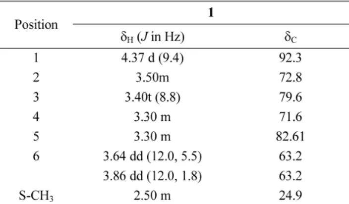
Table 1. 1H and 13C NMR data of 1 in CD3OD (δ in ppm, 700 MHz for 1H and 175 MHz for 13C)
Position 1
δH (J in Hz) δC
1 4.37 d (9.4) 92.3
2 3.50m 72.8
3 3.40t (8.8) 79.6
4 3.30 m 71.6
5 3.30 m 82.61
6 3.64 dd (12.0, 5.5) 63.2
3.86 dd (12.0, 1.8) 63.2
S-CH3 2.50 m 24.9
a1H and 13C NMR data were recorded at 700 and 175 MHz, respectively.
Coupling constants (in Hz) are given in parentheses.
18.9 (C-18); FABMS m/z 313.3 [M+H]
+.
(3 S,5R,6S,7E)-5,6-Epoxy-3-hydroxy-7-megastigmen- 9-one (6) − Colorless gum. : -53.4 ( c 0.01, CHCl
3);
1
H NMR (CDCl
3, 700 MHz): δ 7.05 (1H, d, J = 15.6 Hz, H-7), 6.31 (1H, d, J = 15.6 Hz, H-8), 3.93 (1H, m, H-3), 2.41 (1H, dd, J = 9.2, 5.0 Hz, H-4a), 2.31 (3H, s, CH
3- 10), 1.68 (1H, dd, J = 14.6, 9.2 Hz, H-4b), 1.60 (1H, dd, J = 12.2, 4.2 Hz, H-2a), 1.30 (1H, dd, J = 12.2, 10.2 Hz, H-2b), 1.22 (3H, s, CH
3-13), 1.20 (3H, s, CH
3-11), 1.00 (3H, s, CH
3-12);
13C NMR (CDCl
3, 175 MHz): δ 197.6 (C-9), 142.6 (C-7), 132.8 (C-8), 69.6 (C-6), 67.4 (C-5), 64.2 (C-3), 46.8 (C-4), 40.8 (C-2), 35.3 (C-1), 29.5 (C- 11), 28.5 (C-10), 25.2 (C-12), 20.0 (C-13); FABMS m/z 225.1 [M+H]
+.
Loliolide (7) − Colorless gum. : -32.1 ( c 0.01, CHCl
3);
1H-NMR (CDCl
3, 700 MHz): δ 5.72 (1H, s, H- 7), 4.36 (1H, m, H-3), 2.49 (1H, td, J = 13.8 Hz, H-4α), 2.00 (1H, dd, J=14.4 Hz, H-2α), 1.81 (3H, s, H-9), 1.74 (1 H, d, J = 3.3 Hz, H-4β), 1.60 (1 H, d, J = 3.6 Hz, H- 2 β), 1.50 (3H, s, H-10), 1.30 (3H, s, H-11);
13C-NMR (CDCl
3, 175 MHz): δ 185.6 (C-8), 171.4 (C-6), 112.5 (C- 7), 87.6 (C-5), 65.2 (C-3), 48.3 (C-2), 47.0(C-4), 38.1 (C- 1), 32.5 (C-9), 27.3 (C-10), 27.0 (C-11); FABMS m/z 197.1 [M+H]
+.
β-Sitosterol (8) − White powder, mp. 145
oC;
1H-NMR (CDCl
3, 700 MHz): δ 5.35 (1H, d, J = 5.0 Hz, H-6), 3.53 (1H, m, H-3), 1.06 (3H, s, H-19), 0.95 (3H, d, J = 6.5 Hz, H-21), 0.92 (3H, d, J = 6.5 Hz, H-26), 0.80 (3H, t, J = 6.5 Hz, H-29), 0.76 (3H, s, H-18).
13C-NMR (CDCl
3, 175
MHz): δ 141.7 (C-5), 120.9 (C-6), 73.4 (C-3), 58.0 (C- 14), 56.3 (C-17), 51.2 (C-9), 47.0 (C-24), 46.1 (C-4), 43.5 (C-13), 40.3 (C-12), 38.2 (C-1), 35.1 (C-10), 36.2 (C-20), 34.0 (C-22), 32.8 (C-2), 31.5 (C-8), 29.4 (C-25), 27.9 (C- 16), 26.5 (C-23), 25.8 (C-15), 23.4 (C-28), 20.6 (C-11), 20.0 (C-27), 19.3 (C-26),19.1(C-21), 19.0 (C-19), 12.0 (C-29), 11.9 (C-18); FABMS m/z 415.3 [M+H]
+.
3-Hydroxy-1-(4-hydroxy-3-methoxyphenyl)-1-propa- none (9) − Colorless gum.
1H-NMR (CD
3OD, 700 MHz):
δ 7.55 (1H, dd, J = 8.5, 2.0 Hz, H-6), 7.53 (1H, d, J = 2.0 Hz, H-2), 6.95 (1H, d, J = 8.5Hz, H-5), 4.02 (2H, t, J = 5.5Hz, H-5), 3.96 (3H, s, 3-OCH
3), 3.18 (2H, t, J = 5.5Hz, H-5);
13C-NMR (CD
3OD, 175 MHz): δ 199.3 (C-7), 152.4 (C-3), 148.4 (C-4), 131.0 (C-1), 123.8 (C-6), 115.4 (C-5), 111.9 (C-2), 58.5 (C-9), 56.5 (OCH
3), 50.8 (C-3), 41.6 (C-8); FABMS m/z 197.0 [M+H]
+.
1-Feruloyl-β-D-glucopyranoside (10) − Yellowish gum.
1
H NMR (CD
3OD, 700 MHz): δ 7.72 (1H, d, J = 15.9 Hz, H-8), 7.19 (1H, d, J = 1.9 Hz, H-2), 7.09 (1H, dd, J = 8.2, 1.8 Hz, H-6), 6.82 (1H, d, J = 8.3 Hz, H-5), 6.39 (1H, d, J = 15.9 Hz, H-7), 5.58 (1H, d, J = 7.9 Hz, H-1'), 3.86 (1H, s, 3-OCH
3), 3.83 (1H, m, H-6'), 3.70 (1H, m, H-3'), 3.43 (1H, m, H-5'), 3.31 (1H, m, H-2'), 3.23 (1H, m, H- 4');
13C NMR (CD
3OD,175 MHz): δ 151.1 (C-9), 149.3 (C-4), 147.6 (C-3), 147.4 (C-7), 137.6 (C-1), 133.9(C-6), 120.2 (C-8), 119.9 (C-2), 95.9 (C-1'), 78.9 (C-3'), 78.2 (C- 5'), 74.2 (C-2'), 71.9 (C-4'), 62.3 (C-6'), 56.9 (3-OCH
3);
FABMS m/z 357.1 [M+H]
+.
Pinoresinol-4'-O-β-D-glucopyranoside (11) − Yellowish
α[ ]D23
α [ ]D21
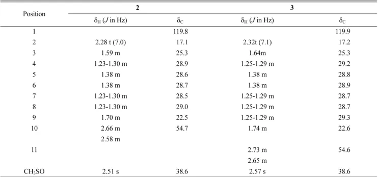
Table 2. 1H and 13C NMR data of 2 and 3 in CDCl3 (δ in ppm, 700 MHz for 1H and 175MHz for 13C)
Position 2 3
δH (J in Hz) δC δH (J in Hz) δC
1 119.8 119.9
2 2.28 t (7.0) 17.1 2.32t (7.1) 17.2
3 1.59 m 25.3 1.64m 25.3
4 1.23-1.30 m 28.9 1.25-1.29 m 29.2
5 1.38 m 28.6 1.38 m 28.8
6 1.38 m 28.7 1.38 m 28.9
7 1.23-1.30 m 28.5 1.25-1.29 m 28.7
8 1.23-1.30 m 29.0 1.25-1.29 m 28.7
9 1.70 m 22.5 1.25-1.29 m 29.3
10 2.66 m 54.7 1.74 m 22.6
2.58 m
11 2.73 m 54.6
2.65 m
CH3SO 2.51 s 38.6 2.57 s 38.6
a1H and 13C NMR data were recorded at 700 and 175 MHz, respectively. Coupling constants (in Hz) are given in parentheses.
gum. : +21.3 ( c 0.01, MeOH);
1H NMR (CD
3OD, 700 MHz): δ 7.14 (1H, d, J = 8.3 Hz, H-5), 7.02 (1H, d, J = 1.9 Hz, H-2), 6.94 (1H, d, J = 1.8 Hz, H-2'), 6.91 (1H, dd, J = 8.3, 1.9 Hz, H-6), 6.81 (1H, dd, J = 8.1, 1.9 Hz, H- 6'), 6.77 (1H, d, J = 8.1 Hz, H-5'), 4.88 (1H, d, J = 7.8 Hz, H-1''), 4.76 (1H, d, J = 4.1 Hz, H-7), 4.70 (1H, d, J = 4.5 Hz, H-7'), 4.24 (2H, m, H-9a, 9'a), 3.86 (3H, s, 3-OCH
3), 3.85 (3H, s, 3'-OCH
3), 3.82 (2H, m, H-9b, 9'b), 3.70 - 3.26 (5H, m, sugar-H), 3.12 (2H, m, H-8, 8');
13C NMR (CD
3OD, 175 MHz): δ 151.1 (C-4), 149.3 (C-4'), 147.6 (C-3'), 147.4 (C-3), 137.6 (C-1), 133.9(C-1'), 120.2 (C-6'), 119.9 (C-6), 118.2 (C-5), 116.2 (C-5'), 111.8 (C-2), 111.2 (C-2'), 102.9 (C-1''), 87.6 (C-7'), 87.2 (C-7), 78.3 (C-5''), 78.0 (C- 3''), 75.1 (C-2''), 72.8 (C-9'), 72.8 (C-9), 71.4(C- 4''), 62.6 (C-6''), 56.9 (3-OCH
3), 56.6 (3'-OCH
3), 55.6 (C- 8'), 55.5 (C-8); FABMS m/z 521.1 [M+H]
+.
Luteolin (12) − Yellowish gum.
1H NMR (CD
3OD, 700 MHz): δ 7.40 (1H, dd, J = 8.4, 2.0 Hz, H-6'), 7.38 (1H, d, J = 2.0 Hz, H-2'), 6.89 (1H, d, J = 8.4 Hz, H-5'), 6.53(1H, s, H-3), 6.43 (1H, s, H-8), 6.19 (1H, s, H-6).;
13C NMR (CD
3OD, 175 MHz): δ 184.1 (C-4), 166.3 (C-7), 166.1 (C-2), 163.2 (C-5), 159.5 (C-9), 151.7 (C-4'), 147.2 (C-3'), 123.1 (C-1'), 120.4 (C-6'), 116.9 (C-5'), 114.3 (C- 2'), 105.4 (C-10), 104.5 (C-3), 100.2 (C-6), 95.1 (C-8);
FABMS m/z 287.0 [M+H]
+.
Quercetin-3-O-β-D-glucopyranoside (13) − Yellowish gum. : -20.3 ( c 0.01, MeOH);
1H NMR (CD
3OD, 700 MHz): δ 7.72 (1H, br s, H-2'),7.59 (1H, d, J = 8.2 Hz, H-6'), 6.87 (1H, d, J = 8.2 Hz, H-5'), 6.37 (1H, s, H-6), 6.19 (1H, s, H-8), 5.24 (1H, s, J = 7.3 Hz, H-1''), 3.72 (1H, dd, J = 14.1, 1.30 Hz, H-6''), 3.58 (1H, dd, J = 11.5, 5.3 Hz, H-3''), 3.50 (1H, td, J = 10.5, 7.1 Hz, H-5''), 3.36 (1H, m, H-2''), 3.23 (1H, m, H-4'');
13C NMR (CD
3OD, 175 MHz): δ 173.6 (C-4), 166.2 (C-7), 163.2 (C-5), 153.1 (C-9), 158.6 (C-2), 150 (C-4'), 146.1 (C-3'), 135.7 (C-3), 123.3 (C-6'), 123.2 (C-1'), 117.7 (C-5'), 116.5 (C-2'), 105.8 (C-10), 104.5 (C-1''), 100.1 (C-6), 94.8 (C-8), 78.5 (C-3''), 78.2 (C-5''), 75.8 (C-2''), 71.3 (C-4''), 62.7 (C-6'');
FABMS m/z 465.0 [M+H]
+.
Luteolin 6-C-β-glucopyranoside (14) − Yellowish gum.
1
H NMR (CD
3OD, 700 MHz): δ 7.36 (2H, br s, H-2', 6'), 6.89 (1H, d, J = 8.3 Hz, H-5'), 6.54 (1H, s, H-3), 6.48 (1H, s, H-8), 4.90 (1H, d, J = 9.9 Hz, H-1''), 4.16 (1H, t, J = 9.1, 8.4 Hz, H-2''), 3.88 (1H, dd, J = 12.1, 1.8 Hz, H- 6''a), 3.74 (1H, dd, J = 12.1, 5.1 Hz, H-6''b), 3.48 (1H, m, H-5''), 3.42 (1H, m, H-4''), 3.41 (1H, m, H-3'');
13C NMR (CD
3OD, 175 MHz): δ 184.1 (C-4),165.1 (C-7), 162.2 (C- 5), 158.8 (C-9), 151.2 (C-4'), 147.2 (C-3'), 123.7 (C-1'), 120.5 (C-6'), 116.9 (C-5'), 116.4 (C-2), 105.3 (C-10), 95.3 (C-8), 82.7 (C-5''), 80.3 (C-3''), 75.5 (C-1''), 72.7 (C-4''),
71.9 (C-2''), 63.1 (C-6''); FABMS m/z 449.1 [M+H]
+. Measurement of nitric oxide production and cell viability − BV 2 cells were plated into a 96-well plate (3 × 10
4cells/well). After 24 h, cells were pretreated with compounds 1 - 14 for 30 min, and then stimulated with 100 ng/ml of LPS for another 24 h. Nitrite, a soluble oxidation product of NO, was measured in the culture media using the Griess reaction. The supernatant was har- vested and mixed with an equal volume of Griessreagent (1% sulfanilamide, 0.1% N-1 napthylethylenediamine dihydrochloride in 5% phosphoric acid). After 10min, the absorbance at 570 nm was measured using a microplate reader. Sodium nitrite was used as a standard to calculate the NO2- concentration. Cell viability was assessed by a 3-[4,5-dimethylthiazol-2-yl]-2,5-diphenyl-tetrazolium bromide (MTT) assay. NG-mono- methyl-L-arginine (L-NMMA, Sigma, St.Louis, MO, USA), a well-known nitric oxide synthase (NOS) inhibitor, was tested as a positive control.
Result and Discussion
Structures of compounds 4 - 14 were determined by comparing
1H,
13C NMR, and MS spectral data with those in the literatures to be 1- O-(lauroyl)glycerol (4),
9phytene- 1,2-diol (5),
10(3 S,5R,6S,7E)-5,6-epoxy-3-hydroxy-7-me- gastigmen-9-one (6),
11loliolide (7),
12β-sitosterol (8),
133- hydroxy-1-(4-hydroxy-3-methoxyphenyl)-1-propanone (9),
141-feruloyl- β-D-glucopyranoside (10),
15pinoresinol- 4'- O-β-D-glucopyranoside (11),
16luteolin (12),
17quercetin- 3- O-β-D-glucopyranoside (13),
18and luteolin 6- C-β-glu- copyranoside (14).
19Compounds 1 - 14 were first isolated from this sources. The following describes the structure elucidation of 1, which was first isolated from natural source, although it was reported as synthetic compound.
20Since NMR spectral data of 2 and 3 have not been reported, the NMR data were explained and assigned (Table 2).
Compound 1 was obtained as a yellowish gum. From the HR-FAB-MS (positive-ion mode) m/z: 265.0178 [M+
Na]
+(calcd for C
7H
14O
5S
2Na, 265.0180), the molecular formula was deduced to be C
7H
14O
5S
2. The IR spectrum of 1 indicated the presence of hydroxyl (3412 cm
−1) and C-O functional groups (1031 cm
−1). The
1H NMR spectrum of 1 showed signals for a methyl group [ δ
H2.50 (3H, s, S- CH
3)], four oxygenated methine proton signals [ δ
H3.50 (1H, m, H-2), 3.40 (1H, t, J = 8.8 Hz, H-3), and 3.69 (2H, m, H-4, 5)] and two oxygenated methylene proton signals [ δ
H3.86 (1H, dd, J = 16.0, 5.5 Hz, H-6a), and 3.64 (1H, dd, J = 16.0, 1.8Hz, H-6b)] and an anomeric proton adjacent to disulfide group [ δ
H4.37 (1H, d, J = 9.4Hz, H-
α[ ]D22
α [ ]D19
1)]. The
13C NMR spectrum showed 7 carbon signals, including one methyl carbon [ δ
C24.9 (S-CH
3)], four oxygenated methine carbons [ δ
C82.6 (C-5), 79.6 (C-3), 72.8 (C-2), and 71.6 (C-4)], an anomeric carbon [ δ
C92.3 (C-1)]. The β-form of D-glucose was confirmed by the large coupling constant (9.4 Hz, 1; 5.4 Hz, α-form; 9.1 Hz, β-form).
21This spectroscopic data were very similar to those of ethyl 1-thio- β-D-glucopyranosyl disulfide
20except that the absence of a methylene group [ δ
H2.86; δ
C33.8]. Based on the above evidences, the structure of 1 was elucidated as methyl-1-thio- β-D-glucopyranosyl disulfide.
Compound 2 was isolated as a colorless gum. The molecular formula was determined to be C
11H
21NOS from HR-FAB-MS (positive-ion mode) m/z: 216.1424 [M +H]
+(calcd for C
11H
22NOS, 216.1422). The IR spectrum of 2 displayed characteristic absorption band of C N (2204 cm
−1). The
1H-NMR spectrum showed the presence of a methylene signals [ δ
H2.66 (1H, m, H-10a), and 2.58 (1H, m, H-10b)] and a methyl group [ δ
H2.51 (3H, s, Me- 12)]. Additionally, eight methylene protons were shown [ δ
H2.28 (2H, t, J = 7.1 Hz, H-2), 1.70 (2H, m, H-9), 1.59 (2H, m, H-3), 1.38 (4H, m, H-5, 6), and 1.23 - 1.30 (6H, m, H-4, 7, 8)]. The
13C NMR spectrum showed 11 carbon signals, including a nitrile carbon at δ
C119.8 (C N), a methyl sulfoxide at δ
C38.6 (CH
3SO), two characteristic methylene carbons [ δ
C54.7 (C-10), and 17.1 (C-2)], and seven methylene carbons [ δ
C29.0 (C-8), 28.9 (C-3), 28.7 (C-6), 28.6 (C-5), 28.5 (C-7), 25.3(C-3), and 22.5 (C-9)].
These spectroscopic data was very similar to those of 9- (methylsulphinyl)-nonanenitrile
22except that the presence of a methylene group [ δ
H1.23; δ
C28.6]. Thus, the structure of 2 was elucidated as 10-methylsulphinyl-decanenitrile.
Compound 3 was isolated as a colorless gum. The molecular formula was determined to be C
12H
23NOS from HR-FAB-MS (positive-ion mode) m/z: 230.1580 [M +H]
+(calcd for C
12H
24NOS, 230.1579). The IR spectrum of 3 displayed C N (2306 cm
−1) functional group. Its
1H and
13C-NMR data were very similar to those of 2 except for an additional methylene group [ δ
H1.25; δ
C28.7].
Thus, the structure of 3 was elucidated as11-methylsul- phinyl-undecanenitrile.
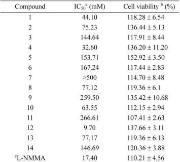
22Anti-inflammatory activity of the isolates (1 - 14) was evaluated by measuring the levels of nitric oxide (NO) production in lipopolysaccharide (LPS)-activated microglia BV-2 cells. Compound 12 significantly inhibited NO levels with IC
50value of 9.70 μM better than positive control (17.40 μM). Compounds 1 and 4 showed moderate NO product inhibitory (44.10 μM and 32.60 μM, respec- tively), but the other compounds showed little effects (Table 3).
Acknowledgments
This research was supported by the Basic Science Research Program through the National Research Foun- dation of Korea (NRF) funded by the Ministry of Education, Science and Technology (2012R1A5A2A 28671860). We are thankful to the Korea Basic Science Institute (KBSI) for the measurements of NMR and MS spectra.
References
(1) Lee, Y. N.; Flora of Korea; Kyohaksa: Korea, 1996; p250.
(2) Song, N.; Xu, W.; Guan, H.; Liu, X.; Wang, Y.; Nie, X. Asian. J.
Tradit. Med. 2007, 2, 218-222.
(3) Grosso, C.; Vinholes, J.; Silva, L. R.; de Pinho, P. G.; Gonçalves, R.
F.; Valentão, P.; Jäger, A. K.; Andrade, P. B. Braz. J. Pharmacog. 2011, 21, 635-644.
(4) Selenu, M. B.; Carrus, F.;Bonsignore, L. Boll. Chim. Farm. 2005, 144, 66-78.
(5) Park, C. J.; Park, C. B.; Hong, S. S.; Lee, H. S.; Lee, S. Y.; Kim, S.
C. Plant Mol.Biol. 2000, 44, 187-197.
(6) Kuroda, K.; Akao, M. Jpn. J. Cancer. Res. 1981, 72, 777-782.
(7) Kuroda, K.; Akao, M. Jpn. J. Cancer. Res. 1975, 66, 461-462.
(8) Cha, J. M.; Suh, W. S.; Lee, T. H.; Subedi, L.; Kim, S. Y.; Lee, K. R.
Molecules 2017, 22, 1023.
(9) Reif, D. W.; McCreedy, S. A. Arch. Biochem. Biophys. 1995, 320, 170-176.
≡
≡
≡
Table 3. Effects of compounds 1 - 14 on NO production in LPS- activated BV-2 cells
Compound IC50a (mM) Cell viability b (%)
1 44.10 118.28± 6.54
2 75.23 136.44± 5.13
3 144.64 117.91± 8.44
4 32.60 136.20± 11.20
5 153.71 152.92± 3.50
6 167.24 117.44± 2.83
7 >500 114.70± 8.48
8 77.12 119.36± 6.1
9 259.50 135.42± 10.68
10 63.55 112.15± 2.94
11 266.61 107.41± 2.63
12 9.70 137.66± 3.11
13 77.17 119.36± 6.13
14 146.69 120.36± 3.88
cL-NMMA 17.40 110.21± 4.56
aThe IC50 value of each compound was defined as the concentra- tion (μM) that caused 50% inhibition of NO production in LPS- activated BV-2 cells; bCell viability after treatment with 20μM of each compound was determined by the MTT assay and is expressed as a percentage (%). The results are averages of three independent experiments, and the data are expressed as mean± SD; c L-NMMA was used as a positive control
(10) Batovska, D., I.; Tsubota, S.; Kato, Y.; Asano, Y.; Ubukata, M.
Tetrahedron 2004, 15, 3551-3559.
(11) Wong, H. F.; Brown, G.D. J. Chem. Res. 2002, 5, 30-33.
(12) Duan, H.; Takaishi, Y.; Momota, H.; Ohmoto, Y.; Taki, T.
Phytochemistry 2002, 59, 85-90.
(13) Kimura, J.; Maki, N. J. Nat. Prod. 2002, 65, 57-58.
(14) Chang, Y. C.; Chang, F. R.; Wu, Y. C. J. Chin. Chem. Soc. 2000, 47, 373-380.
(15) Achenbach, H.; Stöcker, M.; Constenla, M. A. Phytochemistry 1988, 27, 1835-1841.
(16) Kim, J. S.; Kwon, Y. S.; Sa, Y. J.; Kim, M. J. J. Agric. Food.
Chem. 2011, 59, 138-144.
(17) Kim, D. K.; Lim, J. P.; Kim, J. W.; Park, H. W.; Eun, J. S. Arch.
Pharm. Res. 2005, 28, 39-43.
(18) Li, Y. L.; Li, J.; Wang, N. L.; Yao, X. S. Molecules 2008, 13, 1931- 1941.
(19) Kajjout, M.; Rolando, C. Tetrahedron 2011, 67, 4731-4741.
(20) Rayyan, S.; Fossen, T.; Nateland, H. S.; Andersen, Ø. M.
Phytochem. Anal. 2005, 16, 334-341.
(21) Gamblin, D. P.; Garnier, P.; van Kasteren, S.; Oldham, N. J.;
Fairbanks, A. J.; Davis, B. G. Angew. Chem. Int. Ed. 2004, 43, 828-833.
(22) Angles d'Ortoli, T.; Widmalm, G. Tetrahedron 2016, 72, 912-927.
(23) Rep ak, M.; Imrich, J.; Pihlaja, K.; Kal'atová, M. Phytochemisty 1998, 47, 1219-1221.
Received February 7, 2018 Revised April 2, 2018 Accepted April 2, 2018 c
ê