Alveolar bone thickness around maxillary central incisors of different inclination assessed with cone-beam computed tomography
Objective: To assess the labial and lingual alveolar bone thickness in adults with maxillary central incisors of different inclination by cone-beam computed tomography (CBCT). Methods: Ninety maxillary central incisors from 45 patients were divided into three groups based on the maxillary central incisors to palatal plane angle; lingual-inclined, normal, and labial-inclined. Reformatted CBCT images were used to measure the labial and lingual alveolar bone thickness (ABT) at intervals corresponding to every 1/10 of the root length. The sum of labial ABT and lingual ABT at the level of the root apex was used to calculate the total ABT (TABT). The number of teeth exhibiting alveolar fenestration and dehiscence in each group was also tallied. One-way analysis of variance and Tukey’s honestly significant difference test were applied for statistical analysis.
Results: The labial ABT and TABT values at the root apex in the lingual-inclined group were significantly lower than in the other groups (p < 0.05). Lingual and labial ABT values were very low at the cervical level in the lingual-inclined and normal groups. There was a higher prevalence of alveolar fenestration in the lingual-inclined group. Conclusions: Lingual-inclined maxillary central incisors have less bone support at the level of the root apex and a greater frequency of alveolar bone defects than normal maxillary central incisors. The bone plate at the marginal level is also very thin.
[Korean J Orthod 2015;45(5):245-252]
Key words: Maxillary central incisors, Inclination, Alveolar bone thickness, Cone- beam computed tomography
Yu-lou Tian
aFang Liu
bHong-jing Sun
aPin Lv
aYu-ming Cao
aMo Yu
aYang Yue
aa
Department of Orthodontics, School of Stomatology, China Medical University, Shenyang, China
b
Department of Orthodontics, Shenyang Stomatology Hospital, Shenyang, China
Received January 11, 2015; Revised April 30, 2015; Accepted May 6, 2015.
Corresponding author: Yu-lou Tian.
Professor, Department of Orthodontics, School of Stomatology, China Medical University, 117#, Nanjing North Street, Shenyang 110002, Liaoning Province, China.
Tel +86-24-22894332 e-mail [email protected]
© 2015 The Korean Association of Orthodontists.
The authors report no commercial, proprietary, or financial interest in the products or companies described in this article.
This is an Open Access article distributed under the terms of the Creative Commons Attribution Non-Commercial License (http://creativecommons.org/licenses/by-nc/4.0) which permits unrestricted non-commercial use, distribution, and reproduction in any medium, provided the original work is properly cited.
pISSN 2234-7518 • eISSN 2005-372X
http://dx.doi.org/10.4041/kjod.2015.45.5.245
INTRODUCTION
The dense cortical plates at the apical region of the incisors are regarded as orthodontic walls during orthodontic tooth repositioning, and efficient ortho- dontic tooth movement can only be ensured with ade- quate alveolar bone support.
1Upright positioning of the teeth in the center of the alveolus contributes to stable occlusion and better periodontal condition. As the number of adult orthodontic patients increases, orthodontists must pay closer attention to periodontal problems that could be aggravated during treatment.
A thorough evaluation of the alveolar bone thickness (ABT) of the maxillary incisors to detect the limits of tooth movement helps orthodontists to secure proper torque control and provide safe treatment that can protect patients from iatrogenic bone loss and maintain healthy periodontal condition. Appropriate tooth torque is necessary to achieve better occlusion, facial esthetics, and stability. It is clinically important to correct and control the torque of maxillary anterior teeth.
Cephalometric radiographs are mid-sagittal projections.
Thus, the bone width may be overestimated.
1,2Three- dimensional analysis of specific regions by cone-beam computed tomography (CBCT) may be a better method of assessing bone support. Compared with traditional radiographs, CBCT provides sensitive high-definition true-to-scale three-dimensional images without distor- tions or superimposition of structures. The high accuracy
of CBCT has been reported.
3-6Considering these advan- tages, CBCT is the preferred technique for precise evalu- ation of alveolar bone dimensions.
Previous studies have reported CBCT evaluation of bone morphology in certain malocclusions, volume changes of alveolar bone before and after orthodontic retraction, etc.
7,8Thus, the aim of this study was to apply CBCT for the assessment of ABT of maxillary in- cisors with different labial-lingual inclinations.
MATERIALS AND METHODS
This study was developed according to established precepts of China Medical University Ethics Commi- ttee and included 45 patients (90 maxillary central incisors) aged 18 to 30 years with no prior orthodontic treatment who presented for maxillary CBCT at the Department of Oral Radiology and Or thodontics, School of Stomatology, China Medical Uni versity for general examination before orthodontic or other oral treatment and were recruited by questionnaire. Informed consent was obtained from all the included patients.
The exclusion criteria were missing or decayed teeth, prosthetic crowns, crowding more than 3 mm or spacing more than 1 mm in the maxillary anterior alveolar segment, noticeable periodontal disease, diagnosed systemic disease, and craniofacial dysmorphology.
Three-dimensional CBCT images (NewTom TG; QR s.r.l., Verona, Italy) with a voxel size of 0.3 mm were
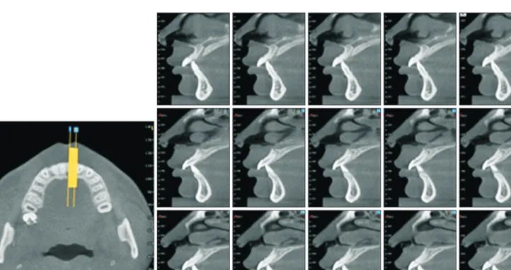
Figure 1. Three-dimensional reconstructions of cross-sectional and sagittal slices.
acquired with an exposure time of 20 s at 0.75A, 110 kV, with axial slice thickness 0.3 mm. Reconstruction of the data was performed with NNT software (QR s.r.l.) to create three-dimensional projections of the images with maximum intensity for making linear measurements.
The slices were generated from the assumed point of greatest labio-lingual distance (Figure 1). Any site showing no cortical bone around the root in at least three sequential slices was defined as an alveolar defect.
When the alveolar defect was more than 2 mm from the cementoenamel junction (CEJ), it was defined as
dehiscence. A fenestration was identified as an opening through the alveolar bone exposing parts of the root surface but not involving the alveolar crest (Figure 2).
9The root length of each tooth was defined as the distance from the CEJ to the apex and was divided into 10 intervals of equal length in selected sagittal slices (level 0 represented the CEJ junction and level 10 repre- sented the root apex) (Figure 3). The labial and lingual thicknesses of the alveolar bone perpendicular to the tooth axis were measured linearly at each level (Figure 2).
A total of 90 teeth were evaluated. The angle between the axis of the maxillary central incisors and the palatal plane (U1-PP) was determined, and the tomographs were divided into three groups of 30 teeth on the basis of U1-PP angle.
10The lingual-inclined group (U1-PP ≤ 110.1
o) comprised 7 men and 8 women, mean age 22.3
± 4.3 years; the normal group (110.1
o< U1-PP ≤ 121.5
o) comprised 6 men and 9 women, mean age 21.6 ± 4.5 years; and the labial-inclined group (U1-PP > 121.5
o) comprised 7 men and 8 women, mean age 22.4 ± 4.2 years (Figure 4 and 5).
The prevalence of fenestration and dehiscence in each group was also evaluated by careful observation of each image set.
Statistical analysis
All measurements were repeated after 2 weeks by the same investigator. Statistical calculations were performed with Statistical Package for the Social Sciences (SPSS) version 13.0 (SPSS Inc., Chicago, IL, USA). The data were found to be normally distributed with homo- geneity of variance among groups by Shapiro-Wilks normality test and Levene’s variance homogeneity test. Statistical comparisons of alveolar thickness in Figure 2. Determination of measurement levels.
7.9 mm 5.8 mm 5.0 mm 4.5 mm 3.7 mm 3.1 mm 2.6 mm 2.2 mm 1.5 mm 0.9 mm 1.5 mm
1.7 mm 1.3 mm
1.3 mm 1.1 mm
1.1 mm 1.1 mm
1.7 mm 1.9 mm
3.6 mm
Figure 3. Schematic illustration of labial and lingual bone thickness measurements. Level 0, cementoenamel junction; level 10, root apex area. ABT, Alveolar bone thi- ckness.
Lingual ABT
Tooth long axis Level 0
Labial ABT Fenestration
Level 10
Canal Incisive
Figure 4. Measuring the angle between the axis of the maxillary central incisors and the palatal plane (U1-PP) in the median sagi ttal view.
109.8
the different inclination groups were conducted by one-way analysis of variance (ANOVA) and Tukey’s ho- nestly significant difference (HSD) test. The level of probability for statistical significance was set at p = 0.05. Chi-square test was used for the evaluation of the prevalence of fenestrations and dehiscence. Intraclass correlation coefficients were used to determine intra- rater agreement for the measurements. The intraclass correlation coefficients were between 0.881 and 0.992.
Intraclass correlation coefficients of 0.75 or above are usually considered to be good, above 0.9 is excellent.
RESULTS
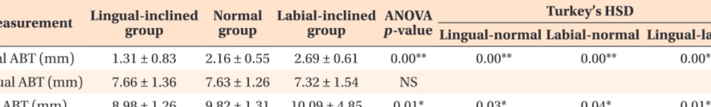
The results of the labial and lingual ABT measurements of the maxillary central incisors are listed in Tables 1, 2, and 3. Statistically significant differences of ABT among
the groups were found only at the root apex (level 10).
At this level, the labial ABT (p < 0.01) and TABT (p < 0.05) values in the lingual-inclined group were significantly lower than those of the other groups. The labial ABT values around the maxillary central incisors in the lin- gual-inclined group were also lower than in the other groups from levels 1 to 9, but these differences were not significant, and there were no differences in lingual ABT values among the groups.
The ABT measurements of both the labial and lingual aspects of level 1 in the lingual-inclined and normal groups revealed that the bone at this level was very thin.
Lingual ABT gradually increased from the CEJ to the apex. The labial ABT measured at the cervical segment in the normal group increased from the CEJ, remained constant in the middle third of the root, and then show- ed a continuous increase in the apical third.
Figure 5. Representative images of three groups classi- fied by different incisor incli- nation. A, Lingual-inclined group; B, normal group; C, la- bial-inclined group. The unit of the numberal data in the figure is millimeters.
9.0
2.7
8.2 5.1
5.8
A B C
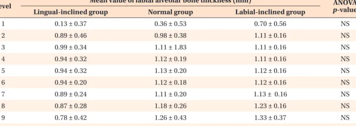
Table 1. Comparison of mean labial alveolar bone thickness values at ten root levels by analysis of variance (ANOVA)
Level Mean value of labial alveolar bone thickness (mm) ANOVA
p-value Lingual-inclined group Normal group Labial-inclined group
1 0.13 ± 0.37 0.36 ± 0.53 0.70 ± 0.56 NS
2 0.89 ± 0.46 0.98 ± 0.38 1.11 ± 0.16 NS
3 0.99 ± 0.34 1.11 ± 1.83 1.11 ± 0.16 NS
4 0.94 ± 0.32 1.12 ± 0.19 1.11 ± 0.16 NS
5 0.94 ± 0.32 1.13 ± 0.20 1.12 ± 0.16 NS
6 0.94 ± 0.20 1.12 ± 0.18 1.12 ± 0.16 NS
7 0.89 ± 0.24 1.11 ± 0.20 1.13 ± 0.16 NS
8 0.87 ± 0.28 1.18 ± 0.26 1.23 ± 0.16 NS
9 0.78 ± 0.42 1.26 ± 0.43 1.33 ± 0.37 NS
10 1.31 ± 0.83 2.16 ± 0.55 2.69 ± 0.61 0.00**
Values are presented as mean ± standard deviation.
**p < 0.01.
NS indicates no statistical significance among the three groups.
Fenestrations were observed on the labial bone surface of the root in seven central incisors (Figure 6) in the lingual-inclined group, one central incisor in the normal group, and none in the labial-inclined group (Table 4).
One central incisor in the normal group exhibited de- hiscence at the marginal bone plate (Figure 7).
DISCUSSION
A thorough assessment of the ABT of the maxillary incisors can help orthodontists improve outcomes and make right treatment decisions. Labial-lingual movement of the anterior teeth to improve the sagittal relationship of the maxillary and mandibular arches is mandatory to achieve a more harmonious profile. However, excessive tooth movement can cause iatrogenic sequelae including root resorption, gingival recession, and alveolar bone loss. Apart from esthetics, periodontal health and the alveolar bone boundaries are important factors in orthodontic treatment. According to our research, there is a significant association between labial-lingual
0.0