Introduction
The principle purpose of root canal treatment is to reduce discomfort and pain, eliminate bacteria from the root canal and prevent reinfection. A definite un- derstanding of root morphology and canal anatomy is essential to achieving success, and perfect obtu- rated root canal system.
The variation of root canal system morphology, especially multi-rooted teeth represents a continuous challenge to endodontic diagnosis and therapeutics.
Anatomic characteristics of permanent maxillary molars are described as a group of teeth with three roots, one palatal and two buccal, each root with one root canal. Most of the clinical reports deal with fourth mesiobuccal canal of maxillary molar, but this is also common to other roots.
Case reports of maxillary molars with four or more canals also describe maxillary molars with three
buccal roots maxillary molars with five canals, and maxillary molars with two canal in the palatal root.1
An anomalous root morphology which occurs infre- quently, such as two palatal roots on maxillary molar, is rarely mentioned in textbooks. In 1952, Diamond2 shows two extracted teeth that are identified as first molars and have two widely divergent palatal roots.
Slowey3 also reported the treatment of first maxil- lary molar with two palatal roots and showed a sec- ond maxillary molar with four independent roots.
Libfeld and Rostein4 examined 1,200 molars and found a 0.4% incidence of maxillary molar with four roots. Stone and Stroner1 examined more than 500 extracted molars and found less than a 2% incidence of more than one palatal canal when both anomalies were included.
Radiographs are an important and necessary aid in root canal treatment. Therefore an accurate radio- graphic techniques and proper interpretation are es-
*Correspondence to: Byeongcheol Song
Department of Dentistry, Seoul Veterans Hospital, 53, Jinhwangdo-ro 61-gil, Gangdong-gu, Seoul, 05368, Republic of Korea
Tel: +82-2-2225-1467, Fax: +82-2-2225-1659, E-mail: [email protected] Received: September 17, 2015/Last Revision: March 3, 2016/Accepted: March 4, 2016
Detection of maxillary second molar with two palatal roots using cone beam computed tomography: a case report
Jeong-Hee Kim, Byeongcheol Song*
Department of Dentistry, Seoul Veterans Hospital, Seoul, Republic of Korea
The purpose of this clinical report was to show anatomical variations in permanent maxillary second molar using computed tomography (CT). This case report describes the application of CT to detect the unusual root anatomy of maxillary second molar with 2 separate palatal roots for successful endodontic treatment procedures. The use of cone beam computed tomography (CBCT) can overcome the limitation of the periapical standard radiography caused by the overlap of buccal and secondary palatal roots. (J Dent Rehabil Appl Sci 2016;32(1):87-92)
Key words: root canal anatomy; two palatal root; cone beam computed tomography (CBCT)
Copyright© 2016 The Korean Academy of Stomatognathic Function and Occlusion.
It is identical to Creative Commons Non-Commercial License.
cc
ISSN 2384-4353 eISSN 2384-4272
sential for accurate diagnosis and treatment. The use of preoperative radiograph is the best way to detect and evaluate tooth canal morphology and anatomy.
Further radiographs should be taken at different angles to confirm any variation, in the case of, com- puted tomography (CT) is useful.
The purpose of this clinical report is to describe the unusual anatomy of maxillary molar with 2 sepa- rate palatal roots and the application of CT for suc- cessful endodontic treatment.
Case Report
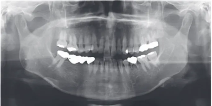
A 54-year-old woman visited Seoul Veterans Hos- pital for a dental examination. The medical history was noncontributory. The chief complaint of the pa- tient is discomfort of left maxillary molar area. Dur- ing clinical examination, left maxillary first molar and second molar were found to be restored with gold crown. In radiographic examination, left maxillary first molar seemed to be in incomplete endodontic treatment state and diagnostic radiograph revealed radiolucency in the periapical area (Fig. 1). The pa- tient was diagnosed with chronic periapical periodon- titis. After removing previously existed bridge and clinical examination, severe secondary decay on the left maxillary first molr was detected (Fig. 2) and the left maxillary first molar was extracted.
At the second appointment, left maxillary second molar was anesthetized and isolated with cotton roll.
The access to the pulp chamber was achieved us- ing a round diamond bur (ISO 801001016, Komet,
Lemgo, Sweitzerland). Three orifices were located, and working lengths were determined by electronic apex locator (Root ZX, Morita, Tokyo, Japan) then confirmed by a radiograph (Fig. 3). The root canals were initially instrumented with stainless steel hand K-file and Gate Glidden drill under irrigation with 2.5% NaOCl. All canal enlarged to a size #40 using K3 Ni-Ti file (K3 file, Sybronendo, Orange, USA).
The root canals were dried with sterile paper points, and calcium hydroxide was applied as an intracanal medicament.
Fig. 1. Preoperative radiograph.
Fig. 3. Working length with 3 roots.
Fig. 4. Postoperative radiograph of maxillary second molar filled with guttapercha and sealer.
Fig. 2. Radiograph showing severe secondary decay after old bridge removal.
The patient returned in 10 days with no symptoms.
The root canals were filled using the continuous wave technique with Elements Obturation Unit (Sybronendo) (Fig. 4). The tooth was restored with a resin core and gold crown. After two weeks, Cone Beam Computed Tomography (Kavo 3D Exam, Icat, Hatfield, USA) was taken for implant surgery on left maxillary first molar.
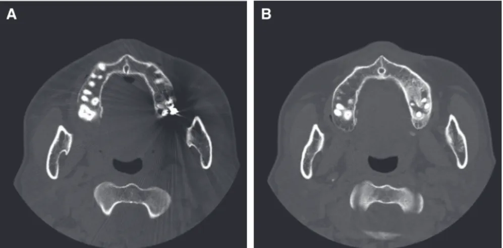
CT examination revealed a 2 palatal roots two sep- arated foramina at the apex by chance (Fig. 5). After implantation of left maxillary first molar in OroMax- illaFacialSurgery (OMFS), further radiograph was taken at different angles to confirm two separated palatal canals (Fig. 6). The gold crown and resin core were removed and access was gained using a round diamond bur. Pulp extirpation was carried out and the 4th root canal orifice were clearly identified (Fig.
7). The working length was determined by electronic apex locator (Root ZX, Morita, Tokyo, Japan) then confirmed by a radiograph (Fig. 8). The 4th canal was cleaned and prepared by hand with nickel titanium files. The 4th canal was filled using the continuous wave technique with Element Obturation Unit (Fig.
9).
After completion of root canal treatment, the left maxillary second molar was restored with resin core (Luxacoresmartmix dual, CE, Hamburg, Germany) and gold crown. During a 5-month follow-up, the
Fig. 6. Periapical standard radiograph was taken at different angles to confirm two separated.
Fig. 5. Cone beam computed tomography showing secondary palatal root. (A, B) Revealed a 2 palatal roots two separated foramina at the apex of maxillary second molar.
A B
Fig. 7. 4th root canal orifice was clearly identified.
patient remained asymptomatic (Fig. 10).
The patient used the treated teeth for 5 years with- out any symptoms and follow-up check after treat- ment (Fig. 11).
Discussion
Root canal anatomy can vary in number. Christie et al.5 reported 16 cases of maxillary molars with two palatal roots found during 40 years of daily clinical practice. Christie classified these teeth according to the shape and root separation as type I, II, III. Type I maxillary molars have two widely divergent palatal roots, which often are long and tortuous. The buccal roots often are “cow-horn” shaped and less diver- gent. Four separate root apices are seen on the radio- graph. Type II maxillary molars have four separate roots, but the roots often are shorter, run parallel, have buccal and lingual root morphology, and have blunt root apices. Type III maxillary molars also are constricted in root morphology with the mesiobuc- cal, mesiopalatal, and distopalatal canal engaged in a web of root dentin. The distobuccal root in these Fig. 8. Working length of 4th canal. Fig. 9. 4th canal filled with guttapercha and sealer.
Fig. 10. (A) Periapical standard radiograph: 5-month follow-up after 4th canal filling, (B) Cone beam computed tomography: 5-month follow-up after 4th canal filling.
A
B
Fig. 11. 5-years follow-up after 4th canal filling.
cases seems to stand and may even diverge to the distobuccal.
Recently, Shin et al.6 and Barbizam et al.7 reported case of unusual root canal anatomy in permanent maxillary molars using microscope. Tomazinho et al.8 reported maxillary first molar showing unusual anatomy (four roots and six root canals).
This clinical report covered type I maxillary molar.
Conventional radiography do not show four roots.
After three canal endodontic treatment, for pre- surgical assessment of maxillary left first molar for implant, CT revealed fourth root by accident. In pre- surgical assessment, CT showed conventional type I maxillary molar and resin core was removed. The fourth palatal canal was treated with conventional endodontic treatment. After finishing the root canal treatment, at 3-month routine check-up, the patient remained asymptomatic.
The management of endodontic problem is reliant on radiographs to assess the anatomy of the tooth and its surrounding anatomy.9 Recently, most of this core information would be obtained from conven- tional radiographs. However, such images have inher- ent limitation. The lack of three-dimensional infor- mation and masking of areas of interest by overlying anatomy are of particular relevance in endodontics.
Cone beam computed tomography reconstructed scans are invaluable for assessing teeth with unusual anatomy, such as teeth with an unusual number of root, dilacerated teeth and dens in dente. The exact location and anatomy of root canal system can be assessed, allowing successful management of the case.10
Conclusion
The variation of root canal system morphology, especially multi-rooted teeth represents a continuous challenge to endodontic diagnosis and therapeutics.
An accurate radiographic techniques and proper in- terpretation are essential for accurate diagnosis and treatment. The use of cone beam computed tomog- raphy (CBCT) can overcome the limitation of the periapical standard radiography.
References
1. Stone LH, Stroner WF. Maxillary molars demon- strating more than one palatal root canal. Oral Surg Oral Med Oral Pathol 1981;51:649-52.
2. Diamond M. Dental anatomy including anatomy of the head and neck. 3rd ed. New York; MacMillan;
1952. p. 203-5.
3. Slowey RR. Radiographic aids in the detection of extra root canals. Oral Surg Oral Med Oral Pathol 1974;37:762-72.
4. Libfeld H, Rotstein I. Incidence of four rooted maxillary second molars: literature review and ra- diographic survey of 1200 teeth. J Endod 1989;15:
129-31.
5. Christie WH, Peikoff MD, Fogel HM. Maxillary molar with two palatal roots: a retrospective clinical study. J Endod 1991;17:80-4.
6. Shin SJ, Park JW, Lee JK, Hwang SW. Unusual root canal anatomy in maxillay second molar: two case reports. Oral Surg Oral Med Oral Pathol Oral Ra- diol Endod 2007;104:e61-5.
7. Barbizam JV, Ribeiro RG, Tanomaru Filho M. Un- usual anatomy of permanent maxillary molars. J Endod 2004;30:668-71.
8. Tomazinho FS, Baratto-Filho F, Zaitter S, Leonardi DP, Gonzaga CC. Unusual anatomy of a maxillary first molar with two palatal roots: a case report. J Oral Science 2010;52:149-53.
9. Patel S, Dawood A. The use of cone beam comput- ed tomography in the management of external cer- vical resorption lesions. Int Endod J 2007;40:730-7.
10. Cotton TP, Geisler TM, Holden DT, Schwartz SA, Schindler WG. Endodontic applications of cone- beam volumetric tomography. J Endod 2007;33:
1121-32.
*교신저자: 송병철
(05368)서울시 강동구 진황도로61길 53 중앙보훈병원 치과병원 치과보존과 Tel: 02-2225-1467|Fax: 02-2225-1659|E-mail: [email protected] 접수일: 2015년 9월 17일|수정일: 2016년 3월 3일|채택일: 2016년 3월 4일
두개의 구개측 치근을 갖는 상악 제2대구치에서 cone beam computed tomography 활용: 증례보고
김정희, 송병철*
중앙보훈병원 치과병원 치과보존과
이 임상증례의 목적은 상악 제2대구치의 해부학적 형태를 computed tomography (CT)를 이용해 확인하여 근관치료에 활용한 증례이다. 이번 증례에서는 두개의 구개치근을 갖는 상악 제 2대구치에서 성공적인 근관치료를 위해서 CT를 활용하여 해부학적 형태를 확인하여 활용하였다. Cone beam computed tomography (CBCT)의 사용은 구개치근과 협
측치근이 겹쳐보이는 치근단 방사선 사진의 한계를 극복할 수 있게 한다.
(구강회복응용과학지 2016;32(1):87-92)
주요어: 근관형태; 제2구개치근; conebeam CT