Ovarian Clear Cell Carcinoma Sub-Typing by ARID1A Expression
8
0
0
전체 글
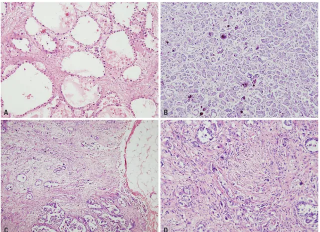
(2) ARID1A-Positive Ovarian Clear Cell Carcinoma. noted in ovarian cancers,13 which may also be involved in ARID1A loss-dependent O-CCC pathophysiology. Of its downstream signaling, SWI/SNF complex regulates cell adhesion proteins CD44 and E-cadherin.14 Thereby, loss of CD44 and/or E-cadherin could be a sign that ARID1A loss-dependent carcinogenic process is activated. Overexpression of HNF1β is another recently found O-CCC molecular feature. Positive expression of HNF1β is noted in 90– 95% of O-CCCs, contrary to less than 10% of positivity in other epithelium-origin ovarian cancers.15 HNF1β seems to work in the maintenance of E-cadherin expressing epithelial phenotype of O-CCC16 and, thereby, associated with good prognosis.17 However, the functional implications of HNF1β overexpression, particularly in relation to ARID1A loss in O-CCC pathophysiology, have yet to be determined. O-CCC is often resistant to therapies. If found at an early stage, it can be effectively managed by oophorectomy.18 In contrast, advanced O-CCCs exhibit heterogeneous behavior, being unpredictable with known clinic-pathological information, such as tumor grade, histologic subtype, stage, etc.19 In this situation, the expressional status of ARID1A and other biomarkers, including ER β, E-cadherin, and HNFβ, may provide further prognostic values.17,20 In this study, we analyzed the molecular. profiles and prognostic factors of O-CCC, focusing on the different clinical and molecular characteristics of ARID1A-positive and ARID1A-negative tumors.. MATERIALS AND METHODS Patients The study subjects were patients diagnosed with O-CCC at Severance Hospital. Medical charts were retrospectively reviewed to compare clinicopathological features including prognosis. For 87 patients from the Yonsei University Health System archived files, O-CCC diagnoses were confirmed in the past 5 years. All of the tumors were examined retrospectively by double-blind test for accuracy of histological subtyping and grading. With exclusion of ambiguous histology or inappropriate immunoprofiles, 70 were eligible for this study (29–67 years of age, median 49 years of age). Four cases with mixed subtypes (endometrioid carcinoma and serous carcinoma), and thirteen cases with an incompatible and distinct immunoprofile were excluded. All of the patients provided written informed consent, and the Research Ethics Board Committee of Yonsei University Health Medicine approved the study. Evalu-. A. B. C. D. Fig. 1. Histologic grades of O-CCC. (A and B) Low grade O-CCC. (A) tubulocystic or alveolar patterns are the most common patterns seen in low grade tumor. Abortive small tubules are less differentiated (H-E, ×100). (B) Psammomatous calcifications (H-E, ×100). (C and D) High grade O-CCC. (C) Diffuse, solid areas are frequently seen in high grade tumor. Desmoplasia is frequently found in association with infiltrative cells (H-E, ×40). (D) highly cellular spindle cells are parallel with infiltrating tumor cells (H-E, ×200). O-CCC, clear cell carcinoma of ovary; H-E, hematoxylin-eosin.. 60. https://doi.org/10.3349/ymj.2017.58.1.59.
(3) Jae Yoon Choi, et al.. ated parameters included patient age, parity, follow-up periods, tumor size and FIGO stage,21 associated pathology (endometriosis and adenofibroma), histologic grade, and oncologic outcomes (disease recurrence, distant metastasis, and cancerrelated death). Histologic grade was based on cellular atypia and mitotic indices of the major parts of the tumors. Low grade was defined as minimal or no atypia in focal areas less than one high-powered field (HPF), a low mitotic index (MI) less than 5/10 HPF, and/or primarily tubular or papillary patterns (Fig. 1A and B), whereas high grade was defined as severe atypia in multifocal areas more than one HPF, a high MI more than 5/10 HPF, and/or a primarily solid pattern (Fig. 1C and D).. Immunohistochemistry using tissue microarray preparation and interpretation Macroscopic inspection of whole mount tumor sections was performed to determine whether they were homogeneous or heterogeneous in nature. A core needle was used to make 2-mm holes in recipient blocks. Based on prior inspection, three adjacent areas of ovarian carcinomas from matching donor blocks were transplanted into recipient blocks using a 2-mm core needle. The 4-μm tissue sections were placed on silane-coated slides, deparaffinized, immersed in phosphate-buffered saline containing 0.3% (v/v) hydrogen peroxide, and processed in a microwave oven for 15 min at 700 W in 10 mM sodium citrate buffer (pH 6.5). After blocking for 30 min with 1% (w/v) bovine serum albumin, sections were incubated for 16 h at 4°C with biotin-labeled rabbit antibodies. The primary antibodies included ARID1A (C-terminal, 1:50, Abcam, Cambridge, MA, USA), HNF1β (SPA002083, 1:400, Sigma-Aldrich, St. Louis, MI, USA), p53 (DO-7, 1:100, Dako, Santa Clara, CA, USA), p38 (Phospho T180+Y182, 1:100, Abcam, Cambridge, MA, USA), E-cadherin (H-108, 1:200, Santa Cruz, Dallas, TX, USA), ERβ (1:50, Proteintech, Rosemonta, IL, USA), and Survivin (EP2880 1:100, Abcam, Cambridge, MA, USA). Streptavidin-conjugated peroxidase was used as the secondary antibody (1:10000). Normal goat serum and subtype-matched normal mouse IgG were used as negative controls. The final reaction product was visualized upon the addition of 0.03% (w/v) of 3, 3’-diaminobenzidine tetrachloride (DAB) for 5 to 20 min. ARID1A1 expression was interpreted as “loss” if less than 5% of the tumor cells showed nuclear staining. If staining was modest (between 5% and 50% of the tumor cells), expression was interpreted as “focal loss.” When more than 50% of the cells were positive, ARID1A was evaluated as “intact.” For HNF1β expressions, two-tiered grading was applied: grade 1 in patch staining (focal loss) or grade 2 for diffuse and strong expression. ERβ expressions were graded from 0 to 3+ according to the American Society of Clinical Oncology/College of American Pathologists (ASCO/CAP) guidelines and grade 1, 2, and 3 were considered “positive” (2). E-cadherin membrane staining was graded as grade 0, indicating diffuse loss of more than 10%; grade 1, indicating focal loss of less than 10%; and grade 2, indihttps://doi.org/10.3349/ymj.2017.58.1.59. cating no loss. All of the antibodies were initially applied to whole slides followed by tissue microarray.. Statistical analyses Demographic data were analyzed using the parametric, independent two-sample t-test and the chi-square (or Fisher’s exact test) for continuous and categorical variables, respectively. Categorical variables are reported as frequencies, and continuous variables are reported as means±standard deviations. To compare cancer-specific survival, Kaplan-Meier survival curves were generated for ARID1A-positive and negative tumors. Mean survival times of ARIDA-positive and negative tumors were compared by using log-rank test. All p-values <0.05 were considered to be statistically significant. All analyses were performed with SAS (version 9.2, SAS Institute Inc., Cary, NC, USA) and R statistical software (version 3.1.1, www. R-project.org).. RESULTS Table 1 summarizes patient characteristics. Tumors that were removed were of variable sizes ranging from 2–25 cm (median 8 cm). The majority of tumors were low stage [stage I: 36 (52%); stage II: 5 (7%)] (Table 1). Mean follow-up period were 40±36 months. The numbers of recurrences, metastasis, and deaths due to disease were 4 (3%), 19 (13%), and 10 (14%) cases, respectively. Table 1. Patient Characteristics No. (%) Age, mean±SD, yrs 48.4±9.2 0–39 yrs 17 (24) 40–49 yrs 23 (33) 50 yrs or older 30 (43) Childbirth 0 (nullipara) 24 (34) 1 or more 46 (66) Follow-up periods, mean±SD, months 40±36 Primary tumor size, mean±SD, cm 10.9±11.2 FIGO stage Stage I 36 (52) Stage II 5 (7) Stage III 20 (29) Stage IV 8 (12) Associated pathology Endometriosis 33 (47) Adenofibroma 6 (9) Oncologic outcome Recurrence 4 (3) Distant metastasis 19 (13) Cancer-related death 10 (14) SD, standard deviation; FIGO, International Federation of Gynecology and Obstetrics.. 61.
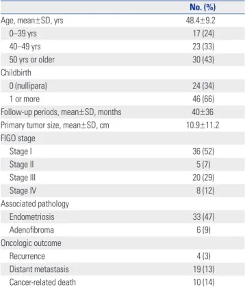
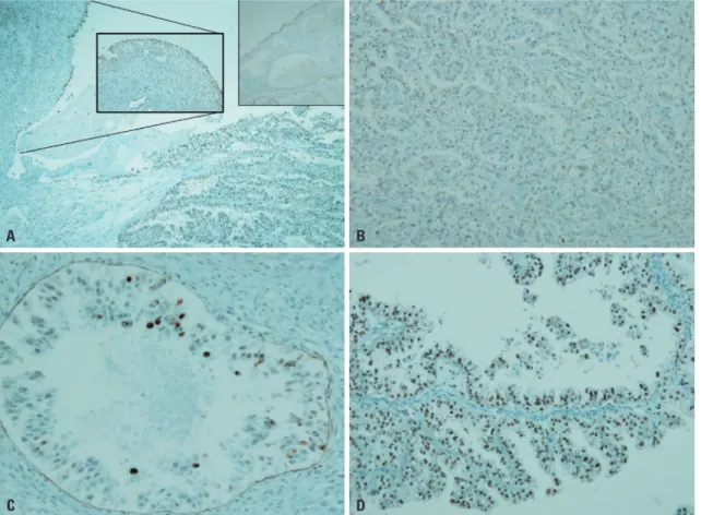
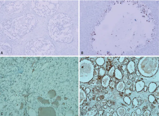
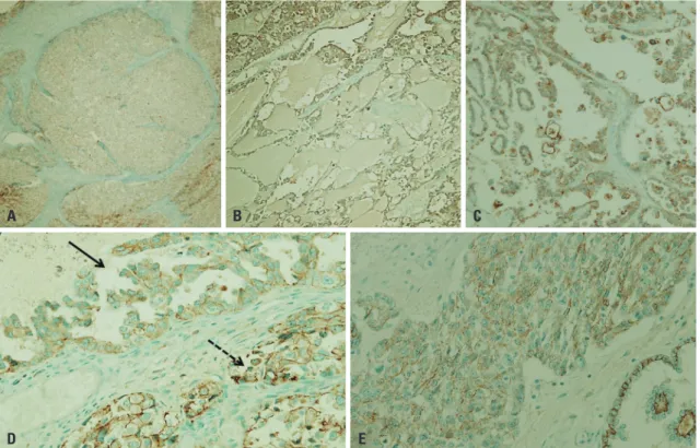
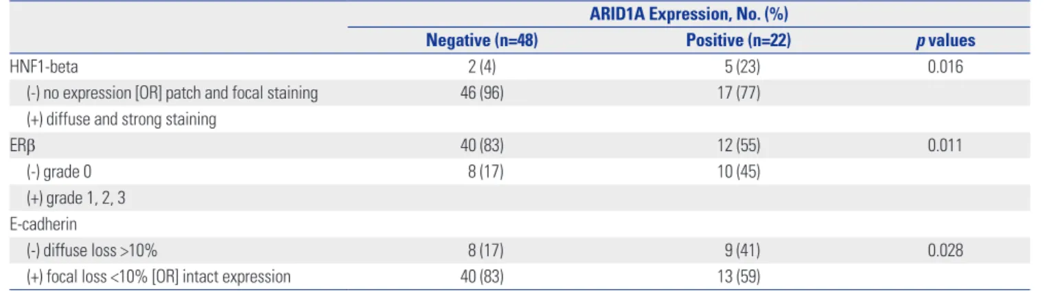
(4) ARID1A-Positive Ovarian Clear Cell Carcinoma. Immunohistochemical profiles of O-CCCs We analyzed expressional characteristics of several molecules, including HNF1β, ERβ, and E-cadherin, along with ARID1A. In CCC-associated endometriosis, nuclear staining of ARID1A occurs along the epithelial lining, although CCC shows complete loss of ARID1A (Fig. 2A). Fig. 2B-E shows varying degrees of ARID1A loss in O-CCC, from total expressional loss (Fig. 2B) and focal expression (Fig. 2C) to intact staining (Fig. 2D). Complete loss of HNF1β was rarely found (5.7%) in O-CCCs (Fig. 3A), and most of these O-CCCs were negative for p53 or ERβ and loss of ARID1A. In HNF1β-positive cells, HNF1β expression was higher in cells closer to the luminal side (Fig. 3B). Complete loss of ERβ was rare in O-CCCs (Fig. 3C). Luminal protruding hobnail cells were positive for ERβ (Fig. 3D). Loss of E-cadherin or CDH1 was seen in 21.4% of O-CCCs, and E-cadherin loss was variable ranging from complete loss (Fig. 4A and B) to aberrant expression, such as fragmentation and broken linear forms (Fig. 4C and D) or cytoplasmic staining (Fig. 4E).. Characteristics of ARID1A-negative and ARID1A-positive O-CCCs The expressions of ARID1A “complete loss,” “focal loss,” and “intact” were noted in 48 (66%), 12 (17%), and 10 (14%) cases, re-. spectively. We grouped “focal loss” or “intact” expressions in to ARID1A-positive tumors and “complete loss” to ARID1Anegative tumors. Table 2 compares their clinical and pathological characteristics. Patient age, parity, tumor size, and distribution of cancer stage were not statistically different between ARID1A-negative and positive O-CCCs. In contrast, the ratio of histologic high and low grades were significantly different; most (90%) of the ARID1A-negative tumors were low grade, while 41% of ARID1A-positive tumors were high grade (p=0.003) (Table 2). Along with this finding, immunohistochemical profiles of ARID1A-positive and negative tumors were further analyzed (Table 3). To simplify comparisons, the expression status of each molecule was categorized as (-) or (+). ARID1A-negative tumors exhibited a mostly homogenous expression profile, HNF1β-positive (96%), ERβ-negative (83%), and E-cadherin positive (83%); whereas ARID1A-positive tumors exhibited rather heterogeneous immunohistochemical profiles, HNF1βnegativity (23%); ERβ-positivity (55%), and E-cadherin loss (41%).. Association with oncologic outcomes To compare cancer-speficic survival, Kaplan-Meier survival. A. B. C. D. Fig. 2. ARID1A expressions in O-CCC. (A) In endometriosis, nuclear staining occurs along the epithelial lining (inlet), but CCC shows complete loss of ARID1A (DAB, ×40). (B) Total loss of ARID1A in a branched tubular pattern (DAB, ×100). (C) Focal expression (nearly total loss) in an alveolar pattern (DAB, ×200). (D) Moderate preservation of AR1D1A expression in a cystic papillary pattern (DAB, ×200). ARID1A, AT-rich DNA-interacting domain 1A; O-CCC, clear cell carcinoma of ovary; DAB, 3’-3’ diaminobenzidine.. 62. https://doi.org/10.3349/ymj.2017.58.1.59.
(5) Jae Yoon Choi, et al.. curves were generated for ARID1A-positive and negative tumors (Fig. 5). Mean survival times of ARIDA-positive and negative tumors were 74 months (95% CI 53–95) and 118 months (95% CI 105–131) respectively, and the difference was not statistically significant (p=0.054, log-rank test).. DISCUSSION To date, two significant mutational/expressional molecular characteristics of O-CCC have been found, ARID1A loss and HNF1β-overexpression.2,15 However, their functional roles in cancer are still largely unknown, and the relationship between those two proteins has not yet been determined. For ARID1A, its loss is suggested as a critical event in endometriosis (EM)derived O-CCC tumorigenesis.22 Indeed, ARID1A loss has been noted in O-CCC associated atypical EM tissues, as well.3 Since ARID1A is a major component of the human SWI/SNF chromatin-remodeling complex,6 its oncogenic role may be related with remodeling of chromatin and histone re-arrangements4,9,23 or with modulation of estrogenic action.9 We found that ARID1A-loss in O-CCC is linked to a specific immunohistochemical profile: ERβ loss, intact E-cadherin, and HNF1β. overexpression. ERβ expressional loss occurs frequently across all epithelial ovarian cancers,24 and ARID1A is important in carrying steroid hormone signaling to the SWI/SNF-induced transcriptional activations.12 Since E-cadherin is one of the downstream activators of the SWI/SNF complex,14 we initially thought that ARID1A loss would accompany E-cadherin loss, which turned out to be not true, as ARID1A-lost tumors were more-likely to have intact E-cadherin expression than ARID1Aexpressing tumors. This is in contrast with a previous study that stated that, in gastric cancer, reduced ARID1A expression down-regulates E-cadherin transcription.25 We suspect a hidden significance of HNF1β overexpression here.26 The functional role of HNF1β overexpression in O-CCC has not yet been derived,15 although it may also be mediated by E-cadherin: knock down of HNF1 has been shown to reduce E-cadherin expression and promote epithelial-mesenchymal transition.16 Hypothetically, the uniformly-expressed HNF1β in ARID1A-loss tumors in our study might have served a protective role by maintaining tumor cell E-cadherin expression. Thereafter, we argue that this signaling axis, ERβ/ARID1A/Ecadherin along with HNF1β, requires further molecular functional studies to elucidate the mechanism of O-CCC development.. A. B. C. D. Fig. 3. HNF1β and ERβ expressions in O-CCC. (A and B) HNF1β expression. (A) Complete loss of HNF1-β is rarely found in CCC. However, negative expression of p53 or ER with loss of ARID1A is common (DAB, ×100). (B) Luminal hobnail cells express HNF1-β. More HNF1-β expression occurs in cells closer to the luminal side (DAB, ×200). (C and D) ERβ expression. (C) complete loss of ERβ is common (DAB, ×100). (D) Luminal protruding hobnail cells express ERβ (DAB, ×100). ERβ, estrogen receptor beta; O-CCC, clear cell carcinoma of ovary; ARID1A, AT-rich DNA-interacting domain 1A; DAB, 3’-3’ diaminobenzidine. https://doi.org/10.3349/ymj.2017.58.1.59. 63.
(6) ARID1A-Positive Ovarian Clear Cell Carcinoma. Using readily available clinical samples and immunohistochemical tools, we sought to characterize ARID1A-negative OCCCs in comparison with ARID1A-intact (or positive) O-CCCs.. A. While ARID1A-negative tumors exhibited a homogenous immunoprofile, the immunoprofiles of ARID1A-positive tumor were rather heterogeneous: HNF1β-positive rate was slightly. B. D. C. E. Fig. 4. E-cadherin expressions in O-CCC. Loss of CDH1, or E-cadherin was manifested variably. (A) Complete loss of CDH1 (DAB, ×10). (B) Partial and weak staining (DAB, ×100). (C) Aberrant CDH1 expression. Aberrant cytoplasmic expression with fragmented or broken linear forms instead of membrane staining is an indicator of E-cadherin disruption (DAB, ×100). (D) Within the same tumor, CDH1 loss is heterogeneous. The solid arrow points to a region of partial CDH1 loss as seen by weak staining along the membrane. The broken arrow points to disrupted fragments in the membrane (DAB, ×200). (E) Images taken at a higher magnification show aberrant staining, such as and non-continuous, fragmented membrane, or cytoplasmic staining (DAB, ×200). O-CCC, clear cell carcinoma of ovary; DAB, 3’-3’ diaminobenzidine. Table 2. Clinico-Pathologic Characteristics of ARID1A-Expressing Ovarian Clear Cell Carcinoma Negative (n=48) 47.4±8.8. Age, mean±SD, yrs Childbirth 0 (nullipara) 15 (31) 1 or more 33 (69) Endometriosis Absent 23 (48) Present 25 (52) Primary tumor size, mean±SD, cm Histologic grade 11.0±13.9 Low grade 43 (90) High grade 5 (10) Cancer stage Stage I 26 (55) Stage II 4 (9) Stage III 13 (28) Stage IV 4 (9) ARID1A, AT-rich DNA-interacting domain 1A. t-test for continuous variables, chi-square test for non-continuous variables.. 64. ARID1A expression, No. (%) Positive (n=22) 50.1±9.7. p value 0.162 0.429. 9 (41) 13 (59) 0.221 14 (64) 8 (36) 10.9±6.0 13 (59) 9 (41). 0.976 0.003. 0.593 10 (46) 1 (5) 7 (32) 4 (18). https://doi.org/10.3349/ymj.2017.58.1.59.
(7) Jae Yoon Choi, et al.. Table 3. Immunohistochemical Profiles of ARID1A-Expressing Ovarian Clear Cell Carcinoma Negative (n=48) HNF1-beta 2 (4) (-) no expression [OR] patch and focal staining 46 (96) (+) diffuse and strong staining ERβ 40 (83) (-) grade 0 8 (17) (+) grade 1, 2, 3 E-cadherin (-) diffuse loss >10% 8 (17) (+) focal loss <10% [OR] intact expression 40 (83) ARID1A, AT-rich DNA-interacting domain 1A; ERβ, estrogen receptor beta. Chi-square test for non-continuous variables. Survival functions 100. Cancer-specific survival, %. 80. 60 Log-rank test, p=0.054 40 ARID1A Negative 20. Positive Negative, censored Positive, censored. 0 0. 25. 50. 75. 100. 125. Follow-up period, months. Fig. 5. Kaplan-Meier curve of cancer-specific survival for O-CCC, stratified by ARID1A expression status. O-CCC, clear cell carcinoma of ovary; ARID1A, AT-rich DNA-interacting domain 1A.. low, and the rates of ERβ-negativity and E-cadherin positivity were around 50%. Overall, the frequency of ARID1A loss in our series was slightly higher (69%) than those of previous studies.2,3,20 Nevertheless, this still implies that for about half of O-CCC tumors show intact ARID1A, and their tumorigenic processes require further explanation.26 HNF1β-overexpression and intact E-cadherin are associated with better prognosis in O-CCC,17,27 while the prognostic significance of ERβ expression in O-CCC has not been validated. The prognostic effect of ARID1A loss in O-CCC is debatable, some suggesting a poor prognsosis while others reporting no prognostic impacts.17,20,28 In our study, ARID1A expression status did not have a significant impact on survival. This indifference in survival is noticible since most ARID1A-negative tumors https://doi.org/10.3349/ymj.2017.58.1.59. ARID1A Expression, No. (%) Positive (n=22) 5 (23) 17 (77). p values 0.016. 12 (55) 10 (45). 0.011. 9 (41) 13 (59). 0.028. in our study were low grade (90%), whereas many of ARID1Apositive tumors were high grade (41%), and histologic grade is directly linked to survival.29 The human SWI/SNF complex regulates gene transcription mainly by modulating DNA methylation status.14 Interestingly, O-CCCs is characterized by wide-spread CpG island promoter hypermethylation.30 In particular, both HNF and ER pathways are frequently methylated in O-CCCs.31,32 Moreover, promoter methylation-related expressional changes can lead to disease progression in epithelial ovarian cancers. For instance, methylation status changes result in overexpression of Mucin 13 and carbonic anhydrase 9, both of which contribute to aggressive behavior of affected cancer cells.33,34 Therefore, it is probable that epigenetic dysregulations play a significant role in ARID1Anegative O-CCC pathophyology, especially if it depends on HNF and ER pathways. In this study, we used a modified two-tier histologic grading system based on nuclear atypia and MI. For histological grading of O-CCC, the Shimizu-Silverberg or the International Federation of Gynecology and Obstetrics grading systems can be used. However, a recent study criticized the significances of both systems, which were not effective enough to predict clinical outcomes.35 Along with previous reports, our study may provide a base for development of further molecular subtyping of OCCC, presumably relying on ARID1A expression status. In conclusion, ARID1A-positive tumors and negative tumors differed in gross histology and immunoprofiles. This study proposes that ARID1A expression status can be utilized for molecular subtyping of O-CCC. Further study is mandatory to enlighten the molecular events underlying O-CCC, particularly an ARID1A-independent pathway.. ACKNOWLEDGEMENTS This study was supported by the 2014 medical student research grant from the Yonsei University College of Medicine and by a grant from the Korea Health Technology R&D Project through the Korea Health Industry Development Institute. 65.
(8) ARID1A-Positive Ovarian Clear Cell Carcinoma. (KHIDI), funded by the Ministry of Health & Welfare, Republic of Korea (grant number: HI13C0858).. REFERENCES 1. Ahmed Q, Alosh B, Bandyopadhyay S, Ali-Fehmi R. Gynecologic cancers: molecular updates. Clin Lab Med 2013;33:911-25. 2. Jones S, Wang TL, Shih IeM, Mao TL, Nakayama K, Roden R, et al. Frequent mutations of chromatin remodeling gene ARID1A in ovarian clear cell carcinoma. Science 2010;330:228-31. 3. Wiegand KC, Shah SP, Al-Agha OM, Zhao Y, Tse K, Zeng T, et al. ARID1A mutations in endometriosis-associated ovarian carcinomas. N Engl J Med 2010;363:1532-43. 4. Yamamoto S, Tsuda H, Takano M, Tamai S, Matsubara O. Loss of ARID1A protein expression occurs as an early event in ovarian clear-cell carcinoma development and frequently coexists with PIK3CA mutations. Mod Pathol 2012;25:615-24. 5. Chene G, Ouellet V, Rahimi K, Barres V, Provencher D, Mes-Masson AM. The ARID1A pathway in ovarian clear cell and endometrioid carcinoma, contiguous endometriosis, and benign endometriosis. Int J Gynaecol Obstet 2015;130:27-30. 6. Reisman D, Glaros S, Thompson EA. The SWI/SNF complex and cancer. Oncogene 2009;28:1653-68. 7. Nagl NG Jr, Zweitzig DR, Thimmapaya B, Beck GR Jr, Moran E. The c-myc gene is a direct target of mammalian SWI/SNF-related complexes during differentiation-associated cell cycle arrest. Cancer Res 2006;66:1289-93. 8. Samartzis EP, Noske A, Dedes KJ, Fink D, Imesch P. ARID1A mutations and PI3K/AKT pathway alterations in endometriosis and endometriosis-associated ovarian carcinomas. Int J Mol Sci 2013;14: 18824-49. 9. Tanase Y, Yamada Y, Shigetomi H, Kajihara H, Oonogi A, Yoshizawa Y, et al. Modulation of estrogenic action in clear cell carcinoma of the ovary (review). Exp Ther Med 2012;3:18-24. 10. LaGrenade A, Silverberg SG. Ovarian tumors associated with atypical endometriosis. Hum Pathol 1988;19:1080-4. 11. Prefumo F, Todeschini F, Fulcheri E, Venturini PL. Epithelial abnormalities in cystic ovarian endometriosis. Gynecol Oncol 2002;84: 280-4. 12. Inoue H, Furukawa T, Giannakopoulos S, Zhou S, King DS, Tanese N. Largest subunits of the human SWI/SNF chromatin-remodeling complex promote transcriptional activation by steroid hormone receptors. J Biol Chem 2002;277:41674-85. 13. Lazennec G. Estrogen receptor beta, a possible tumor suppressor involved in ovarian carcinogenesis. Cancer Lett 2006;231:151-7. 14. Banine F, Bartlett C, Gunawardena R, Muchardt C, Yaniv M, Knudsen ES, et al. SWI/SNF chromatin-remodeling factors induce changes in DNA methylation to promote transcriptional activation. Cancer Res 2005;65:3542-7. 15. Tsuchiya A, Sakamoto M, Yasuda J, Chuma M, Ohta T, Ohki M, et al. Expression profiling in ovarian clear cell carcinoma: identification of hepatocyte nuclear factor-1 beta as a molecular marker and a possible molecular target for therapy of ovarian clear cell carcinoma. Am J Pathol 2003;163:2503-12. 16. Tomassetti A, De Santis G, Castellano G, Miotti S, Mazzi M, Tomasoni D, et al. Variant HNF1 modulates epithelial plasticity of normal and transformed ovary cells. Neoplasia 2008;10:1481-92. 17. Ye S, Yang J, You Y, Cao D, Huang H, Wu M, et al. Clinicopathologic significance of HNF-1β, AIRD1A, and PIK3CA expression in ovarian clear cell carcinoma: a tissue microarray study of 130 cases. Medicine (Baltimore) 2016;95:e3003. 18. Okamoto A, Glasspool RM, Mabuchi S, Matsumura N, Nomura H,. 66. Itamochi H, et al. Gynecologic Cancer InterGroup (GCIG) consensus review for clear cell carcinoma of the ovary. Int J Gynecol Cancer 2014;24(9 Suppl 3):S20-5. 19. Lee YY, Kim TJ, Kim MJ, Kim HJ, Song T, Kim MK, et al. Prognosis of ovarian clear cell carcinoma compared to other histological subtypes: a meta-analysis. Gynecol Oncol 2011;122:541-7. 20. Itamochi H, Oumi N, Oishi T, Shoji T, Fujiwara H, Sugiyama T, et al. Loss of ARID1A expression is associated with poor prognosis in patients with stage I/II clear cell carcinoma of the ovary. Int J Clin Oncol 2015;20:967-73. 21. Zeppernick F, Meinhold-Heerlein I. The new FIGO staging system for ovarian, fallopian tube, and primary peritoneal cancer. Arch Gynecol Obstet 2014;290:839-42. 22. Xiao W, Awadallah A, Xin W. Loss of ARID1A/BAF250a expression in ovarian endometriosis and clear cell carcinoma. Int J Clin Exp Pathol 2012;5:642-50. 23. Ayhan A, Mao TL, Seckin T, Wu CH, Guan B, Ogawa H, et al. Loss of ARID1A expression is an early molecular event in tumor progression from ovarian endometriotic cyst to clear cell and endometrioid carcinoma. Int J Gynecol Cancer 2012;22:1310-5. 24. Lin K, Zhan H, Ma J, Xu K, Wu R, Zhou C, et al. Increased steroid receptor RNA activator protein (SRAP) accompanied by decreased estrogen receptor-beta (ER-β) levels during the malignant transformation of endometriosis associated ovarian clear cell carcinoma. Acta Histochem 2014;116:878-82. 25. Yan HB, Wang XF, Zhang Q, Tang ZQ, Jiang YH, Fan HZ, et al. Reduced expression of the chromatin remodeling gene ARID1A enhances gastric cancer cell migration and invasion via downregulation of E-cadherin transcription. Carcinogenesis 2014;35:867-76. 26. Gounaris I, Brenton JD. Molecular pathogenesis of ovarian clear cell carcinoma. Future Oncol 2015;11:1389-405. 27. Faleiro-Rodrigues C, Macedo-Pinto I, Pereira D, Lopes CS. Prognostic value of E-cadherin immunoexpression in patients with primary ovarian carcinomas. Ann Oncol 2004;15:1535-42. 28. Rahman M, Nakayama K, Rahman MT, Katagiri H, Katagiri A, Ishibashi T, et al. Clinicopathologic analysis of loss of AT-rich interactive domain 1A expression in endometrial cancer. Hum Pathol 2013; 44:103-9. 29. Ryu SY, Park SI, Nam BH, Kim I, Yoo CW, Nam JH, et al. Prognostic significance of histological grade in clear-cell carcinoma of the ovary: a retrospective study of Korean Gynecologic Oncology Group. Ann Oncol 2009;20:1032-6. 30. Earp MA, Cunningham JM. DNA methylation changes in epithelial ovarian cancer histotypes. Genomics 2015;106:311-21. 31. Yamaguchi K, Huang Z, Matsumura N, Mandai M, Okamoto T, Baba T, et al. Epigenetic determinants of ovarian clear cell carcinoma biology. Int J Cancer 2014;135:585-97. 32. Shen H, Fridley BL, Song H, Lawrenson K, Cunningham JM, Ramus SJ, et al. Epigenetic analysis leads to identification of HNF1B as a subtype-specific susceptibility gene for ovarian cancer. Nat Commun 2013;4:1628. 33. Sung HY, Park AK, Ju W, Ahn JH. Overexpression of mucin 13 due to promoter methylation promotes aggressive behavior in ovarian cancer cells. Yonsei Med J 2014;55:1206-13. 34. Sung HY, Ju W, Ahn JH. DNA hypomethylation-mediated overexpression of carbonic anhydrase 9 induces an aggressive phenotype in ovarian cancer cells. Yonsei Med J 2014;55:1656-63. 35. Yamamoto S, Kasajima A, Takano M, Yaegashi N, Fujiwara H, Kuzuya K, et al. Validation of the histologic grading for ovarian clear cell adenocarcinoma: a retrospective multi-institutional study by the Japan Clear Cell Carcinoma Study Group. Int J Gynecol Pathol 2011;30:129-38.. https://doi.org/10.3349/ymj.2017.58.1.59.
(9)
수치
+3
관련 문서
Insulin-like growth factor II mRNA-binding protein 3 (IMP3), Ki-67, and p53 expression levels in a clear cell renal cell carcinoma (CC- RCC) patient with metastasis..
In this study, to clarify the roles of BTG1 in ovarian carcinogenesis, BTG1 mRNA and protein expression was evaluated in ovarian carcinoma cell lines; to understand the
Comparison of the mutation of 88 genes in the custom gene panel between clear cell renal cell carcinoma with cystic change (MCRCC) and multi- locular cystic renal neoplasm of
Salivary gland neoplasms associated with a high proportion of clear cells include clear cell mucoepidermoid carcinoma, epithelial- myoepithelial carcinoma, acinic cell carcinoma,
유방에 발생하는 원발성 종양 중, 투명한 세포질을 포함 하는 암종은 각각의 세포질 내에 함유된 성분과 조직학적 유형에 따라 당원성
This study aimed to analyze the clinical features of clear cell carcinoma in relation to endometriosis and to determine an appropriate surveillance strategy for the early detection
Here we report a 54-year-old patient with ovarian clear cell carcinoma with skin metastases on the anterior chest at 11 months after initial diagnosis.. Although she
Expression level of insulin receptor (IR) in clear cell renal cell carcinoma (CCRCC) is associated with tumor grade.. (A) Representative immunoblotting of IR in the low and high
