Immunophenotypes of Glycogen Rich Clear Cell Carcinoma
Sung Eun Kim,* Ja Seung Koo, and Woo-Hee Jung
Department of Pathology, Gangnam Severance Hospital, Yonsei University College of Medicine, Seoul, Korea.
Received: November 10, 2011 Revised: January 17, 2012 Accepted: January 18, 2012
Corresponding author: Dr. Woo-Hee Jung, Department of Pathology,
Gangnam Severance Hospital, Yonsei University College of Medicine, 211 Eonju-ro, Gangnam-gu, Seoul 135-720, Korea.
Tel: 82-2-2019-3541, Fax: 82-2-3463-2103 E-mail: [email protected]
*Current address: Department of Pathology, CHA Gangnam Medical Center, CHA University, Seoul, Korea.
∙ The authors have no financial conflicts of interest.
© Copyright:
Yonsei University College of Medicine 2012 This is an Open Access article distributed under the terms of the Creative Commons Attribution Non- Commercial License (http://creativecommons.org/
licenses/by-nc/3.0) which permits unrestricted non- commercial use, distribution, and reproduction in any medium, provided the original work is properly cited.
Purpose: Glycogen rich clear cell carcinoma (GRCC) of the breast is a rare sub- type of invasive ductal carcinoma and involves a poor prognosis. In the literature, less than 150 cases have been reported. Many researchers have attempted to charac- terize GRCC according to electron microscope, flow cytometry, or clinical data.
However, an organized study of the immunophenotype of GRCC has yet to be re- ported. Materials and Methods: Here, we present three cases of GRCC and their immunohistochemical profiles. Results: Histologically, all three cases contained periodic acid stain (PAS) positive and d-PAS labile granules in their clear cyto- plasm. Case I showed positivity for only estrogen receptor (ER) and c-erbB2. Case II exhibited positivity for progesterone receptor and negativity for ER and c-erbB2.
Case III presented with triple negative invasive carcinoma. The expression pattern of E-cadherin was concordant with epidermal growth factor receptor and c-kit, but discordant with ki-67. Among these three cases, p53-positive cases exhibited a low proliferative index (ki-67: 15%), while p53-negative cases showed a high prolifera- tive index (ki-67: 50-60%). Conclusion: In conclusion, the immunophenotype of GRCC is not uniform, but is similar to that of conventional ductal carcinoma.
Key Words: Breast, carcinoma, clear cell, glycogen, immunohistochemistry
INTRODUCTION
Glycogen rich clear cell carcinoma (GRCC) of the breast is known as a rare sub- type of invasive ductal carcinoma, and involves a poor prognosis. It was first de- scribed by Hull, et al.1 in 1981. Since then, fewer than 150 cases have been report- ed. Many researchers have attempted to characterize GRCC, according to, electron microscope (EM),1,2 flow cytometry,3 or clinical data,4-6 the results of which have been summarized in Table 1.
Hull, et al.1 confirmed intracytoplasmic glycogen of GRCC by EM and stated that the H&E and EM features of GRCC were very similar to the morphology of 13-week old fetal developing mammary gland. In diagnosing GRCC, tumor cells that contain abundant fine glycogen granules in their clear cytoplasm, are stained by periodic acid stain (PAS), but not stained by diastase-periodic acid stain (d- PAS). The incidence of GRCC is estimated to be 1 to 3% of breast carcinoma.
Opinions differ with among researchers concerning the prognosis of GRCC, rang- ing from favorable to unfavorable.2-6 Immunohistochemical (IHC) staining for es- trogen receptor (ER) is usually positive. However, IHC for progesterone receptor
cases found during the last 10 years. One case was lost to follow up after diagnosis by biopsy. The ages at diagnosis were 49 to 69 years old and follow up data were 14 months and 18 months. They were currently disease-free states.
They were treated with total and partial mastectomy fol- lowed by chemotherapy and radiation therapy.
Immunohistochemical (IHC) staining
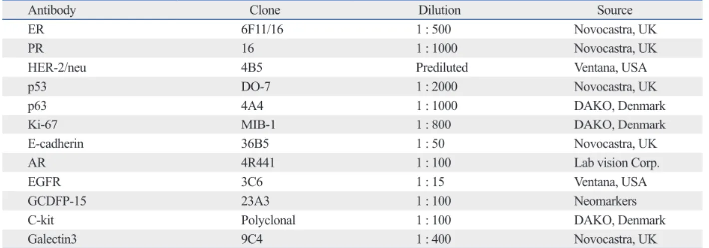
The antibodies used to conduct this study are listed in Table 3. Paraffin blocks were sectioned to a thickness of 4 um and tissues were attached on silane coated slides. The slides (PR) and c-erbB2 has been shown to vary.5-8 Other than
hormone receptors, no organized study concerning the im- munophenotype of GRCC has been published. Here, we present three cases of GRCC and their immunohistochemi- cal profiles.
MATERIALS AND METHODS
Clinical data
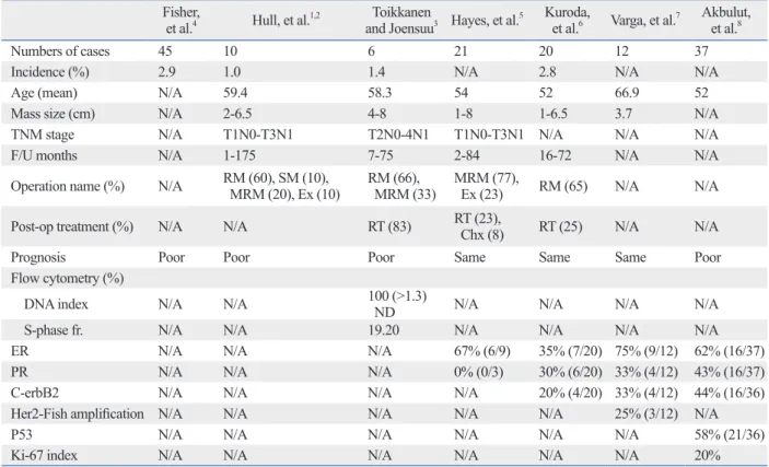
Table 2 summarizes the clinical data. There were only three Table 1. GRCC in Previous Publications
Fisher,
et al.4 Hull, et al.1,2 Toikkanen
and Joensuu3 Hayes, et al.5 Kuroda,
et al.6 Varga, et al.7 Akbulut, et al.8
Numbers of cases 45 10 6 21 20 12 37
Incidence (%) 2.9 1.0 1.4 N/A 2.8 N/A N/A
Age (mean) N/A 59.4 58.3 54 52 66.9 52
Mass size (cm) N/A 2-6.5 4-8 1-8 1-6.5 3.7 N/A
TNM stage N/A T1N0-T3N1 T2N0-4N1 T1N0-T3N1 N/A N/A N/A
F/U months N/A 1-175 7-75 2-84 16-72 N/A N/A
Operation name (%) N/A RM (60), SM (10),
MRM (20), Ex (10) RM (66),
MRM (33) MRM (77),
Ex (23) RM (65) N/A N/A
Post-op treatment (%) N/A N/A RT (83) RT (23),
Chx (8) RT (25) N/A N/A
Prognosis Poor Poor Poor Same Same Same Poor
Flow cytometry (%)
DNA index N/A N/A 100 (>1.3)
ND N/A N/A N/A N/A
S-phase fr. N/A N/A 19.20 N/A N/A N/A N/A
ER N/A N/A N/A 67% (6/9) 35% (7/20) 75% (9/12) 62% (16/37)
PR N/A N/A N/A 0% (0/3) 30% (6/20) 33% (4/12) 43% (16/37)
C-erbB2 N/A N/A N/A N/A 20% (4/20) 33% (4/12) 44% (16/36)
Her2-Fish amplification N/A N/A N/A N/A N/A 25% (3/12) N/A
P53 N/A N/A N/A N/A N/A N/A 58% (21/36)
Ki-67 index N/A N/A N/A N/A N/A N/A 20%
N/A, not available; F/U, follow up; RM, radical mastectomy; SM, simple mastectomy; MRM, modified radical mastectomy; Ex, excision; RT, radiation therapy; Chx, chemotherapy; fr, fraction; ND, non-diploid; ER, estrogen receptor; PR, progesterone receptor; GRCC, glycogen rich clear cell carcinoma.
Table 2. Clinical Findings
Case 1 Case 2 Case 3
Age 57 49 69
Ass. Lesion FCD FCD N/A
Mass size (mm) 23 12 N/A
pTNM T2N0M0 T1N0M0 N/A
Location RUL LUM N/A
Last F/U 2011-11-06 2011-12-16 N/A
F/U mos 18 14 N/A
Op name TM/c SLNB PM/c SLNB N/A
Post-op Tx 6th AC 4th AC+postRT N/A
FCD, fibrocystic change; RUL, right upper lateral; LUM, left upper medial; F/U, follow up; TM/c SLNB, total mastectomy with sentinel node biopsy; PM/c SLNB, partial mastectomy with sentinel node biopsy; AC, cyclophosphamide+adriamycin; RT, radiation therapy; N/A, not available.
receptor (AR), p53, p63, and ki-67; cytoplasmic positivity for gross cystic disease fluid protein 15 (GCDFP-15); nu- clear and cytoplasmic positivity for c-kit and galectin3; and cytoplasmic membrane positivity for c-erbB2, e-cadherin, and epidermal growth factor receptor (EGFR). Additional- ly, a proportional index was evaluated to describe the per- centage of positivity for a few of the antibodies, including hormone receptors (ER, PR, and AR) and p53.
RESULTS
Three patients were diagnosed with invasive glycogen rich clear cell carcinoma from 2000 to 2011 at our institution. On gross examination, GRCCs appeared as well-circumscribed, irregular and solid masses. No difference was found in com- parison to conventional invasive ductal carcinoma. Micro- scopically, GRCCs consisted of solid nests of clear cells with fine meshwork vasculatures. The individual cancer cells showed clear cytoplasm with fine granules and rela- tively low nuclear and cytoplasmic ratio. Nuclei were were baked at 60°C for 60 minutes and washed in xylene
for deparaffinization. For hydration of sections, the slides were bathed in gradient alcohol and distilled water. The anti- gen retrieval step was followed by boiling slides in 10 mM citrate buffer (pH6.0) for 10 minutes. Afterwards, the slides were immersed in 3% H2O2 for 15 minutes to block intrinsic hydro-peroxidase activity. The slides were incubated with primary antibodies for 2 hours at room-temperature, fol- lowed by incubation with secondary antibody (Enzyme-con- jugated polymer, EnVision Detection kit, Denmark) for 15 minutes at room-temperature. A NovaRED substrate kit (VECTOR Laboratory, Burlingame, CA, USA) was ap- plied to develop color following the manufacture’s proto- col. Harris hematoxylin was used for counter staining.
Interpretation of immunohistochemical staining
IHC slides were evaluated by light microscopy. The scoring intensity of IHC staining was based on a three-grade sys- tem; 1 positive (weak), 2 positive (intermediate) and 3 posi- tive (strong). The grade intensity of positive stainig was as- sessed as follows; nuclear positivity for ER, PR, androgen Table 3. Antibodies Used in This Study
Antibody Clone Dilution Source
ER 6F11/16 1 : 500 Novocastra, UK
PR 16 1 : 1000 Novocastra, UK
HER-2/neu 4B5 Prediluted Ventana, USA
p53 DO-7 1 : 2000 Novocastra, UK
p63 4A4 1 : 1000 DAKO, Denmark
Ki-67 MIB-1 1 : 800 DAKO, Denmark
E-cadherin 36B5 1 : 50 Novocastra, UK
AR 4R441 1 : 100 Lab vision Corp.
EGFR 3C6 1 : 15 Ventana, USA
GCDFP-15 23A3 1 : 100 Neomarkers
C-kit Polyclonal 1 : 100 DAKO, Denmark
Galectin3 9C4 1 : 400 Novocastra, UK
ER, estrogen receptor; PR, progesterone receptor; AR, androgen receptor; EGFR, epidermal growth factor receptor; GCDFP-15, gross cystic disease fluid protein 15.
Fig. 1. Histologic features of GRCC. (A) Tumor shows solid nests of clear cells with fine meshwork vasculatures (×100). (B) The individual cancer cells have clear cytoplasm with fine granules, low N-C ratio, wrinkled nuclei, and prominent nucleoli (×400). (C) The granules in tumor cells are stained with PAS (×400 and ×1000 in box). (D) The granules are not stained with d-PAS (×400 and ×1000 in box). GRCC, glycogen rich clear cell carcinoma; PAS, periodic acid stain.
A B C D
three groups. The first group of proteins shared the same im- muno-scoring and were related to cell surface molecules, in- cluding EGFR, c-kit, and E-cadherin. The second group was related to hormone receptors, including ER and AR. The third group was characterized by p53 related proliferation.
EGFR and c-kit are both cell surface receptors important to the PI3K through AKT and RAS to MAPK signal trans- duction pathways, and have been shown to be involved in cell survival and proliferation,9,10 Double expression of EGFR and c-kit in breast carcinoma is known as a predic- tive marker of poor prognoses.11 Among our cases, ki-67, marker of cell proliferation, was also synchronously up- regulated along with up-regulation of EGFR and c-kit. Tak- en together, in predicting poor prognoses, expression of both EGFR and c-kit sould be associated with high mitotic index. In our GRCC cases, case 1 was both EGFR and c-kit negative with a low mitotic index, while the other two (cas- es 2 and 3) were positive for both with a high mitotic index.
The expression pattern of EGFR and c-kit was concordant with E-cadherin and discordant with ki-67. Most studies on E-cadherin have focused on lobular carcinoma.12 The role of E-cadherin in ductal carcinoma is still unknown. The function of E-cadherin is considered related with cell adhe- sion molecules; thus, loss of expression would be associat- ed with a high incidence of invasion and metastasis. A few researchers stated that up-regulation of E-cadherin would be associated with a better prognosis in invasive ductal car- cinoma.13 However, the mechanism is unclear.
P53 is a well known nuclear protein that participates in cell proliferation. In benign conditions, p53 is not detectable by IHC staining. Cancer progression demonstrates accumu- lations of non-functional and non-degraded p53 protein in somewhat wrinkled. Nuclear membranes were irregularly
thickened and their nucleoli were prominent. All of them contained PAS positive and d-PAS labile granules in the cy- toplasm of more than 90% of tumor cells (Fig. 1). Mucin was not detected upon alcian blue and mucicarmin staining.
Tests for GCDFP-15 were negative. The tumors were, there- fore, considered to involve no apocrine differentiation.
The three cases revealed three different immuno-pheno- types (Table 4). Case 1 showed positivity for ER and negativ- ity for PR. Case 2 was positive for PR, but negative for ER.
Case 3 presented with triple negative invasive carcinoma. All three cases were negative for c-erbB2. Androgen receptor was positive in the ER positive case 1. EGFR, E-cadherin, and CD117 (c-kit) showed positivity in two cases (cases 2 and 3). Among these three cases, p53-positive case exhibited a low proliferative index (ki-67: 15%), while p53-negative cases showed high proliferative index (ki-67: 50-60%).
DISCUSSION
GRCC is a very rare entity that exhibits distinctive clear cells with intracytoplasmic glycogen. However, no trials have been conducted to reveal the distinct characteristics of GRCC, including clinical behavior, biologic features, im- munophenotypes, and genetics.2-6,8
Because of the rarity, we accumulated only three cases of GRCC out of more than 2000 cases of carcinoma over the last 10 years. Twelve different antibodies were evaluated in the three cases, the immunophenotypes of which prove not to be uniform. Based on immuno-scoring, we were able to characterize the expression patterns of targeted proteins into Table 4. Immunophenotypes of GRCC
Case 1 Case 2 Case 3
ER 100%/2 pos Neg Neg
PR Neg 60%/2 pos Neg
C-erbB2 1 pos Neg Neg
P53 80%/2 pos Neg Neg
P63 Neg Neg 5%
Ki-67 15% 40% 60%
E-Cad No loss Gain (3 pos) Gain (3 pos)
AR 90%/2 pos Neg Neg
EGFR Neg 3 pos 2 pos
GCDFP-15 Neg Neg Neg
C-kit Neg 2 pos 1 pos
Galectin3 2 pos 1 pos Neg
ER, estrogen receptor; PR, progesterone receptor; E-Cad, E-cadherin; AR, androgen receptor; EGFR, epidermal growth factor receptor; GCDFP-15, gross cystic disease fluid protein 15; pos, positive; neg, negative.
Glycogen-rich clear cell carcinoma of the breast: a light and elec- tron microscopic study. Cancer 1981;48:2003-9.
2. Hull MT, Warfel KA. Glycogen-rich clear cell carcinomas of the breast. A clinicopathologic and ultrastructural study. Am J Surg Pathol 1986;10:553-9.
3. Toikkanen S, Joensuu H. Glycogen-rich clear-cell carcinoma of the breast: a clinicopathologic and flow cytometric study. Hum Pathol 1991;22:81-3.
4. Fisher ER, Tavares J, Bulatao IS, Sass R, Fisher B. Glycogen-rich, clear cell breast cancer: with comments concerning other clear cell variants. Hum Pathol 1985;16:1085-90.
5. Hayes MM, Seidman JD, Ashton MA. Glycogen-rich clear cell carcinoma of the breast. A clinicopathologic study of 21 cases.
Am J Surg Pathol 1995;19:904-11.
6. Kuroda H, Sakamoto G, Ohnisi K, Itoyama S. Clinical and patho- logical features of glycogen-rich clear cell carcinoma of the breast.
Breast Cancer 2005;12:189-95.
7. Varga Z, Zhao J, Ohlschlegel C, Odermatt B, Heitz PU. Preferen- tial HER-2/neu overexpression and/or amplification in aggressive histological subtypes of invasive breast cancer. Histopathology 2004;44:332-8.
8. Akbulut M, Zekioglu O, Kapkac M, Erhan Y, Ozdemir N. Fine needle aspiration cytology of glycogen-rich clear cell carcinoma of the breast: review of 37 cases with histologic correlation. Acta Cytol 2008;52:65-71.
9. Sutton LM, Han JS, Molberg KH, Sarode VR, Cao D, Rakheja D, et al. Intratumoral expression level of epidermal growth factor re- ceptor and cytokeratin 5/6 is significantly associated with nodal and distant metastases in patients with basal-like triple-negative breast carcinoma. Am J Clin Pathol 2010;134:782-7.
10. De Miguel MP, Cheng L, Holland EC, Federspiel MJ, Donovan PJ. Dissection of the c-kit signaling pathway in mouse primordial germ cells by retroviral-mediated gene transfer. Proc Natl Acad Sci U S A 2002;99:10458-63.
11. Nalwoga H, Arnes JB, Wabinga H, Akslen LA. Expression of EGFR and c-kit is associated with the basal-like phenotype in breast carcinomas of African women. APMIS 2008;116:515-25.
12. Koo JS, Jung W. Clinicopathlogic and immunohistochemical characteristics of triple negative invasive lobular carcinoma. Yon- sei Med J 2011;52:89-97.
13. Mahler-Araujo B, Savage K, Parry S, Reis-Filho JS. Reduction of E-cadherin expression is associated with non-lobular breast carci- nomas of basal-like and triple negative phenotype. J Clin Pathol 2008;61:615-20.
14. Petitjean A, Achatz MI, Borresen-Dale AL, Hainaut P, Olivier M.
TP53 mutations in human cancers: functional selection and impact on cancer prognosis and outcomes. Oncogene 2007;26:2157-65.
nuclei, resulting in immuno-positivity of p53 protein. Mutat- ed p53 protein cannot suppress genetic errors leading to can- cer development. Nevertheless, p53 over-expression has been considered a poor prognostic marker and accompanied by a high mitotic index in invasive ductal carcinoma.14 Our three cases demonstrate just the opposite. Case 1 showed strong positivity for p53 with low mitotic index, while cas- es 2 and 3 showed negativity for p53 with high mitotic in- dex. These findings support the possibility of an association of cell proliferation with the signal transduction of surface molecules (EGFR, c-kit and E-cadherin), not with mutation of p53 protein, in GRCC.
Case 3 presented as a triple negative carcinoma and also expressed p63 protein. Case 3 was a basal-like breast can- cer and was predicted to involve a poor prognosis. Increased proliferative activity as determined by ki-67 (60%) and over- expression of EGFR supported the possibility of a poor prognosis. The diagnosis of this case was confirmed only 3 months prior to the preparation of this manuscript; there- fore, follow up data are currently not available. Based on IHC results, this case will require continued attention for future treatment. Presently, researchers agree that if GRCC is comparable to the conventional ductal carcinoma of the same TNM stage, the prognosis of the two types of carcino- mas would be the same.7
As only three cases were available for study, the drawing of any conclusions would be limited. However, these cases did reveal different IHC characteristics for GRCC. We be- lieve that the cases presented herein substantiate a need to re-examine the general features of GRCC. While the immu- nophenotypes of GRCC were not uniform, they appeared to be very similar to conventional ductal carcinoma.
REFERENCES
1. Hull MT, Priest JB, Broadie TA, Ransburg RC, McCarthy LJ.