CASE REPORT
거대세포바이러스 장염에 중복감염된 궤양성 대장염 환자에서 발생한 폐동맥 색전증에 대한 새로운 경구 항응고제 치료 1예
김석환, 장선희1, 성예규1, 박준규1, 박연정1, 윤진탁, 강상범
가톨릭대학교 의과대학 대전성모병원 소화기내과, 가톨릭대학교 의과대학 가톨릭중앙의료원 내과1
Use of Novel Oral Anticoagulant to Treat Pulmonary Thromboembolism in Patient with Ulcerative Colitis Superinfected Cytomegalovirus Colitis
Seok-Hwan Kim, Sunhee Jang1, Yegyu Sung1, Jun Kyu Park1, Yunjung Park1, Jintak Yun and Sang-Bum Kang
Division of Gastroenterology, Department of Internal Medicine, Daejeon St. Mary’s Hospital, College of Medicine, The Catholic University of Korea, Daejeon, Department of Internal Medicine, Catholic Medical Center, College of Medicine, The Catholic University of Korea1, Seoul, Korea Crohn’s disease and ulcerative colitis are the two major types of inflammatory bowel disease, and affect mainly the gastrointestinal tract but also have extraintestinal sequelae, such as arterial and venous thromboembolism. Thromboembolic complications, partic- ularly pulmonary thromboembolism, can be life threatening and require prompt management with anticoagulants. Conventional vita- min K antagonists have been used for the treatment of thromboembolic complications, but the development of novel oral anti- coagulants has shifted the paradigm. We report a case of a 42-year-old female with ulcerative colitis who experienced an acute flare-up due to cytomegalovirus superinfection with pulmonary thromboembolism. She was treated with oral mesalamine, intravenous steroid and ganciclovir and low-molecular-weight heparin, followed by rivaroxaban, a novel oral anticoagulant. Her symptoms resolved after treatment, and no recurrence was noted during a 6-month post-treatment follow-up. (Korean J Gastroenterol 2017;70:44-49) Key Words: Inflammatory bowel disease; Ulcerative colitis; Pulmonary thromboembolism; Rivaroxaban; Cytomegalovirus
Received April 26, 2017. Revised July 3, 2017. Accepted July 11, 2017.
CC This is an open access article distributed under the terms of the Creative Commons Attribution Non-Commercial License (http://creativecommons.org/licenses/
by-nc/4.0) which permits unrestricted non-commercial use, distribution, and reproduction in any medium, provided the original work is properly cited.
Copyright © 2017. Korean Society of Gastroenterology.
교신저자: 강상범, 34943, 대전시 중구 대흥로 64, 가톨릭대학교 의과대학 대전성모병원 소화기내과
Correspondence to: Sang-Bum Kang, Division of Gastroenterology, Department of Internal Medicine, Daejeon St. Mary’s Hospital, College of Medicine, The Catholic University of Korea, Daejeon 34943, Korea. Tel: +82-42-220-9501, Fax: +82-42-252-6807, E-mail: [email protected]
Financial support: None. Conflict of interest: None.
INTRODUCTION
Crohn’s disease (CD) and ulcerative colitis (UC) are the two major types of inflammatory bowel disease (IBD), which af- fect mainly the gastrointestinal tract, but can also exhibit ex- traintestinal involvement, such as arterial and venous thromboembolism.1 Although arterial and venous throm- boembolism can be devastating to IBD patients, they are fre- quently overlooked by physicians.2
The mainstay of therapy for thromboembolism is anti-
coagulation, and vitamin K antagonists (VKAs) are frequently used as the standard treatment.3 The recently developed novel oral anticoagulants (NOACs) may replace VKAs in thromboembolic treatment.3-6 However, no report of treat- ment of UC with thromboembolism using NOACs has been re- ported to date, and treatment guidelines have not been established.2,7 We report here a case of a patient with severe acute UC, deep vein thrombosis and pulmonary throm- boembolism, together with cytomegalovirus colitis, who was successfully treated using the NOAC rivaroxaban.
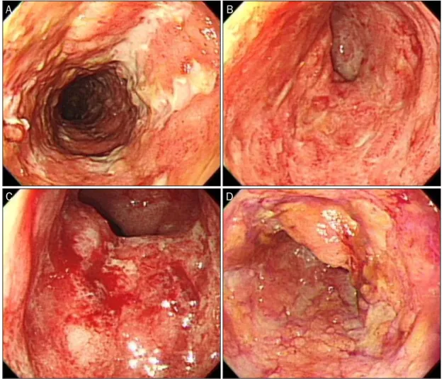
Fig. 1. Multiple longitudinal and geographic ulcerations with some denuded colonic mucosa and easy touch bleeding were noted from the distal descending colon to the rectum. CMV PCR of the tissue biopsy confirmed CMV colitis. (A) Descending colon. (B) Proximal sigmoid colon. (C) Distal sigmoid colon. (D) Rectum. CMV, cytomegalovirus; PCR, polymerase chain reaction.
CASE REPORT
A 42-year-old female patient presented to the clinic with complaints of fever and chest discomfort of 1-week duration.
She had been diagnosed with UC pancolitis 4 years prior and treated with oral mesalamine and episodic oral steroid.
Recently, she had experienced a disease flare-up, stool fre- quency more than 10 times per day with rectal bleeding, and received infliximab as an induction therapy at another hospital. This treatment did not control her rectal bleeding, abdominal pain or fever; moreover, the patient developed new symptoms, such as left calf pain and chest discomfort, 1 week prior to visiting our clinic. She did not have any dis- eases other than UC, and was neither a smoker nor an alcohol drinker.
An initial physical examination showed dull lower abdomi- nal pain, left calf swelling with pain, and chest discomfort
without signs of dyspnea. Her systolic and diastolic blood pressure was 100/60 mmHg with heart rate of 136 beats per minute. The body temperature was 38.0oC. Laboratory ex- amination revealed anemia (hemoglobin 9.2 g/dL, hema- tocrit 29.2%) and elevated ESR (106 mm/hr), C-reactive pro- tein (10.56 mg/dL) and d-dimer (5.85 μg FEU/mL) levels.
Blood gas analysis showed pH 7.5, pCO2 38 mmHg, pO2 92 mmHg, HCO3 29.6 mmol/L, and O2 saturation 98%. Fecal cal- protectin level was more than 400 mg/kg. Factor V Leiden, lupus antibody, antiphospholipid antibody, anticardiolipin antibody, protein C and S activity, homocysteine and comple- ment levels were within the normal range.
Initial sigmoidoscopy revealed multiple longitudinal ulcer- ations with a friable mucosa and easy touch bleeding in all colonic fields (Fig. 1). Random biopsies and tissue cytome- galovirus (CMV) polymerase chain reaction (PCR) were performed. The results indicated acute and chronic colitis, to-
A B
C D
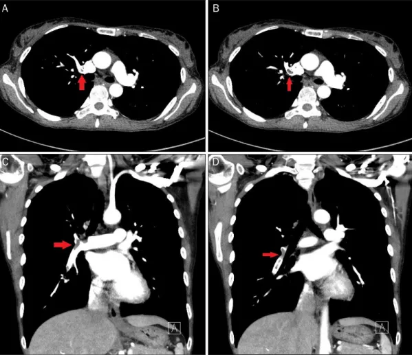
Fig. 2. Chest dynamic computed tomography showed a filling defect in the lobar and segmental branches of the right pulmonary artery (arrows). (A, B) Transverse view. (C, D) Coronal view.
Fig. 3. Lower extremity computed tomography showed diffuse filling defect from the left common iliac vein to the calf veins of the lower left leg (arrows), and normal vessels of the lower right leg (arrowhead). (A) Iliac crest level. (B) Symphysis pubis level. (C) Popliteal level.
gether with negative CMV immunohistochemistry, but pos- itive CMV PCR. CMV IgG was positive and CMV real-time PCR using whole blood yielded a viral load of 633 copies/mL
(19,900 IU/mL). The patient had a Mayo score of 12 points.
Initial chest dynamic computed tomography (CT) and low- er-extremity CT showed diffuse deep vein thrombosis from
A B
C D
A B C
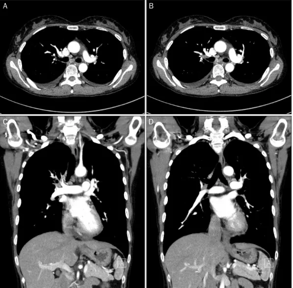
Fig. 4. Follow up chest computed tomography after 2 months of rivaroxaban treatment. The previously noted filling defect in the lobar and segmen- tal branches of the right pulmonary artery had disappeared. (A, B) Transverse view. (C, D) Coronal view.
the left common iliac vein to the calf veins of the lower left leg and filling defects in the lobar and segmental branches of the right lung, suggesting pulmonary thromboembolism (Fig. 2, 3).
We started oral mesalamine and azathioprine and intra- venous hydrocortisone and then switched to oral pre- dnisolone as management for severe acute UC. Ganciclovir 5 mg/kg bid was administered for CMV colitis, and low-mo- lecular-weight heparin for 2 weeks was given for pulmonary thromboembolism, followed by a switch to oral rivaroxaban.
There are many data supporting the efficacy in patient treat- ed with NOACs in pulmonary thromboembolism. Moreover, her veins were very vulnerable, drawing blood and giving in- jections have caused her a great physical and psychological trauma. For these reasons, we have decided to use oral rivar- oxaban rather than conventional anticoagulation with VKAs,
which does not have to draw blood every time to check the tar- get international normalized ratio (INR) level. After 6 weeks, infliximab was administered as a third induction dose. After treatment initiation, the hematochezia ceased on day 17 of hospitalization, and the stool frequency decreased to three times per day on day 39 of hospitalization. Her Mayo score had decreased to 9 points at the time of discharge.
For the pulmonary thromboembolism, chest discomfort subsided on day 3 of hospitalization, and the left leg swelling and pain subsided on day 6 of hospitalization. The patient was discharged on day 40 of hospitalization without complications.
After rivaroxaban treatment for 2 months, a follow-up chest CT was performed (Fig. 4). The previously noted pulmonary embolism at the lobar and segmental pulmonary arteries of the right lung had disappeared. The patient was maintained
A B
C D
on anticoagulation therapy with oral rivaroxaban for 6 months. She regularly visited the clinic after cessation of anti- coagulation treatment for 6 months and maintained deep re- mission with infliximab and oral azathioprine.
DISCUSSION
Deep vein thrombosis (DVT) and pulmonary thromboemb- olism (PTE) are important extraintestinal complications of IBD. In comparison with healthy individuals, patients with UC and CD show a threefold higher incidence of thrombosis, and up to a 16-fold increase during disease flare-ups.8 A study in an Asian population reported 1.98- and 1.80-fold increases in the incidence of DVT and PTE, respectively.9 In the general population, the 30-day case fatality rates of DVT and PTE are 11-30%,10,11 and these data also correlate with IBD patients which can be life threatening.12 Therefore, prompt manage- ment is necessary to reduce the morbidity and mortality of these patients. Thromboembolism is caused by changes in the composition of the blood, stasis of the blood, and changes in the vessel wall, as postulated by Virchow.9 In IBD patients, localized and systemic inflammation can activate the coagu- lation cascade, resulting in increased levels of coagulation factors (such as fibrinogen, thrombocytes, factor V, and fac- tor VIII) and decreased levels of inhibitors of blood clotting ac- tivation (such as antithrombin and protein C and S).13 These changes promote hypercoagulability in IBD patients, which in turn increases the risk of DVT and PTE.
An association between CMV infection and thromboembo- lism has been reported in immunocompromised patients, particularly those infected with human immunodeficiency vi- rus or who have undergone transplantation.14 Other studies have shown a similar relationship in immunocompetent patients. Atzmony et al. reported that 6.4% of patients with acute CMV infection develop thrombosis, irrespective of oth- er risk factors of thrombotic events,15 while Schimanski et al.
reported a higher rate of 9.1%.16 These findings suggest that CMV infection itself may increase the risk of thromboembolic events. Our patient did not have predisposing factors for throm- boembolism other than UC, and the CMV infection could have induced the flare-up, increasing the risk of thromboembolism.
Indeed, the infection itself might enhance the risk. It is thus im- portant to identify CMV coinfection in IBD patients who show flare-up or thromboembolic complications.
Conventionally, VKAs have been used to treat DVT and PTE.3 VKAs are effective for preventing recurrent venous thromboembolism, as evidenced by an 85% decrease in the relative risk.17 However, VKAs increase the risk of major bleeding events by 1.6-2.6% and the case-fatality rate by 11.0% during the 6 months of venous thromboembolism treatment.4 Therefore, the INR should be monitored. Use of NOACs abolishes the need to monitor the INR, with similar ef- ficacy and a lower incidence of bleeding complications.3-6 The patient’s body mass index was 14.0 and her peripheral veins were very vulnerable that every time we gave injections or took blood samples, the patient suffered great pain. The use of rivaroxaban in place of conventional treatment with VKAs yielded an excellent outcome with no complications, and the decreased frequency of blood draws satisfied the patient.
Many cases of pulmonary thromboembolism in IBD pa- tients have been reported,18,19 but none has involved the use of NOACs, with the exception of one case of cerebral venous thrombosis in CD.20 Our patient was treated with rivaroxaban for 6 months and did not experience recurrence during that time. We believe that in the near future, NOACs should re- place VKAs for treatment of thrombotic complications in IBD patients.
REFERENCES
1. Owczarek D, Cibor D, Głowacki MK, Rodacki T, Mach T. Inflammatory bowel disease: epidemiology, pathology and risk factors for hypercoagulability. World J Gastroenterol 2014;20:53-63.
2. Papa A, Gerardi V, Marzo M, Felice C, Rapaccini GL, Gasbarrini A. Venous thromboembolism in patients with inflammatory bow- el disease: focus on prevention and treatment. World J Gastroenterol 2014;20:3173-3179.
3. van der Hulle T, Kooiman J, den Exter PL, Dekkers OM, Klok FA, Huisman MV. Effectiveness and safety of novel oral anti- coagulants as compared with vitamin K antagonists in the treat- ment of acute symptomatic venous thromboembolism: a sys- tematic review and meta-analysis. J Thromb Haemost 2014;12:
320-328.
4. Connolly SJ, Ezekowitz MD, Yusuf S, et al. Dabigatran versus war- farin in patients with atrial fibrillation. N Engl J Med 2009;
361:1139-1151.
5. Giugliano RP, Ruff CT, Braunwald E, et al. Edoxaban versus war- farin in patients with atrial fibrillation. N Engl J Med 2013;
369:2093-2104.
6. Granger CB, Alexander JH, McMurray JJ, et al. Apixaban versus warfarin in patients with atrial fibrillation. N Engl J Med 2011;
365:981-992.
7. Choi CH, Kim YH, Kim YS, et al. Guidelines for the management of ulcerative colitis. Korean J Gastroenterol 2012; 59:118-140.
8. Grainge MJ, West J, Card TR. Venous thromboembolism during active disease and remission in inflammatory bowel disease: a cohort study. Lancet 2010;375:657-663.
9. Chung WS, Lin CL, Hsu WH, Kao CH. Inflammatory bowel disease increases the risks of deep vein thrombosis and pulmonary em- bolism in the hospitalized patients: a nationwide cohort study.
Thromb Res 2015;135:492-496.
10. Cohen AT, Tapson VF, Bergmann JF, et al. Venous thromboembo- lism risk and prophylaxis in the acute hospital care setting (ENDORSE study): a multinational corss-sectional study. Lancet 2008;371:387-394.
11. Cushman M, Tsai AW, White RH, et al. Deep vein thrombosis and pulmonary embolism in two cohorts: the longitudinal inves- tigation of thromboembolism etiology. Am J Med 2004;117:19-25.
12. Talbot RW, Heppell J, Dozois RR, Beart RW Jr. Vascular complica- tions of inflammatory bowel disease. Mayo Clin Proc 1986;
61:140-145.
13. Rosendaal FR. Venous thrombosis: a multicausal disease. Lancet
1999;353:1167-1173.
14. Justo D, Finn T, Atzmony L, Guy N, Steinvil A. Thrombosis asso- ciated with acute cytomegalovirus infection: a meta-analysis. Eur J Intern Med 2011;22:195-199.
15. Atzmony L, Halutz O, Avidor B, et al. Incidence of cytomegalovi- rus-associated thrombosis and its risk factors: a case-control study. Thromb Res 2010;126:e439-e443.
16. Schimanski S, Linnemann B, Luxembourg B, et al. Cytomegalovirus infection is associated with venous thromboembolism-a case- control study. J Thromb Haemost 2009;7 Suppl 2:418.
17. Hutten BA, Prins MH. Duration of treatment with vitamin K antag- onists in symptomatic venous thromboembolism. Cochrane Database Syst Rev 2006;(1):CD001367.
18. Chung C, Kang DH, Kim EA, Hong SH, Kong HS, Park HC. A case of pulmonary embolism in recurrent ileal crohn’s disease after to- tal colectomy. Korean J Gastrointest Endosc 2001;22:111-115.
19. Chung ES, Kim JH, Jung JH, et al. A case of pulmonary embolism in crohn’s disease. Tuberc Respir Dis 2009;66:370-373.
20. Cho YH, Chae MK, Cha JM, et al. Cerebral venous thrombosis in a patient with crohn’s disease. Intest Res 2016;14:96-101.