INTRODUCTION
Beh et’s disease is a chronic systemic inflammatory disease involving mucous membranes, skin, eyes, gastrointestinal tract, joints, vessels, and neurologic systems (1). It occurs endemically in the Middle East and the Mediterranean re- gions. It is also distributed in the Central and East Asian countries including Korea (2), Japan (3), and China (4). The
patients with Beh et’s disease have been increasing in Korea and the epidemiological and clinical features of the disease have been reported by some institutes (2). However, the nationwide studies have not been performed yet.
The Korean Study Group for Beh et’s disease was found- ed in 1999 and the first nationwide multicenter survey on epidemiological and clinical features of Beh et’s disease in Korea was performed.
Dongsik Bang1, Ju Hee Lee1, Eun-So Lee2, Sungnack Lee2, Jong Soo Choi3, Young Keun Kim4, Baik Kee Cho5, Jai Kyoung Koh6, Young Ho Won7, Nack In Kim8, Seok Don Park9, Hong Jun Ahn10, Yoon Woo Lee11, Han Young Wang12, Won Woo Lee13, Hee Chul Eun14, Eun Sup Song15, Sung Won Lee16, Chang Woo Lee17, Chong Ju Lee18, Jae Ho Park19, Yeong Wook Song20, Sang Tae Kim21, Chong Yeol Kim22, Jang Kyu Park23, Kyung Sool Kwon24
1Department of Dermatology, Yonsei University College of Medicine; 2Department of Dermatology, Ajou University School of Medicine; 3Department of Dermatology, College of Medicine, Yeungnam University; 4Department of Dermatology, College of Medicine, Inha University; 5Department of Dermatology, St. Mary’s Hospital, The Catholic Univer- sity of Korea; 6Department of Dermatology, Asan Medical Center, University of Ulsan College of Medicine; 7Department of Dermatology, Chonnam National University Medical School; 8Department of Dermatology, College of Medicine, Kyung Hee University; 9Department of Dermatology, Wonkwang University School of Medicine; 10Department of Internal Medicine, Seoul Paik Hospital, Inje University School of Medicine; 11Department of Rheumatology, Ilsan Paik Hospital, Inje University School of Medicine; 12Department of Dermatology, Pusan Paik Hospital, Inje University School of Medicine; 13Department of Dermatology, Maryknoll Hospital; 14Department of Dermatology, Seoul National Uni- versity College of Medicine; 15Department of Dermatology, Chonju Presbyterian Medical Center; 16Department of Rheumatology, Dong-A Medical College; 17Department of Dermatology, Hanyang University Medical College; 18Department of Dermatology, Kangdong Sacred Heart Hospital, Hallym University Medical College; 19Department of Rheumatology, Keimyung University Dongsan Medical Center; 20Department of Rheumatology, Seoul National University College of Medicine; 21Department of Dermatology, Kosin Medical University & Gaspel Hospital; 22Department of Oral Medicine, Yonsei University College of Dentistry; 23Department of Dermatology, Chungnam National Univer- sity College of Medicine; 24Department of Dermatology Pusan National University College of Medicine, Korea
(Authors’affiliations are listed below) Received : 16 March 2001 Accepted : 20 June 2001 Address for correspondence Dongsik Bang, M.D.
Department of Dermatology, Yonsei University College of Medicine, 134 Shinchon-dong, Seodaemun-gu, Seoul 120-752, Korea Tel : +82.2-361-5720, Fax : +82.2-393-9157 E-mail : [email protected]
*This article was presented at the 9th International Conference on Behcet’s disease on May 27-29, 2000.
615 J Korean Med Sci 2001; 16: 615-8
ISSN 1011-8934
Copyright � The Korean Academy of Medical Sciences
Epidemiologic and Clinical Survey of Behcet's Disease in Korea:
the First Multicenter Study
The prevalence of Behcet’s disease is the highest in the East Asian and the Medi- terranean countries. Behcet’s disease is also distributed in the Asian countries, but the nationwide survey has not been performed in Korea yet. The Korean Study Group for Behcet’s Disease, founded in 1999, conducted a multicenter, retrospective survey on epidemiologic and clinical features of the patients with Behcet’s disease from 20 hospitals around the nation from 1997 to 1999. Of 3,497 patients, 1,527 were classified into complete or incomplete type of Behcet’s dis- ease according to the revised Shimizu’s classification. The sex ratio was 1:1.75 with the female predominance. Geographical distribution showed the highest fre- quency in Seoul (38.5%). Clinically, 98.8% had oral ulcers, 83.2% had genital ulcers, 84.3% had skin lesions and 50.9% had ocular lesions. As for the minor clinical manifestations, articular symptoms were the most frequent. The pathergy test showed positive in 15.4% of patients and revealed a higher positive rate in males (20.2%) than in females (12.7%). In conclusion, we performed the first multicenter study on Behcet’s disease in Korea and revealed the female predomi- nance, higher frequency of ocular lesions, and lower positivity of pathergy test in the patients.
Key Words : Behcet’s Disease; Multicenter Studies, Korea
’
’
’
’
’
’
’
616 D. Bang, J.H. Lee, E.-S. Lee, et al.
PATIENTS & METHODS
The multicenter study was performed retrospectively with the current data from January 1997 to December 1999.
Twenty-four departments from twenty hospitals in Korea participated to this survey. Typical major features including recurrent oral ulcer, skin lesions, eye lesions, and genital ulcers were evaluated in the patients. The patients with four major features diagnosed as complete type, the patients with three major features, two major plus two minor features or typical ocular symptom plus one major or two minor features diag- nosed as incomplete type, and the patients with two major features or one major plus two minor diagnosed as suspect- ed type according to the revised Shimizu’s classification (5).
A total of 3,497 patients who visited the hospitals in the period were included as having Beh et’s disease and were checked by the following inquiry from the hospital records:
patient’s name, date of birth, sex, geographic distribution, clinical manifestations, durations of the symptoms, and pos- itivity to pathergy test. Durations of symptoms were deter- mined as the period from the beginning of at least one of the diagnostic symptom criteria to the patient’s first visit.
Geographic distribution was based on the location where the symptom first occurred. Of 3,497 patients, 1,527 fulfilled the Diagnostic Criteria of International Study Group for Beh et’s disease (6) and/or the revised Shimizu’s classifica- tion (5).
Pathergy test was performed with oblique insertion of 26 gauge needle under sterile condition and read by a physician at 24 hr. Formation of papule or pustule was interpreted as a positive result.
Statistical analysis was performed using SPSS version 9.0 (SPSS Inc. Chicago, U.S.A.). Kolmogorov-Smirnov test was applied to evaluate the normality of the age and sex distri- bution. Chi-square test was applied for comparing the sex difference of pathergy test. Mann-Whitney test was applied to evaluate the age distributions of onset and diagnosis of the disease between males and females. A value of p<0.05 was regarded as statistically significant.
RESULTS
Of 3,497 patients, 1,527 were classified as complete or incomplete type of Beh et’s disease. According to the Shi- mizu’s classification, incomplete type was the most frequent type (32.8%), followed by suspected type (32.3%), possible type (24.0%) and complete type (10.9%), in order of decreas-
At the first visit Age of onset
Age (yr)
Female
Male Total (%) Male Female Total (%)
Under 9 0 1 1 (0.07) 5 8 13 (0.85)
10-19 5 8 13 (0.85) 45 79 124 (8.12)
20-29 48 104 152 (9.95) 139 262 401 (26.26)
30-39 181 280 461 (30.19) 217 398 615 (40.28)
40-49 230 396 626 (41.0) 107 161 268 (17.55)
50-59 63 138 201 (13.16) 35 53 88 (5.76)
60-69 28 39 67 (4.39) 8 8 16 (1.05)
Over 70 1 5 6 (0.39) 0 2 2 (0.13)
Total 556 971 1,527 (100) 556 971 1,527 (100)
Table 2.Age and sex distributions of 1,527 patients with Beh et’s disease Classification Number of patients %
Complete 382 10.9
Incomplete 1,145 32.8
Suspected 1,130 32.3
Possible 840 24.0
Table 1.Classification of 3,497 patients with Beh et’s disease according to the Shimizu’s criteria
Fig. 1.Geographical distribution of 1,527 patients with Beh et’s disease in Korea.
Kyonggido
(27.9%)
∙
Seoul (38.5%)
∙
Chungchong namdo (3.3%)
∙
Chollabukdo
(3.0%)
∙
Chollanamdo
(4.1%)
∙
Chejudo (0.2%)
∙
Kyongsang bukdo (11.7%)
∙
Chungchong bukdo (1.7%)
∙
∙ ∙ ∙
∙ ∙
∙∙ ∙ ∙ ∙ ∙ ∙ ∙ ∙∙ ∙ ∙ ∙ ∙ ∙ ∙ ∙ ∙ ∙ ∙ ∙ ∙ ∙ ∙ ∙ ∙ ∙ ∙ ∙
∙ ∙ ∙ ∙ ∙
∙
∙
∙ ∙ ∙
∙ ∙ ∙
∙ ∙ ∙ ∙ ∙ ∙ ∙ ∙ ∙ ∙ ∙ ∙
∙ ∙ ∙ ∙ ∙ ∙ ∙ ∙ ∙ ∙ ∙ ∙ ∙ ∙ ∙ ∙ ∙ ∙ ∙ ∙ ∙ ∙ ∙ ∙ ∙ ∙ ∙ ∙ ∙ ∙ ∙ ∙ ∙ ∙ ∙ ∙ ∙ ∙ ∙ ∙ ∙ ∙ ∙ ∙ ∙ ∙ ∙
Kangwondo (1.8%)
∙
Kyongsangnamdo (7.7%)
∙
Epidemiologic and Clinical Survey of Beh et’s Disease in Korea 617
ing frequency (Table 1).
Table 2 summarizes age and sex distributions of the data.
The median age of onset was 33 yr (range 1-75) and the medi- an age of diagnosis was 41 yr (range 5-75). The male to female ratio was 1:1.75, indicating female predominance. The age of onset was most commonly in their thirties, followed by twenties (26.3%), forties (17.6%), and teens (8.1%). The age at diagnosis was most commonly in the forties (41%).
There was no differences in the age distributions of onset (p=0.444) and diagnosis (p=0.453) of the disease between males and females.
Geographical distribution showed that 38.5% of total patients with Beh et’s disease was in Seoul with the highest frequency (Fig. 1). The order of frequencies of geographical distribution was Kyonggido (27.9%), Kyongsangbukdo (11.7%), Kyongsangnamdo (7.7%), Chollanamdo (4.1%), Chungchongnamdo (3.3%), Chollabukdo (3.0%), Kang- wondo (1.8%), Chungchongbukdo (1.7%) and Chejudo (0.2%).
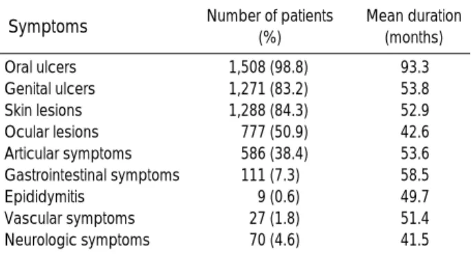
Major and minor clinical manifestations are summarized in Table 3 and were oral ulcers (98.8%), skin lesions (84.3%), genital ulcers (83.2%), ocular lesions (50.9%), articular symp- toms (38.4%), gastrointestinal symptoms (7.3%), neurolog- ic symptoms (4.6%), vascular symptoms (1.8%), and epi- didymitis (0.6%), in order of decreasing frequency. The lon- gest mean duration of clinical manifestation was 7 yr and 9 months with oral ulcer.
The results of pathergy test were available in 715 patients with Beh et’s disease and showed positive in 110 patients (15.4%) and revealed higher positive rates in males (20.2%) than in females (12.7%) (p=0.008) (Table 4).
DISCUSSION
The prevalence of Beh et’s disease is known to be high in Japan, China, Turkey, Tunisia, and the Mediterranean and the Middle Eastern countries. The prevalence of Beh et’s disease in Korea also has been reported as high as the coun- tries mentioned above. However, Beh et’s disease is rarly
seen in the Northern Europe or in the United States.
The frequency of Beh et’s disease in this study demon- strated similar results with the previous report in 1997 per- formed by Bang et al. with the results as follows (2): com- plete type 16.1%, incomplete type 38.2%, suspected type 25.4%, and possible type 20.3%.
The result of sexual distribution in our studies was 1:1.75 with the female predominance, which is in line with the previous study. However, in contrast to those studies, Zouboulis et al. (7) reported the male predominance in classical Japanese and Turkish patients with Beh et’s disease and yet, there is gradual increase of female patients, which leads to more of 1:1 male-to-female ratio. Saylan et al. (8) also noted the male predominance in almost all middle eastern countries such as Turkey, Iran, Lebanon, Iraq, Jordan, Israel, and Egypt. The countries with the female predominance are Korea, China, the United States and Britain. The difference in sexual dis- tribution among the countries indicates some potential envi- ronmental and genetic influences on the pathogenesis of the disease.
The age of onset and the age at diagnosis of Beh et’s dis- ease in the present study were slightly older than those in the previous studies. The proprtion of group with the age of onset under 20 was 8.97%, indicating the rarity of Beh et’s disease in young ages.
The geographical distribution revealed higher proportion in Seoul and Kyonggido. This result may be due to the dif- ferences in the accessibility to the hospitals and population density according to the region. The reports in Japan exhib- ited a higher prevalence in northern area than southern Japan (9). Nonetheless, our study could not determine distribu- tional differences by the north versus south or by east versus west.
As in other studies, the positive rates of major symptoms revealed that the most frequent major symptom was oral ulcers. As for the minor clinical manifestations, articular symptoms were most frequent. Moreover, the positive rate of ocular lesions in the present study was much higher than that in the previous studies in Korea. The increase in fre- quency of ocular lesions may be due to the increase in rou- tine tests for the eyes since the ocular lesions are considered to be one of the most important prognostic factors. Further- more, the exclusion of the possible type in this study might have contributed to the higher frequency of ocular and artic- ular symptoms.
Sex Number of patients
Positive (%) Negative (%)
Male (n=257) 52 (20.2) 205 (79.8)
Female (n=458) 58 (12.7) 400 (87.3)
Total (%) 110 (15.4) 605 (84.6)
Table 4.Result of pathergy test in 715 patients with Beh et’s disease
Symptoms Number of patients (%)
Mean duration (months)
Oral ulcers 1,508 (98.8) 93.3
Genital ulcers 1,271 (83.2) 53.8
Skin lesions 1,288 (84.3) 52.9
Ocular lesions 777 (50.9) 42.6
Articular symptoms 586 (38.4) 53.6
Gastrointestinal symptoms 111 (7.3) 58.5
Epididymitis 9 (0.6) 49.7
Vascular symptoms 27 (1.8) 51.4
Neurologic symptoms 70 (4.6) 41.5
Table 3.Major and minor symptoms of 1,527 patients with Beh et’s disease
618 D. Bang, J.H. Lee, E.-S. Lee, et al.
The positivity of the pathergy test in the present study (15.4%) was significantly lower than that of Japan (43.8%) and China (62.2%) (4, 10). This observation may be attribut- ed to the differences in the method of pathergy test or to the racial differences. The positivity of pathergy test is especial- ly high in the Middle Eastern countries, which made the test as a crucial parameter of diagnosis of Beh et’s disease.
In contrast, the low positive rate of pathergy test in the pre- sent study suggests a limitation of its use as a diagnostic criteria in Korea. Yazici et al. (11) reported a higher positive rate of pathergy test in males, which was also observed in the present study.
In conclusion, we performed the first multicenter study on Beh et’s disease in Korea and revealed that the female predominance, higher positive rate of ocular lesions and lower positivity of pathergy test were the significant features in Korean patients. Thus, the necessity of close observations on the eyes and further investigation of prevalences through the central registration system should be emphasized.
ACKNOWLEDGMENT
This study was supported by a grant (#HMP-97-M-2-0041) of the 1997 Good Health R&D Project, Ministry of Health
& Welfare, Korea.
REFERENCES
1. Shimizu T, Ehrlich GE, Inaba G, Hayashi K. Beh et’s disease. Semin
Arthritis Rheum 1979; 8: 223-60.
2. Bang D, Yoon KH, Chung HG, Choi EH, Lee ES, Lee S. Epidemi- ological and clinical features of Beh et’s disease in Korea. Yonsei Med J 1997; 38: 428-36.
3. Ohno S. Beh et’s disease in the world. In: Lehner T, Barnes CG, eds. Recent advances in Beh et’s disease. London: Royal Society of Medicine Services, 1986: 181-6.
4. Dong Y, Ming QX, Zhang NZ, Li CH, Wu QY. Testing different diagnostic criteria of Beh et’s syndrome in Chinese patients. In:
O’Duffy JD, Kokmen E, eds. Beh et’s disease: basic and clinical aspects. New York: Marcel Dekker, Inc. 1991: 55-9.
5. Mizushima Y, Inaba G, Mimura Y, Ohno S. Diagnostic criteria for Beh et’s disease in 1987, and guideline for treating Beh et’s disease.
Saishin Igaku 1988; 43: 391-3.
6. International study group for Behcet’s disease. Criteria for diagno- sis of Beh et’s disease. Lancet 1990; 335: 1078-80.
7. Zouboulis ChC, Djawari D, Kirch W. Adamantiades-Beh et’s dis- ease in Germany. Data of the German Registry in 1996. In Hamza M, ed. Beh et’s disease. Tunis: Pub Adhoua, 1997: 180-5.
8. Saylan T, Mat C, Fresko I, Melikoglu M. Beh et’s disease in the Middle East. Clin in Dermatol 1999; 17: 209-23.
9. Yamamoto S, Toyokawa H, Matsubara J. A nationwide survey of Beh et’s disease in Japan. Jpn J Ophthalmol 1974; 18: 282-90.
10. Nakae K, Masaki F, Hashimoto T, Inaba G, Mochizuki M, Sakane T. Recent epidemiological features of Beh et’s disease in Japan. In:
Godeau P, Wechsler B, eds. Beh et’s disease. Amsterdam: Excerp- ta Medica, Elsevier Science Publishers B.V., 1993: 145-51.
11. Yazici H, Tuzun Y, Tanman AB, Yurdakul S, Serdaroglu S, Pazarli H, Muftuoglu A. Male patients with Beh et’s syndrome have stronger pathergy reactions. Clin Exp Rheumatol 1985; 3: 137-41.