Received:April 10, 2017, Revised:May 8, 2017, Accepted:May 11, 2017
Corresponding to:Jong Hwan Jung, Division of Nephrology, Department of Internal Medicine, Wonkwang University School of Medicine and Hospital, 895 Muwang-ro, Iksan 54538, Korea. E-mail:[email protected]
Copyright ⓒ 2017 by The Korean College of Rheumatology. All rights reserved.
This is a Open Access article, which permits unrestricted non-commerical use, distribution, and reproduction in any medium, provided the original work is properly cited.
Renal Involvement in Rheumatic Diseases
Seon-Ho Ahn, Jong Hwan Jung
Division of Nephrology, Department of Internal Medicine, Wonkwang University School of Medicine and Hospital, Iksan, Korea
Most rheumatic diseases are chronic inflammatory diseases. Kidney-related symptoms of rheumatic diseases are often present, which increase mortality and morbidity of patients with rheumatic diseases. When patients with rheumatic diseases show signs or symptoms of renal involvement, management for primary rheumatic diseases should be more aggressive. In general, the risk and severity of renal involvement in patients with rheumatic diseases depend on the type of primary rheumatic diseases.
Rheumatic disease itself, chronic use of immunosuppressive agents and non-steroidal anti-inflammatory drugs, and comorbid- ities, such as diabetes, hypertension, and cardiovascular complications, are the main causes of renal involvement in patients with rheumatic diseases. Many studies have reported the predominant features of renal involvement in most rheumatic diseases. We have attempted to summarize the relationships between rheumatic diseases and renal diseases, and clinical or pathophysiological features of renal involvement resulting from primary rheumatic diseases except systemic lupus erythematosus. Review for renal involvement, particularly in relation to early diagnosis and management of renal involvement in rheumatic diseases, is clinically significant because renal involvement in rheumatic diseases generally implies a bad prognosis. (J Rheum Dis 2017;24:174-184)
Key Words. Kidney diseases, Rheumatic diseases, Inflammation
INTRODUCTION
Renal involvement in patients with several rheumatic diseases including rheumatoid arthritis (RA), systemic lupus erythematosus, and vasculitis is multifactorial.
Most patients with chronic rheumatic diseases have co- morbidities, such as diabetes mellitus, hypertension, and several cardiovascular diseases. These comorbidities are associated with the development of chronic kidney dis- ease (CKD) and increased mortality resulting from the development of CKD, particularly in rheumatic diseases [1]. Chronic inflammation is a common pathophysio- logical mechanism of most rheumatic diseases and may result in the development of cardiovascular complica- tions and CKD [2]. Chronic use of rheumatic drugs in- cluding non-steroidal anti-inflammatory drugs (NSAIDs) and disease-modifying anti-rheumatic drugs (DMARDs), such as methotrexate, bucillamine, and tumor necrosis factor-alpha (TNF-α) inhibitor, can cause renal dysfunc-
tion by developing glomerulonephritis or tubulointer- stitial nephritis (TIN) [3-6]. The factors, such as co- morbidities, chronic inflammation, and long-term use of nephrotoxic drugs in patients with rheumatic diseases, are important in relation to renal involvement of rheu- matic disease. In addition, the increase in human lifespan and harmful environmental factors associated with chronic inflammation results in increased comorbidities and the development of CKD in patients with rheumatic disease [7].
Renal involvement of rheumatic diseases clinically var- ies from severe glomerulonephritis to urinary abnormal- ity without renal dysfunction. In detail, renal involve- ment of rheumatic diseases manifests in different forms of clinical manifestation, depending on which part of the nephron is predominantly involved. For example, pa- tients with RA may have renal amyloidosis, which mani- fests by accumulating amyloid fibril mainly in glomeruli or tubulointerstitium. Renal involvement in systemic lu-
pus erythematosus is expressed as lupus nephritis which mainly involves the glomeruli. TIN is mainly expressed in renal involvement of primary Sjӧgren’s syndrome (pSS).
Rapid progressive glomerulonephritis (RPGN) can be of- ten expressed in patients with small vasculitis associated with antineutrophil cytoplasm antibody (ANCA) [8].
On the contrary, long-standing kidney diseases may cause rheumatic diseases, such as dialysis-related amyloi- dosis and gout. Dialysis-related amyloidosis mainly re- sults from beta-2 microglobulin accumulation in several sites including bones, joints, and periarticular tissues.
Chronic arthritis, such as degenerative osteoarthropathy, and spondyloarthropathy, occurs due to dialysis-related amyloidosis. Whether hyperuricemia itself is the cause of renal dysfunction remain unclear, but decreased urinary excretion of uric acid in patients with CKD and hyper- uricemia resulting from it may lead to chronic gout [9].
Since the therapeutic approach to secondary rheumatic diseases differs from the treatment of primary rheumatic disease, the differential diagnosis of primary rheumatic diseases from secondary rheumatic diseases caused by CKD has a particularly great clinical significance in the di- agnosis and treatment of chronic diseases.
In this review, we focused on the clinical or pathological features of renal involvement in patients with rheumatic diseases.
MAIN SUBJECTS
Renal involvement in RA
The prevalence of RA generally varies from 0.1% to 5%
[7,10]. In contrast to systemic lupus erythematosus, re- nal involvement in RA is not frequent [11]. However, re- nal involvement in patients with RA is an important in- dicator of poor prognosis [12]. According to previous lit- erature, the prevalence of renal involvement in patients with RA varies between 5% and 50% [12,13]. Renal in- volvement including glomerulonephritis and renal tubu- lar dysfunction in RA often occurs due to chronic in- flammation, which is the main pathophysiologic mecha- nism of RA. To date, the main causes of this renal involve- ment in RA were speculated as components such as chronic inflammatory disease itself or comorbidities re- sulting from chronic inflammation, and nephrotoxic drugs including DMARDs and NSAIDs, which are used for the management of chronic autoimmune inflammatory reaction. However, the chronic use of anti-rheumatic drugs rather than chronic inflammation itself of RA is the
clinically more frequent cause of renal involvement in pa- tients with RA [8]. Long use of DMARDs, such as metho- trexate, leflunomide, and TNF-α inhibitor in RA may re- duce the incidence of several comorbidities including car- diovascular diseases by effective control of chronic in- flammation [14]. However, conventional drugs, includ- ing NSAIDs, methotrexate, and bucillamine, can often re- sult in renal involvement, such as renal TIN, mem- branous nephropathy, and mesangial proliferative glo- merulonephritis (mspGN) [11,12,15].
Regardless of the causes, renal abnormalities in patients with RA may be clinico-pathologically present as glomer- ular forms. These abnormalities include asymptomatic urinary abnormality, mspGN, membranous nephropathy, minimal change disease, renal amyloidosis, and even ex- tra-capillary proliferative glomerulonephritis or TIN.
According to a previous literature, the frequency of glo- merulonephritis and amyloidosis in RA is about 60% and 20%∼30%, respectively. Chronic or acute TIN is not rela- tively frequent rather than the glomerular involvement [16]. Although the prevalence is different based on sev- eral studies, mspGN and membranous nephropathy are the most frequent types of renal involvement in patients with RA [11,12,15]. The most common feature of direct renal invasion is mspGN with mesangial infiltration of mainly immunoglobulin M and G although a direct renal involvement of RA is uncommon [17]. However, mem- branous nephropathy is mainly associated with chronic use of NSAIDs and previous DMARDs, particularly gold compounds, D-penicillamine rather than direct renal in- volvement of RA [16]. Renal amyloidosis is the most common pattern of renal involvement combined with de- creased renal function in patients with RA [12]. By con- trast, renal dysfunction is relatively less frequent in pa- tients with RA and renal involvement such as mem- branous nephropathy and mspGN. Therefore, the devel- opment of renal amyloidosis in patients with RA in- creases morbidity and renal mortality and is a major cause of patients with RA requiring dialysis [12,16].
Proteinuria on urinalysis can be shown different accord- ing to the histologic pattern of renal involvement.
Proteinuria caused by renal involvement, such as mem- branous nephropathy, mspGN, and minimal change dis- ease, mainly consists of albuminuria. On the other hand, proteinuria composed of relatively low-molecular weight proteins, such as immunoglobulin, tubular protein, and light chain, is common in patients with RA combined with renal amyloidosis and TIN. Generally, dipstick test is
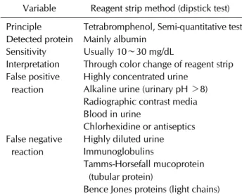
Table 1. Characteristics of urinary dipstick test
Variable Reagent strip method (dipstick test) Principle Tetrabromphenol, Semi-quantitative test Detected protein Mainly albumin
Sensitivity Usually 10∼30 mg/dL
Interpretation Through color change of reagent strip False positive
reaction
Highly concentrated urine Alkaline urine (urinary pH >8) Radiographic contrast media Blood in urine
Chlorhexidine or antiseptics False negative
reaction
Highly diluted urine Immunoglobulins
Tamms-Horsefall mucoprotein (tubular protein)
Bence Jones proteins (light chains) a basic diagnostic tool used to determine urinary abnor- malities including proteinuria, hematuria, and pyuria.
However, proteinuria composed of only other proteins without albumin cannot be identified by urinary dipstick test. Besides this false-negative reaction, false-positive reaction of dipstick test can also be shown in urinary dip- stick test (Table 1). Thus, if proteinuria via urinary dip- stick test is seen or suspected in patients with RA, rheu- matologists or nephrologists consider quantitative uri- nary tests, such as 24-hour urinalysis, and spot urine pro- tein-creatinine ratio (UPCR) or spot urine albumin-crea- tinine ratio (UACR). As previously mentioned, renal in- volvement in RA increases morbidity and mortality par- ticularly in patients with RA [13]. However, renal in- volvement of RA may be present as urinary abnormalities (only proteinuria, only hematuria, proteinuria combined hematuria) without renal dysfunction. Therefore, peri- odic laboratory tests including urinalysis, serum crea- tinine, and electrolytes should be initially performed in patients with rheumatic diseases. If proteinuria is de- tected, quantitative urinalyses, such as 24-hour uri- nalysis, UPCR, and UACR should also be considered.
Managements of renal diseases in RA should be in- dividualized according to the causes and clinical forms of renal involvement. In case of membranous nephropathy or mspGN due to nephrotoxic drugs, such as NSAIDs or DMARDs, avoiding or changing the offending drugs while carefully monitoring disease activity of RA should be attempted. Renal amyloidosis caused by RA usually presents with amyloid A amyloidosis rather than with light-chain amyloidosis. Since the development of renal
amyloidosis shows a positive correlation with disease ac- tivity of RA, effective control of RA itself is important in the development and management of renal amyloidosis.
If patients with RA show rapidly progressive renal dys- function without a definite cause, such as nephrotoxic agents, renal biopsy should be considered. Although clin- ically rare, if crescentic glomerulonephritis or RPGN is confirmed by renal biopsy, the use of pulse-therapy of sys- temic steroid combined with immunosuppressants, in- cluding cyclophosphamide and rituximab, should be considered.
Renal involvement in pSS
pSS is a progressive autoimmune disease, which is mainly characterized by lymphocytic infiltration and mal- function of exocrine glands. The prevalence rate of this disease in women is 9 times higher than that of men [18].
However, the exact prevalence of pSS due to continuous changes in diagnostic guidelines and differentiation from secondary pSS is difficult to determine [19]. pSS can in- volve multiple organs including the kidney, joints, lungs, hematologic system, vascular system, and peripheral nervous system as well as exocrine glands [20].
The prevalence of reported renal involvement in patient with pSS ranges from 5% to 20% according to previous re- ports [20-23]. Renal involvement in patients with pSS is mainly reported in elderly rather than younger patients.
The prognosis of the renal involvement of pSS is relatively good, and CKD progression is clinically rare [21,24].
The renal involvement of pSS can also be expressed in various forms, such as asymptomatic urinary abnormal- ities, renal tubular acidosis (RTA), Fanconi’s syndrome, diabetic insipidus, renal calculi, glomerulonephritis, and TIN [20]. Similarly, the renal involvement of pSS is main- ly composed of renal tubular dysfunction rather than glo- merular dysfunction. According to most clinical or clin- icopathological studies related to the renal involvement of pSS, TIN is the most common pathologic finding in the renal involvement of pSS. However, glomerulonephritis, such as membranoproliferative glomerulonephritis and membranous nephropathy, is usually less frequent in pa- tients with pSS [20,21,24].
The symptoms and signs of TIN may not be clinically relevant. Microscopic hematuria and proteinuria rather than the clinical signs, such as azotemia, hypertension, and electrolytes imbalance, are well developed in TIN. In addition, an isolated proteinuria in TIN is difficult to de- tect with only urinary dipstick test because proteinuria
from the renal tubular injury is mainly composed of tubu- lar proteins with small molecular weight than the rela- tively large albumin [20,25]. Thus, the renal involvement in pSS may be underdiagnosed. This underdiagnosis may be attributed to the limitation of the urinary dipstick test that only detects albumin rather than tubular proteins with small molecular weight.
According to several reports, renal tubular dysfunctions are primarily expressed in a distal RTA form in patients with pSS with histologically acute or chronic TIN [19-21,24].
Distal RTA is the most common acid-base disturbance with electrolyte imbalance particularly in the renal in- volvement of pSS. The distal RTA usually results from the impairment of urine acidification by reducting net hydro- gen ion secretion on the collecting duct. Urine pH is gen- erally over 5.5 due to an inability to acidify urine like men- tioned above. Metabolic acidosis can result in decreased proximal fluid reabsorption, which eventually leads to volume contraction and activation of the renin-angio- tensin-aldosterone system (RAAS). The increase in distal tubular sodium delivery and serum aldosterone by RASS activation and the decrease in proximal fluid reabsorption finally leads to hypokalemia. Beside hypokalemia, meta- bolic acidosis, and RASS activation, nephrolithiasis or hy- percalciuria can be developed in patients with distal RTA by pSS [26]. The increase in urinary calcium excretion is mainly caused by bone buffering against persistent meta- bolic acidosis [27]. In addition, a previous literature showed that distal RTA is well correlated with the degree of hypergammaglobulinemia in pSS [24]. In comparison to distal RTA, proximal RTA, Fanconi’s syndrome can de- velop in patients with pSS, but its incidence is relatively low. The most common presentation of pSS is TIN, which is clinically expressed as signs including hypokalemia, hy- percalciuria, and metabolic acidosis, similar to previous mentioned. However, the relationship between renal dys- function and the clinical signs remains unclear [20].
If the renal involvements of pSS present with clinical signs, such as severe electrolytes imbalance, and sig- nificant renal dysfunction, a renal biopsy should be per- formed to evaluate a possibility of treatment and overall prognosis [20,21]. An identification of the chronicity and severity of renal tubulointerstitial inflammation through renal biopsy will facilitate to determine the therapeutic effect of steroid-based immunosuppressants and the prognosis of the overall disease.
The best treatment option of TIN in patients with pSS is corticosteroids. The initial dose of corticosteroids usually
ranges from 30 mg to 60 mg (approximately ≥0.5 mg/kg of corticosteroid per day) [20,21]. If patients with TIN combined with glomerulonephritis, other immunosup- pressants, such as cyclophosphamide, mycophenolate mofetil, and rituximab, can be added according to specific findings of renal biopsy [21].
The response of first-line therapy, corticosteroid for TIN in patients with pSS according to previous retrospective studies is very good [21,24]. No progression to end-stage renal disease (ESRD) was found in most patients with TIN and pSS, which were adequately treated with cortico- steroids or other immunosuppressants.
In summary, the renal prognosis in patients with renal involvement, such as TIN, glomerulonephritis, and tubu- lar dysfunction caused by pSS, is relatively good. However, the chronicity and severity of TIN, including severe in- filtration of inflammatory cells, interstitial fibrotic changes, and tubular atrophy, should also be considered. The prev- alence of renal involvement in patients with pSS ranges from 5% to 20%, but prompt and adequate treatments in patients with pSS with renal involvement are important for renal prognosis. Therefore, rheumatologists or neph- rologists should perform adequate screening tests, in- cluding quantitative urinalysis via 24-hour urine collec- tion, urinary pH and osmolarity, serum creatinine, serum bicarbonate, serum potassium, and serum phosphate at least twice a year in patients with pSS who are suspected with renal involvement (Table 2) [20]. In addition, if signs of renal involvement are evident, renal biopsy should be immediately considered.
Renal involvement in ANCA-associated vasculitis The kidney is a main target organ of systemic vasculitis.
A renal vasculitis can manifest in one form of the follow- ing disease entities: immunoglobulin A vasculitis (He- noch-Schӧnlein purpura nephritis), cryoglobulinemic vasculitis, and pauci-immune vasculitis mainly involved small-sized vessels. The clinical manifestations of sys- temic vasculitis usually present with nonspecific symp- toms or signs, such as fever, chills, malaise, myalgia, gen- eralized weakness, and arthralgia. The manifestations can also vary depending on involved tissue, disease activ- ity, and disease severity [28]. Small-sized vessel pau- ci-immune vasculitis is usually related to autoantibodies, such as myeloperoxidase (MPO) or proteinase 3 (PR3)–
ANCA [29]. The diagnosis and classification of ANCA-as- sociated vasculitides (AAV) has been continuously devel- oped, and classification is mainly based on histology and
Table 2. Renal screening tests in patients with primary Sjögren’s syndrome
Variable Every 6 months in pSS patients with renal abnormalities Every 1 year in all pSS patients Urinalysis Dipstick test: urine pH, osmolality, glycosuria
24 hour urinalysis: protein, albumin, creatinine, citrate, calcium, culture
Dipstick: urine pH, osmolality, glycosuria UPCR, UACR
Serologic tests Creatinine, potassium, chloride, bicarbonate, calcium, phosphate, uric acid
Creatinine, potassium, chloride, bicarbonate
Imaging tests Kidney ultrasonography -
pSS: primary Sjögren’s syndrome, UPCR: spot urine protein to creatinine ratio, UACR: spot urine albumin to creatinine ratio, -:
none.
Table 3. Frequency of systemic involvement in ANCA-associated small vessel vasculitis
Systemic organ Frequency of involvement
MPA GPA EGPA
Kidney 90 80 45
Skin 40 40 50
Lung 50 90 90
ENT 35 90 50
Musculoskeletal 60 60 50
Neurologic 30 50 60
Values are presented as percentage. ANCA: antineutrophil cytoplasm antibody, MPA: microscopic polyangiitis, GPA:
granulomatosis with polyangiits (Wegener’s), EGPA:
eosinophilic granulomatosis with polyangiits, ENT: ear, nose, throat.
clinical manifestation. According to Chapel Hill Consensus Conference in 2012 [30], AAV is mainly composed of mi- croscopic polyangiitis (MPA), granulomatosis with poly- angiitis (GPA), and eosinophilic granulomatosis with polyangiitis (EGPA). According to a previous literature [31], ANCA positivities of AAV were about 60% in GPA and about 5% to 8% in EGPA. ANCA positivity of MPA was prominently higher compared with other vasculitis (approximately 70% to 90%).
Renal involvement of AAV is frequently observed and al- so reflects a poor prognostic factor for mortality and mor- bidity [29,31]. Renal involvement occurs more frequently in GPA and MPA compared to in EGPA (Table 3) [32]. We can clinically recognize the presence of renal involvement of AAV by identifying hematuria, proteinuria, azotemia, edema, and hypertensive symptoms [30]. Although glo- merular filtration rate (GFR) at onset time of renal vascu- litis associated with AAV reflects a renal outcome, renal histologic feature is also an important predictor for dis- ease outcome particularly in the renal vasculitis asso-
ciated with AAV [29,33].
In a histologic sense, severity of acute lesions, such as glomerular crescents and fibrinoid necrosis, rather than chronic lesion-like glomerular sclerosis is well correlated with renal outcome and responsiveness of immunosup- pressants in several renal vasculitides associated with AAV [29,33,34]. Classification of ANCA-associated glo- merulonephritis based on renal biopsy results has been developed for the prognosis of renal vasculitis [29,33,34].
‘Focal’ lesion represents a relatively preserved renal func- tion and ‘crescentic’ lesion shows a good responsiveness for immunosuppressant therapy. ‘Mixed’ and ‘sclerotic’
lesions represent intermediate and high risks of ag- gravation of renal function, respectively (Table 4) [29,34].
Tubular atrophy and interstitial fibrosis are well-known indicators of poor prognosis for all of renal diseases. In addition to glomerulonephritis, interstitial nephritis and tubular damages, such as tubulitis and tubular atrophy, may also be seen in renal involvement of AAV. The tubu- lar inflammation in ANCA-associated renal vasculitis can be associated with inflammatory cells such as CD4 pos- itive T-cells [29,35]. Thus, T and B cell target therapies may also be used in the treatment of ANCA-associated re- nal vasculitis. The activation of alternative complement pathway can also be related to pathological mechanism of AAV according to a previous study [36].
Clinically, vasculitis may occur even if the serum level of ANCA is the normal range, and this serum level is not correlate with disease activity or severity of vasculitis.
There is also a similar relationship for ANCA-associated renal vasculitis. As mentioned earlier, the renal outcome of ANCA-associated renal vasculitis is mainly related to GFR at onset time of renal disease and specific pathologic findings including several glomerular damages, inter- stitial fibrosis, and tubular atrophy rather than the serum ANCA level. However, according to several studies
Table 4. Classification for ANCA-associated glomerulonephritis
Class Inclusion criteria
Focal >50% normal glomeruli
Crescentic >50% glomeruli with cellular crescents Mixed <50% normal,<50% crescentic, and
<50% globally sclerotic glomeruli Sclerotic >50% globally sclerotic glomeruli ANCA: antineutrophil cytoplasm antibody.
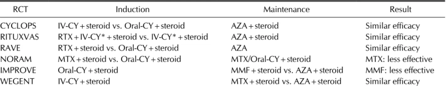
Table 5. Several randomized controlled trials (RCTs) in treatment of ANCA-associated vasculitis
RCT Induction Maintenance Result
CYCLOPS IV-CY+steroid vs. Oral-CY+steroid AZA+steroid Similar efficacy
RITUXVAS RTX+IV-CY*+steroid vs. IV-CY*+steroid AZA+steroid Similar efficacy
RAVE RTX+steroid vs. Oral-CY+steroid AZA Similar efficacy
NORAM MTX+steroid vs. Oral-CY+steroid MTX/Oral-CY+steroid MTX: less effective
IMPROVE Oral-CY+steroid MMF+steroid vs. AZA+steroid MMF: less effective
WEGENT IV-CY+steroid MTX+steroid vs. AZA+steroid Similar efficacy
ANCA: anti-neutrophilic cytoplasmic antibody, IV: intravenous, CY: cyclophosphamide, AZA: azathioprine, RTX: rituximab, MTX:
methotrexate, MMF: mycophenolate mofetil. *Low-dose cyclophosphamide (2 doses).
[33,34,37], less severe pathological involvement, such as focal class, is mainly shown in patients with PR3-ANCA-as- sociated renal vasculitis than in patients with MPO- ANCA-associated renal vasculitis. ANCA type rather than serum titer may be an important factor of treatment response. For example, patients with MPO-ANCA-asso- ciated renal vasculitis have much poorer renal outcome than those with PR3-ANCA-associated renal vasculitis [33,34,37].
The treatment of ANCA-associated renal vasculitis is mainly composed of high-dose glucocorticoid and sys- temic cyclophosphamide for three months or six months in induction therapy. Several randomized controlled trials for the treatment of AAV have been performed over the past several decades, and systemic immunosuppressants, such as cyclophosphamide, methotrexate, and rituximab combined with high-dose glucocorticoid, are very effec- tive drugs in induction therapy, particularly for control- ling acute inflammation (Table 5) [29]. Among these im- munosuppressants, methotrexate should not be used in patients with decreased GFR. After the induction therapy, maintenance therapy with low-dose glucocorticoid com- bined with azathioprine or methotrexate should be also continued to prevent relapse. Clinically, the duration of induction therapy and the intensity of maintenance ther- apy should be decreased as much as possible to reduce
toxic side effects. We can use the intravenous cyclo- phosphamide rather than oral cyclophosphamide to re- duce drug toxicity, such as leukopenia and bladder cancer.
However, we need to recognize that long-term follow-up demonstrates longer time to relapse in the oral cyclo- phosphamide-treated group [38].
In addition, plasma exchange may be a good option in patients with life-threatening pulmonary hemorrhage or severe renal disease which emergent dialysis is needed [29,39,40]. However, plasma exchange did not result in increase of survival rate, especially in patients with severe renal disease due to ANCA-associated renal vasculitis [41]. Recently, several studies have been continued for newer therapeutic agents, including bortezomib (protea- some inhibitor), gusperimus, and alemtuzumab (an- ti-CD52 pan lymphocyte-depleting antibody), to manage AAV effectively [29].
In conclusion, treatment of ANCA-associated renal vas- culitis is largely composed of induction and maintenance therapies, and effective immunosuppressants can be chosen according to disease severity (Figure 1) [32].
Renal involvement associated with systemic sclerosis Systemic sclerosis (SSc) is a rare connective tissue dis- ease even if the prevalence is quite different according to country or area [42]. SSc consists of limited and diffuse systemic sclerosis. It is mainly characterized by vasculop- athy that involves from peripheral to renal vessels and fi- brotic change of connective tissue by chronic auto- immune reaction.
Renal involvement caused by SSc can clinically vary from mild urinary abnormality without renal dysfunction to systemic renal crisis (SRC). Patients with renal involve- ment of SSc often show a decreased GFR. However, mild-to-moderate renal impairment (GFR <90 mL/min/
1.73 m2 and GFR <60 mL/min/1.73 m2, respectively)
Figure 1. Treatment of ANCA-associated renal vasculitis. IV methylprednisolone usually begins with 7 mg/kg per day of methyl- prednisolone for 3 days and followed by oral prednisone 1 mg/kg per day. IV cyclophosphamide usually begins with 0.5 g/m2 per month- ly of cyclophosphamide. Oral cyclophosphamide usually begins with 2 mg/kg per day of cyclophosphamide and the dose can be reduced based on renal function or age. Oral prednisone should be tapered slowly during 3 to 6 months. Oral azathioprine usually begin with 2 mg/kg per day of azathioprine. ANCA: anti-neutrophilic cytoplasmic antibody, IV: intravenous, PE: plasma exchange.
usually occurs more frequently than severe renal dysfunc- tion (GFR <30 mL/min/1.73 m2) or ESRD [43].
SRC is a clinical condition accompanied by malignant hypertension and acute renal dysfunction in SSc. The SRC occurs in less than about 5% of patients with mainly dif- fuse SSc, but it is the most serious complication among renal involvement of SSc [42,44,45]. The pathogenesis of SRC has not been elucidated [42]. However, a specific vascular lesion-like “onion-skin” and luminal narrowing resulting from intimal and medial proliferation of renal vessels, such as arcuate and interlobular arteries on histo- pathology, show a significant relationship between SRC and renal vasculopathy. A progressive systemic sclerosis may often occur in ischemic nephropathy such as renal in- farction and subcapsular hemorrhage [46]. In addition, patients with SRC or renal dysfunction related to SSc usu- ally show decreased blood flow.
Clinically, angiotensin-converting enzyme inhibitor (ACEI) or angiotensin II receptor blocker (ARB) is com- monly used because the vascular change and activation of the RAAS caused by decreased renal blood flow in pa-
tients with SSc with renal involvement. Whether the use of ACEI may be associated with reduced GFR in patients with SSc is not certain. Thus, using ACEI or ARB is gen- erally better in patients with SSc, even if they have renal insufficiency [42,46]. On the contrary, hypovolemic con- ditions caused by several risk factors including sepsis, congestive heart failure, cardiac arrhythmia, and dehy- dration and drugs, such as cyclosporin, corticosteroid, and NSAIDs, are triggering factors of SRC. According to several studies, corticosteroid can increase the serum lev- els of endothelin-1 and endothelin receptors [42,47,48].
The effects of corticosteroid on several factors, such as prostacyclin, blood pressure, and volume, are also related to aggravation of SRC caused by corticosteroid use.
However, determining whether the cause of renal dys- function in patients with SSc is due to the development of SRC or acute kidney injury caused by the aforementioned risk factors is clinically difficult. Clinical signs or symp- toms related to renal involvement of SSc may be often absent. Therefore, rheumatologists and nephrologists should attempt to identify the triggering factors or causa-
tive factors.
Laboratory data, such as proteinuria, microscopic hema- turia, hemolytic anemia, thrombocytopenia, and anti- nuclear antibodies including anti-RNA polymerase III an- tibodies, may be also helpful for SRC diagnosis. A pre- vious cohort study revealed that anti-RNA polymerase III antibodies are positively correlated with the development of SRC [49]. There is also a report that anti-RNA poly- merase III antibody is closely associated with renal in- sufficiency and diffuse SSc [42]. However, a compre- hensive consideration of clinicopathological factors should be given with regard to the definite diagnosis of SRC. In Korea, SRC is very rare, and SRC related with anti-RNA polymerase III antibody is also extremely rare [50]. In general, SRC shows the following symptoms or signs in- cluding severe hypertension accompanied by neurologic symptoms, acute renal dysfunction, thrombotic micro- angiopathy, proteinuria, and microscopic hematuria.
Renal biopsy is often not required in clinical situations where SRC is suspected, but it should be considered when an aggressive deterioration of renal function or atypical renal manifestation by SSc is present [42].
Early diagnosis and management of SRC in patients with SSc is very important. When renal involvement of SSc is suspected, glucocorticoid use should be firstly discarded. As previously mentioned, high-dose cortico- steroid (>15 mg/day of prednisolone) may be a trigger- ing factor of SRC because the characteristics of cortico- steroid influence several factors such as endothelin, pros- tacyclin, and blood volume or pressure [51]. In addition, ACEI or ARB is the main drug used for the renal involve- ment of SSc including SRC. The use of ACEI is better to be continued in patients undergoing dialysis or not and in those who recovered from renal function and has hyper- tension. Besides ACEI or ARB, anti-hypertensive agents, such as calcium channel blockers, minoxidil, and prazo- cin, can be added to control hypertension adequately, but beta blockers should be avoided. As for the relationship between endothelin and SRC, endothelin receptor block- ers may be considered in SRC. However, a large study or a randomized control study about the use of endothelin receptor blockers for SRC is needed [42]. When patients with renal involvement of SSc have uremic symptoms or signs, physicians should consider emergent dialysis.
Dialysis is usually required about half of patients with SRC according to a previous report [42].
Renal involvement associated with gout or hyperuricemia
A long-standing hyperuricemic condition may lead to progressive formation of uric acid crystal within renal tu- bules, which may result in CKD. According to previous studies, hyperuricemia itself may be associated with sev- eral pathologies such as metabolic syndrome, afferent ar- teriolopathy of kidney, activation of RAAS, and endothe- lial dysfunction characterized by inhibition of nitric oxide system [9,52-54].
These factors, caused by hyperuricemia itself, lead to re- nal involvement, such as acute TIN, and ischemic nephr- opathy and eventually lead to progression to CKD. On the contrary, Latif et al. [55] reported that the antioxidant ef- fect of uric acid may decrease all-cause mortality and car- diovascular mortality in patients with ESRD requiring dialysis. Therefore, the causal relationship between hy- peruricemia itself and renal dysfunction has been not def- initely clarified. In addition, clinical factors, such as hy- pertension associated with hyperuricemia, nephrotoxic drugs such as NSAIDs in patients with gout and hyper- urmicemia, and underlying vasculopathy in hyperuricemic patients can influence the reduction of renal function [9].
The prevalence of renal involvement in patients with gout is relatively high. In particular, the prevalence of hy- pertension or moderate CKD (lesser than GFR <60 mL/min) in patients with gout was reported to be over 70% by Zhu et al. [56]. In addition, CKD patients includ- ing ESRD patients undergoing dialysis may also often ex- perience gout. Thus, the relationship between gout and renal involvement is bi-directional.
Clinically, the therapeutic target of serum uric acid level in gout patients regardless of CKD is below 6 mg/dL. For the appropriate reduction of serum uric acid in patients with gout, many physicians use xanthine oxidase in- hibitors, such as allopurinol and febuxostat. However, the use of allopurinol in CKD patients requires consid- erable care. In particular, allopurinol hypersensitivity syndrome, characterized by symptoms or signs such as skin rash, anaphylactic reaction, Stevens-Johnson syn- drome, toxic epidermal necroloysis, and multi-organ dys- function may be easily developed in patients with CKD.
However, febuxostat has a relatively low incidence of ad- verse effects compared to allopurinol and its use is rela- tively flexible even in patients with renal dysfunction. So, many nephrologists and rheumatologists prefer febuxo- stat rather than allopurinol [57].
The use of xanthine oxidase inhibitor and colchicine in
patients with CKD and gout has been debated. However, in patients with advanced CKD, administration of 50 mg/day allopurinol is better, and the dose of the drug should be gradually increased to maintain the target level of serum uric acid. Colchicine should be also started with 50% dose reduction and continued for 3 to 6 months after achieving the target level of serum uric acid depending on the presence or absence of tophi [9,58,59].
Renal involvement associated with nephrotoxic for control of rheumatic diseases
Since most rheumatic diseases are not curable and re- quire the use of anti-inflammatory drugs for a long time, therapeutic drugs, such as NSAIDs, DMARDs, and even biologic agents, may often influence renal dysfunction or renal involvement in rheumatic patients. NSAIDs are mainly associated with renal involvement, such as acute TIN, and minimal change disease. In addition, NSAIDs contract the afferent arteriole of the kidneys by inhibiting prostaglandin production. The influence on renal vascu- lature of NSAIDs can result in decreased GFR. According to few reports, NSAIDs worsen renal function in patients with advanced renal dysfunction (estimated GFR <30 mL/min/1.73 m2), but they do not show adverse effects in patients with normal renal function [5,7].
Recently, previous DMARDs such as gold compounds and D-penicillamine, are rarely used. However, conven- tional DMARDs including methotrexate, sulfasalazine, and lefunomide are widely used in patients with RA to manage chronic inflammation. The nephrotoxicity of these drugs is relatively less than the previous DMARDs mainly resulting in membranous nephropathy. However, conventional DMARDs should also be used cautiously ac- cording to the elimination rate of each drug by the kid- neys [8,60].
Biologic agents, such as TNF-α blockers (adalimumab and etanercept), can also result in membranous nephrop- athy or proliferative glomerulonephritis accompanied by proteinuria via direct invasion of glomerular visceral epi- thelial layer [8]. Thus, when biologic DMARDs or con- ventional DMARDs are used in rheumatic diseases in- cluding RA, a renal surveillance should be regularly per- formed by rheumatologists or nephrologists.
In conclusion, kidney-related diseases related to an- ti-rheumatic drugs, including NSAIDs and DMARDs, are mainly composed of glomerulonephritis such as mem- branous nephropathy and TIN.
CONCLUSION
Kidney diseases and rheumatic diseases demonstrates a close relationship. They can act as causative factors of each other. In particular, renal manifestation or renal in- volvement of rheumatic diseases is clinically significant because of the increase in mortality and morbidity in rheumatic patients with renal dysfunction.
Thus, early diagnosis and proper management of renal involvement in rheumatic diseases may improve overall or renal prognosis of rheumatic patients. The clinical and histologic manifestations of renal involvement in rheu- matic diseases were investigated throughout this review.
Renal involvement can be caused by anti-rheumatic drugs and rheumatic diseases. RA mainly induces glomer- ulonephritis, such as membranous nephropathy, mspGN, and amyloidosis. pSS induces tubular dysfunction includ- ing TIN and RTA. In addition, ANCA-associated vasculi- tis mainly induces RPGN accompanied by acute renal dysfunction. SSc is a relatively rare disease, but renal in- volvement of SSc including SRC may be fatal in view of re- nal prognosis. In relatively frequent patients with gout, renal involvement may be related to hyperuricemia.
In conclusion, a more effective approach for a definite di- agnosis and proper care of rheumatic diseases can be ach- ieved by accurately grasping the clinical characteristics of renal involvement in various rheumatic diseases.
ACKNOWLEDGMENTS
This paper was supported by Wonkwang University in 2017.
CONFLICT OF INTEREST
No potential conflict of interest relevant to this article was reported.
REFERENCES
1. Chiu HY, Huang HL, Li CH, Chen HA, Yeh CL, Chiu SH, et al. Increased risk of chronic kidney disease in rheumatoid arthritis associated with cardiovascular complications - a national population-based cohort study. PLoS One 2015;
10:e0136508.
2. Turesson C. Comorbidity in rheumatoid arthritis. Swiss Med Wkly 2016;146:w14290.
3. Gilani ST, Khan DA, Khan FA, Ahmed M. Adverse effects of low dose methotrexate in rheumatoid arthritis patients. J Coll Physicians Surg Pak 2012;22:101-4.
4. Manabe S, Banno M, Nakano M, Fujii T, Fujiwara M, Kita Y, et al. Bucillamine-induced membranous nephropathy with crescent formation in a patient with rheumatoid arthritis:
case report and literature review. Case Rep Nephrol Dial 2014;5:30-8.
5. Möller B, Pruijm M, Adler S, Scherer A, Villiger PM, Finckh A; Swiss Clinical Quality Management in Rheumatic Diseases (SCQM) Foundation, CH-8048 Zurich, Switzerland.
Chronic NSAID use and long-term decline of renal function in a prospective rheumatoid arthritis cohort study. Ann Rheum Dis 2015;74:718-23.
6. Kaushik P, Rahmani M, Ellison W. Membranous glomer- ulonephritis with the use of etanercept in ankylosing spondylitis. Ann Pharmacother 2011;45:e62.
7. Tokoroyama T, Ando M, Setoguchi K, Tsuchiya K, Nitta K.
Prevalence, incidence and prognosis of chronic kidney dis- ease classified according to current guidelines: a large retro- spective cohort study of rheumatoid arthritis patients.
Nephrol Dial Transplant 2016 Sep 16 [Epub]. DOI:
10.1093/ndt/gfw315.
8. Mittal T, Rathi M. Rheumatological diseases and kidneys: a nephrologist's perspective. Int J Rheum Dis 2014;17:834-44.
9. Gibson T. Hyperuricemia, gout and the kidney. Curr Opin Rheumatol 2012;24:127-31.
10. Carmona L, Cross M, Williams B, Lassere M, March L.
Rheumatoid arthritis. Best Pract Res Clin Rheumatol 2010;24:733-45.
11. Vinicki JP, Pellet SC, De Rosa G, Dubinsky D, Laborde HA, Marini A, et al. Analysis of 65 renal biopsies from patients with rheumatoid arthritis (1976-2015): Change in treat- ment strategies decreased frequency and modified histo- pathological findings. J Clin Rheumatol 2015;21:335-40.
12. Makino H, Yoshinaga Y, Yamasaki Y, Morita Y, Hashimoto H, Yamamura M. Renal involvement in rheumatoid arthri- tis: analysis of renal biopsy specimens from 100 patients.
Mod Rheumatol 2002;12:148-54.
13. Hickson LJ, Crowson CS, Gabriel SE, McCarthy JT, Matteson EL. Development of reduced kidney function in rheumatoid arthritis. Am J Kidney Dis 2014;63:206-13.
14. Naranjo A, Sokka T, Descalzo MA, Calvo-Alén J, Hørslev- Petersen K, Luukkainen RK, et al. Cardiovascular disease in patients with rheumatoid arthritis: results from the QUEST-RA study. Arthritis Res Ther 2008;10:R30.
15. Helin HJ, Korpela MM, Mustonen JT, Pasternack AI. Renal biopsy findings and clinicopathologic correlations in rheu- matoid arthritis. Arthritis Rheum 1995;38:242-7.
16. Icardi A, Araghi P, Ciabattoni M, Romano U, Lazzarini P, Bianchi G. Kidney involvement in rheumatoid arthritis.
Reumatismo 2003;55:76-85.
17. Karie S, Gandjbakhch F, Janus N, Launay-Vacher V, Rozenberg S, Mai Ba CU, et al. Kidney disease in RA pa- tients: prevalence and implication on RA-related drugs management: the MATRIX study. Rheumatology (Oxford) 2008;47:350-4.
18. Ramos-Casals M, Brito-Zerón P, Sisó-Almirall A, Bosch X.
Primary Sjogren syndrome. BMJ 2012;344:e3821.
19. Talal N, Zisman E, Schur PH. Renal tubular acidosis, glo- merulonephritis and immunologic factors in Sjögren's syndrome. Arthritis Rheum 1968;11:774-86.
20. François H, Mariette X. Renal involvement in primary Sjögren syndrome. Nat Rev Nephrol 2016;12:82-93.
21. Maripuri S, Grande JP, Osborn TG, Fervenza FC, Matteson EL, Donadio JV, et al. Renal involvement in primary Sjögren's syndrome: a clinicopathologic study. Clin J Am Soc Nephrol 2009;4:1423-31.
22. Bossini N, Savoldi S, Franceschini F, Mombelloni S, Baronio M, Cavazzana I, et al. Clinical and morphological features of kidney involvement in primary Sjögren's syndrome. Nephrol Dial Transplant 2001;16:2328-36.
23. Goules A, Masouridi S, Tzioufas AG, Ioannidis JP, Skopouli FN, Moutsopoulos HM. Clinically significant and biop- sy-documented renal involvement in primary Sjögren syndrome. Medicine (Baltimore) 2000;79:241-9.
24. Ren H, Wang WM, Chen XN, Zhang W, Pan XX, Wang XL, et al. Renal involvement and followup of 130 patients with primary Sjögren's syndrome. J Rheumatol 2008;35:278-84.
25. Baldini C, Pepe P, Quartuccio L, Priori R, Bartoloni E, Alunno A, et al. Primary Sjogren's syndrome as a multi-or- gan disease: impact of the serological profile on the clinical presentation of the disease in a large cohort of Italian patients. Rheumatology (Oxford) 2014;53:839-44.
26. Evan AP, Lingeman J, Coe F, Shao Y, Miller N, Matlaga B, et al. Renal histopathology of stone-forming patients with dis- tal renal tubular acidosis. Kidney Int 2007;71:795-801.
27. Bonny O, Rubin A, Huang CL, Frawley WH, Pak CY, Moe OW. Mechanism of urinary calcium regulation by urinary magnesium and pH. J Am Soc Nephrol 2008;19:1530-7.
28. Falk RJ, Hogan S, Carey TS, Jennette JC. Clinical course of anti-neutrophil cytoplasmic autoantibody-associated glo- merulonephritis and systemic vasculitis. The Glomerular Disease Collaborative Network. Ann Intern Med 1990;113:
656-63.
29. Furuta S, Jayne DR. Antineutrophil cytoplasm antibody- as- sociated vasculitis: recent developments. Kidney Int 2013;84:244-9.
30. Jennette JC. Rapidly progressive crescentic glomerulone- phritis. Kidney Int 2003;63:1164-77.
31. Kim HW, Song YW. ANCA-associated vasculitis: report from Korea. Clin Exp Nephrol 2013;17:708-11.
32. Johnson RJ, Feehally J, Floege J. Comprehensive clinical nephrology. 5th ed. Philadelphia, Saunders Press, 2014, p.
291-6.
33. Chen YX, Xu J, Pan XX, Shen PY, Li X, Ren H, et al.
Histopathological classification and renal outcome in pa- tients with antineutrophil cytoplasmic antibodies-associated renal vasculitis: a study of 186 patients and metaanalysis. J Rheumatol 2017;44:304-13.
34. Berden AE, Ferrario F, Hagen EC, Jayne DR, Jennette JC, Joh K, et al. Histopathologic classification of ANCA-associated glomerulonephritis. J Am Soc Nephrol 2010;21:1628-36.
35. Abdulahad WH, Lamprecht P, Kallenberg CG. T-helper cells as new players in ANCA-associated vasculitides. Arthritis Res Ther 2011;13:236.
36. Xing GQ, Chen M, Liu G, Heeringa P, Zhang JJ, Zheng X, et al. Complement activation is involved in renal damage in human antineutrophil cytoplasmic autoantibody associated pauci-immune vasculitis. J Clin Immunol 2009;29:282-91.
37. Cornec D, Cornec-Le Gall E, Fervenza FC, Specks U.
ANCA-associated vasculitis - clinical utility of using ANCA specificity to classify patients. Nat Rev Rheumatol 2016;
12:570-9.
38. Harper L, Morgan MD, Walsh M, Hoglund P, Westman K,
Flossmann O, et al. Pulse versus daily oral cyclo- phosphamide for induction of remission in ANCA-asso- ciated vasculitis: long-term follow-up. Ann Rheum Dis 2012;71:955-60.
39. Klemmer PJ, Chalermskulrat W, Reif MS, Hogan SL, Henke DC, Falk RJ. Plasmapheresis therapy for diffuse alveolar hemorrhage in patients with small-vessel vasculitis. Am J Kidney Dis 2003;42:1149-53.
40. Jayne DR, Gaskin G, Rasmussen N, Abramowicz D, Ferrario F, Guillevin L, et al; European Vasculitis Study Group.
Randomized trial of plasma exchange or high-dosage meth- ylprednisolone as adjunctive therapy for severe renal vasculitis. J Am Soc Nephrol 2007;18:2180-8.
41. Walsh M, Catapano F, Szpirt W, Thorlund K, Bruchfeld A, Guillevin L, et al. Plasma exchange for renal vasculitis and idiopathic rapidly progressive glomerulonephritis: a meta-analysis. Am J Kidney Dis 2011;57:566-74.
42. Mouthon L, Bussone G, Berezné A, Noël LH, Guillevin L.
Scleroderma renal crisis. J Rheumatol 2014;41:1040-8.
43. Ostojic P, Stojanovski N. Arterial hypertension treated with angiotensin converting enzyme inhibitors and glucocorti- coids are independent risk factors associated with decreased glomerular filtration rate in systemic sclerosis. Rheumatol Int 2017;37:363-8.
44. Logee KM, Lakshminarayanan S. Scleroderma renal crisis as an initial presentation of systemic sclerosis: a case report and review of the literature. Clin Exp Rheumatol 2015;33(4 Suppl 91):S171-4.
45. Steen VD, Costantino JP, Shapiro AP, Medsger TA Jr.
Outcome of renal crisis in systemic sclerosis: relation to availability of angiotensin converting enzyme (ACE) inhibitors. Ann Intern Med 1990;113:352-7.
46. Fisher ER, Rodnan GP. Pathologic observations concerning the kidney in progressive systemic sclerosis. AMA Arch Pathol 1958;65:29-39.
47. Kobayashi H, Nishimaki T, Kaise S, Suzuki T, Watanabe K, Kasukawa R, et al. Immunohistological study endothelin-1 and endothelin-A and B receptors in two patients with scle- roderma renal crisis. Clin Rheumatol 1999;18:425-7.
48. Vancheeswaran R, Magoulas T, Efrat G, Wheeler-Jones C, Olsen I, Penny R, et al. Circulating endothelin-1 levels in systemic sclerosis subsets--a marker of fibrosis or vascular dysfunction? J Rheumatol 1994;21:1838-44.
49. Okano Y, Steen VD, Medsger TA Jr. Autoantibody reactive with RNA polymerase III in systemic sclerosis. Ann Intern Med 1993;119:1005-13.
50. Kang EH, Im CH, Kim SH, Chung JR, Lee EY, Kim DJ, et al.
A case of renal crisis in a Korean scleroderma patient with anti-RNA polymerase I and III antibodies. J Korean Med Sci 2006;21:1121-3.
51. Desbois AC, Cacoub P. Systemic sclerosis: An update in 2016. Autoimmun Rev 2016;15:417-26.
52. Khosla UM, Zharikov S, Finch JL, Nakagawa T, Roncal C, Mu W, et al. Hyperuricemia induces endothelial dysfunction.
Kidney Int 2005;67:1739-42.
53. Avram Z, Krishnan E. Hyperuricaemia--where nephrology meets rheumatology. Rheumatology (Oxford) 2008;47:
960-4.
54. Mount DB. The kidney in hyperuricemia and gout. Curr Opin Nephrol Hypertens 2013;22:216-23.
55. Latif W, Karaboyas A, Tong L, Winchester JF, Arrington CJ, Pisoni RL, et al. Uric acid levels and all-cause and car- diovascular mortality in the hemodialysis population. Clin J Am Soc Nephrol 2011;6:2470-7.
56. Zhu Y, Pandya BJ, Choi HK. Comorbidities of gout and hy- peruricemia in the US general population: NHANES 2007-2008. Am J Med 2012;125:679-87.e1.
57. Faruque LI, Ehteshami-Afshar A, Wiebe N, Tjosvold L, Homik J, Tonelli M. A systematic review and meta-analysis on the safety and efficacy of febuxostat versus allopurinol in chronic gout. Semin Arthritis Rheum 2013;43:367-75.
58. Khanna D, Fitzgerald JD, Khanna PP, Bae S, Singh MK, Neogi T, et al. 2012 American College of Rheumatology guidelines for management of gout. Part 1: systematic non- pharmacologic and pharmacologic therapeutic approaches to hyperuricemia. Arthritis Care Res (Hoboken) 2012;64:
1431-46.
59. Khanna D, Khanna PP, Fitzgerald JD, Singh MK, Bae S, Neogi T, et al. 2012 American College of Rheumatology guidelines for management of gout. Part 2: therapy and anti- inflammatory prophylaxis of acute gouty arthritis. Arthritis Care Res (Hoboken) 2012;64:1447-61.
60. Schiff MH, Whelton A. Renal toxicity associated with dis- ease-modifying antirheumatic drugs used for the treatment of rheumatoid arthritis. Semin Arthritis Rheum 2000;
30:196-208.