I. 서론
치주치료의 목적은 치주질환으로 상실된 치주조 직을 재생시키는 것이다. 치주조직의 재생은 신생 골, 신생백악질과 함께 새로운 치주인대 섬유가 기능 적으로 삽입, 배열되어 재형성되는 것이며1,2)치주질 환으로 상실된 치주조직을 재생시키기 위해 골이식 술3,4), 골조직유도재생술5~7) 그리고 여러 가지 성장인
자 적용8,9) 등이 제시되어 왔다.
골조직의 재형성에는 골기질에 포함되어 있거나 골흡수 중 방출되는 폴리펩타이드계 성장인자 (polypeptide growth factor)가 관련되며, 성장인자는 골기질에서 발견되고, 골세포에 의해 생성되는 것으 로 알려져 있다10,11), 이러한 성장인자는 창상부위의 치유과정에서 다양한 세포의 이주과정에 관련되어 골조직 대사 조절에 전신적 조절 물질과 같이 중요 한 역할을 하는 것으로 알려져 있다12,13).
성장인자중에서 혈소판유래성장인자(platelet- derived growth factor, 이하 PDGF)로 30kDa의 분자 량을 가지고 있는 조절성 단백질로서14~16)단종 이량 체(PDGF-AA, BB)와 이종 이량체(PDGF-AB)로 존재 함이 발견되어졌다17). PDGF가 합성되고 유리되는 근원은 혈소판으로 알려졌으나 현재 다양한 세포에 서도 유리되고 있다18). PDGF의 역할에 대한 연구로 쥐의 골아세포나 전구골아세포 배양시 PDGF가
DNA와 교원질 합성을 촉진하고19)두개골 배양시 골 기 질 축 적 을 증 가 시 킨 다 고 보 고 되 었 으 나20) Varghese 등21)은 골아유사세포 배양에서 PDGF-BB 를 투여시 교원질 형성을 감소시킨다고 주장하였고, Canalis등22)은 두개골 배양에서 PDGF가 골아세포의 분화를 억제시킨다고 보고하였다.
Glucocorticoid(이하 GC)는 다양한 실험을 통하여 생체내에서는 골형성을 감소시키고 골흡수를 야기
시키지만23~28)골세포 배양 시 적용하면 골세포의 분
화를 촉진시켜 골결절(bone nodule) 형성을 촉진시 키는 것으로 알려져 있다29,30). 이러한 GC 중 long- acting GC인 Dexamethasone(이하 Dex)은 in vitro 실험에서 전구골아세포의 증식을 촉진시키고31), 성 인 골수세포를 골아세포로 분화시키는 것으로 알려 졌으며32), in vivo 실험으로 Sato등33)은 쥐 두정골 천 공후 Dex을 투여한 실험에서는 Dex가 골 재생지연 과 연조직 치유을 지연시킨다고 보고하였다.
Dex는 성장인자와 병용시 세포기능을 조절하는 효과를 나타내어, 섬유아세포에서 섬유아세포성장 인자의 세포증식능을 증가시키는 것으로 보고되었 으나34), 상피성장인자에 의한 세포증식능은 억제시 키는 것으로 알려져 있다35). 또한 in vivo 실험에서 PDGF와 Dex의 병용 시 치주인대조직과 골조직 형 성을 증가시킨다고 보고36)된 바도 있다. 그러나 아직 PDGF와 같은 성장인자나 Dex와 같은 항염증약물
MC3T3-E1 세포의 분화에 PDGF-BB와 Dexamethasone 병용 효과
이재목*·서조영*·김성조**·최점일**
*경북대학교 치과대학 치주과학교실
**부산대학교 치과대학 치주과학교실
대한치주과학회지 : Vol. 30, No. 1, 2000
사용시 골재생에 대한 견해가 다르고 병용시 골세포 의 성장과 분화 과정에 대한 연구가 미진하여, 저자 는 골아유사세포배양에서 PDGF와 Dex 병용시 골아 유사세포의 성장과 분화의 어느 단계에서 어떠한 영 향을 미치는 지 관찰하여 골조직 재생의 임상적용 가능성에 대해 알아보고자 본 실험을 실시하였다
II. 실험재료 및 방법
1. 골아세포의 배양골결절 형성과 세포 증식능을 관찰하기 위해 MC3T3-E1세포를 100 mm dish에 1×10⁵cells를 접종 하고, DNA 합성능과 ALP 활성도 측정을 위해 24 well plate에 1×10⁴cells/well로 10% fetal bovine serum(Gibco, U.S.A. 이하 FBS), 10mM β-glyc- erophosphate, 50㎍/ml의 ascorbic acid(Sigma, U.S.A), 100U/ml penicilline, 100㎍/ml streptomycin 을 함유한 alpha-modified eagle medium(Gibco, U.S.A. 이하α-MEM)으로 37℃, 5% CO2배양기에서 5일, 10일, 15일, 20일, 25일간 배양하였다. 각 해당일 48시간전에 serum free media로 교체한 군을 대조군 으로 하였으며, 24시간 경과후 10-7M Dex(Sigma, U.S.A)적 용 군 을 Dex군 으 로 , 10ng/ml의 PDGF(Genzyme, U.S.A.) 적용한 군을 P군으로, Dex 와 PDGF 병용군을 DP군으로 하여 3일 간격으로 배 양액을 교체하였다.
2. 세포증식능 측정
세포접종 후 대조군과 실험군의 5, 10, 15, 20, 25일 째에 배지를 제거하고 phosphate buffered saline(이 하 PBS)으로 세척한 후 0.05% Trypsin과 0.02%
EDTA가 함유된 용액으로 처리 후 수 분간 배양시킨 다음 세포가 well로 부터 완전히 분리된 뒤 hemocy- tometer를 이용하여 도립현미경(Lomb and Bauch, Germany) 하에서 세포수를 측정하였다.
3. DNA합성 측정
각군의 배양완료 6시간 전에 10㎕mol/L 농도의 5- Bromo-2'-deoxy-uridine(이하 Brdu) 10㎕를 첨가하 여 Brdu가 DNA내로 표지되는 방법37)으로 세포의 DNA 합성능을 측정하였다.
DNA 양을 측정하기 위해 well 당 10% serum이 함 유된 배양액으로 2회 세척하고 세포를 고정한 후 250㎕ 배양액으로 3번 세척하고 well당 100㎕의 nuclease working solution을 첨가한 다음 37℃에서 30분간 CO2가 없는 상태에서 배양하였다. well당 100㎕의 Anti-BrdU-POD, Fab fragment를 첨가하고 37℃에서 30분간 배양한 후 Anti-BrdU-POD, Fab fragment를 제거하고 250㎕의 washing buffer로 3번 세척하고 기질촉진제가 첨가된 100㎕의 peroxidase substrate를 첨가하여 녹색이 보일때까지 실온에 둔 후 spectrophotometer(Titertek, Finland)를 이용하여 405 nm에서 측정하였다.
4. ALP 활성도 측정
세포를 24 well plate에 접종하고 3일 간격으로 배 양액을 교체해 주면서 5, 10, 15, 20, 25일째 각군의 ALP 활성도를 아래와 같은 방법38)으로 측정하였다.
부착된 세포를 PBS로 2회 세척후 세포층에 0.02%
lysis 완충액(Nonidet P-40) 1ml 첨가하여 ultrasoni- cator(Fischer, U.S.A) 에서 15초간 sonication시켜 12,000 g에서 15분간 원심분리하였다. 효소 활성을 측정하기 전에 상층액을 -20℃에 보관후 37℃에서 30분간 cell digestion buffer(1.5M Tris-HCL, 1mM ZnCl2, 1mM MgCl2·6H2O, pH 9.2, containing 1%
Triton X-100)로 처리하며 7mM p-nitrophenyl phos- phate(Sigma, USA)를 기질로 이용하여 410nm에서 흡수도를 측정하였다. 단백질 농도는 BCA protein assay reagent(Pierce, USA)를 사용하여 측정하고 ALP활성도는 nmole/min/mg of protein으로 나타내 었다.
5. Histochemical analysis
골결절 형성을 관찰하기 위해서 세포를 5, 10, 15,
20, 25일 동안 100mm dish에서 배양하여 PBS으로 2 번 세척하고 실온에서 1시간동안 0.1% Alizarin Red S 용액을 적용시킨 후 세포를 순차적으로 0.1%
acetic acid와 absolute ethanol로 처리하여 도립현미 경으로 관찰하였다.
6. 통계처리
대조군과 실험군간의 통계학적 처리는 SAS pro- gram 의 ANOVA 분석을 이용하였다.
III. 연구결과
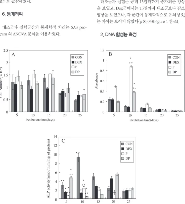
1. 세포증식율 측정대조군과 실험군 공히 15일째까지 증가되는 양상 을 보였고, Dex군에서는 15일까지 대조군보다 감소 양상을 보였으나, 각 군간에 통계학적으로 유의성 있 는 차이는 보이지 않았다(p<0.05)(Figure 1 참조).
2. DNA 합성능 측정
Figure 1 Cell proliferation activities(A), DNA synthesis activities(B) and Alkaline phosphatase activities(C) of MC3T3-E1 cells cul- tured for 5, 10, 15, 20, 25 days in control and experimental group
* : significantly different from control value(p < 0.05) ** : significantly different from dex value(p < 0.05)
*
* *
*
**
* *
*
* *
* *
**
**
*
* *
* *
* * 2.5
2
1.5
1
0.5
0 Cell Number( 106)
CON DEX P DP
5 10 15 20 25
lncubation time(days)
1.2 1 0.8 0.6 0.4 0.2 0
Absorbance
5 10 15 20 25
lncubation time(days)
CON DEX P DP
5 10 15 20 25
lncubation time(days)
CON DEX P DP 14
12 10 8 6 4 2 vity(nmol/mim/mg/ of protein)ALP acti 0
A B
C
대조군과 Dex군에서는 시간경과에 따라 감소 양 상을 보였으나, PDGF군과 Dex와 PDGF병용군에서 는 10일째 대조군과 Dex군에 비해 높은 양상을 보 여 , 통 계 학 적 으 로 유 의 성 있 는 차 이 를 보 였 다.(p<0.05)(Figure 1 참조)
3. ALP 활성도 측정
대조군에서는 10일째까지 증가하다가 이후 감소 하는 것으로 나타났다.Dex군에서는 20일까지 증가 양상을 보였고, 5일째와 10일째는 낮은 양상을 보여 대조군과 비교시 통계학적으로 유의성 있는 차이를 보였다(p<0.05)(Figure 1 참조). PDGF군은 15일째까 지 증가 양상을 보였으며, 5일째에는 대조군과 Dex 군에 비해 낮은 양상을 나타내었고, 10일째는 대조군 보다 낮은 양상을 보였으나 Dex군보다는 높은 양상 을 보여, 통계학적으로 유의성 있는 차이를 보였다 (p<0.05)(Figure 1 참조).
Dex와 PDGF병용군에서는 시간경과에 따라 감소 양상을 보였으나, 5일째 조군과 Dex군에 비해 높은 활성도를 보여, Dex와 PDGF의 상승작용을 관찰할 수 있었다(P<0.05)(Figure 1 참조).
4. 골결절 형성
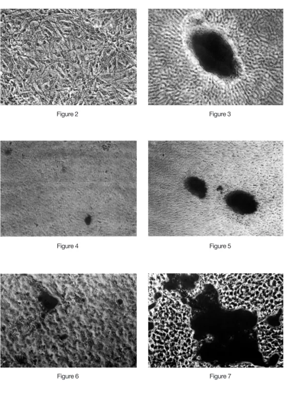
골결절 형성은 대조군, PDGF군, 그리고 Dex와 PDGF병용군은 20일째, 처음 관찰되어 25일째는 다 수의 골 결절 형성이 관찰되었다(Figure 2-7 참조).
Dex군에서는 10일째 처음 관찰되어, 시간이 경과 함에따라 크기가 큰 다수의 골 결절 형성이 관찰되 어 PDGF가 Dex의 기능을 억제 시킨 것으로 관찰되 었다(Figure 2-7 참조).
IV. 총괄 및 고찰
치주조직은 치은, 치주인대, 백악질과 치조골로 되 어 있으며 이 중 골조직의 재형성은 골흡수와 신생골 합성이 동시에 일어나는 과정으로 여기에는 골세포 와 골기질에 포함되어 있는 폴리펩타이드계 성장인
자가 관련되어 지는데 이 중 하나가 PDGF로써 골조 직 대사에 중요한 조절인자라고 알려져 있다.
항 염증 치료제로 이용되고 있는 Dex는 GC중의 하나로 생체내에서는 골형성을 감소시키고 골흡수 를 야기시키지만 골세포 배양시 적용하면 골아세포 의 분화를 촉진시켜 골결절 형성을 촉진시키는 것으 로 알려져 왔다.
본 연구에 사용된 MC3T3-E1 세포는 골아세포 분 화의 정상적인 성장 과정을 거치게 되는데 쥐의 두 개골 기원의 골아세포에서와 유사하게 세포외기질 에 광화된 형태로 관찰되어지고 있다39~41). 이 세포 는 세포증식, 골기질 침착, 광화, 성장인자에 대한 영 향, 형태의 변화와 대사에 관한 실험에 이용되어 왔
으며42,43), 교원질과 골아유사세포 분화에 대한 보고
44)와 MC3T3-E1 세포의 분화 과정에서 골기질 단백 질의 발현 양상에 대해서도 보고 된 바 있다41).
본 연구에 사용된 PDGF-BB는 in vitro 실험에서 PDGF-AA나 PDGF-AB에 비해 골아세포의 생물학적 활성을 자극하는데 더 효과적인 것으로 보고되고 있
는데45,46), 이는 PDGF-AA가α수용기에만 결합하는
것에 비해 PDGF-BB는 α와 β수용체 모두에 결합하 기 때문인 것으로 알려져 있다46).
PDGF-BB의 농도는 태생기 백서 두개골로부터 추 출되어 배양된 골아유사세포에 10, 100 ng/ml의 PDGF-BB 투여시 DNA 합성 증가 효과를 보고한 Canalis 등22)의 결과와 골아유사세포에 0.01~100 ng/ml 농도의 PDGF를 투여했을 때 10 ng/ml에서 최 대의 DNA 합성과 골아유사세포, 섬유아세포의 증식 능의 증가효과를 각각 밝힌 Kasperk 등47), Davidai 등48), Rutherford 등49)의 보고를 바탕으로 본 실험에 서는 PDGF-BB의 투여농도를 10 ng/ml로 하였다.
또한 Dex의 농도는 골아유사세포의 분화를 촉진 시켜 골결절 형성을 촉진시키는 것으로 알려져30,31) 있는 10-7 M 를 선택하였다.
골아유사세포의 성장과 분화과정에서 ALP는, 칼 슘과 인 대사에 관여하는 효소로서 Whyte50) 등이 유 기인산 기질에서 무기인산을 분리해 낼 수 있는 효 소라고 보고하였고, Siffert51) 등은 ALP가 칼슘과 인 이온이 결정화되기 이전의 골기질 형성에 주로 관여
된다고 보고하였다.
또한 Stein 등52)은 골아세포의 표지인자로서 ALP 활성도를 측정해 보아야 하며 높은 ALP 활성도는 골 세포 분화의 척도라고 보고한 것을 기초로 MC3T3- E1 세포의 ALP 활성을 측정하였다.
대조군에서는 ALP활성도가 최고에 도달한 후 골 결절이 관찰되어 Choe 등40)의 보고와 같은 양상을 보었으나, Dex군은 대조군보다 낮은 ALP 활성을 보 였음에도 골 결절은 가장 빠르게 관찰되어, Dex가 골세포의 성장과정에서 다른 광화인자에 영향을 미 치는 것으로 생각된다.
PDGF군에서의 ALP 활성도는 Centrella19)등, Yu53) 등 그리고 Canalis 등54)의 보고에서와 같이 전반적으 로 낮은 양상을 보였는데, 이는 PDGF가 골아유사세 포에 작용하는 다른 다양한 성장인자의 효과를 조절 하거나 혹은, 골아유사세포의 초기분화 과정에 관여 하는 골기질 단백질의 기능을 억제시킨 결과로 생각 된다.
Dex와 PDGF 병용 시 골아유사세포의 성장초기 단계의 세포 증식능과 DNA 합성에서는 PDGF 적용 군보다 감소 양상을 보여 Dex가 PDGF의 작용을 억 제시키는 것으로 생각된다.
ALP 활성도에서 5일째 가장 증가된 양상을 보여, 대조군 그리고 Dex군과 비교해 볼 때 큰 차이를 보 였는데, 이것은 골아유사세포의 성장과 분화 초기에 PDGF가 Dex와 함께 상승작용을 하여, 골아유사세 포의 ALP 활성을 증가시키고, 다른 골기질단백질 형 성과정에도 깊이 관여하는 것으로 생각된다.
골결절 형성에서는 Dex와 PDGF 병용군이, 대조 군과 PDGF군과 같이 처음 관찰된 시기가 Dex군보 다 늦게 나타나, PDGF가 Dex의 작용을 억제시켜 골 결절 형성을 지연시킨 것으로 추정된다. 이것은 PDGF가 다른 골기질 단백질이나 성장인자에 직접 적으로 영향을 미쳤거나, Dex와 PDGF-BB에 영향을 받은 ALP가 다른 성장인자나 골기질 단백질에 영향 을 미친 결과로 볼 수 도 있을 것으로 생각된다.
이상의 실험에서 Dex와 PDGF-BB의 골아유사세 포에 대한 결과를 근간으로, 다른 성장인자와 병용시 골아유사세포의 분화와 다양한 다양한 골기질 단백
질 발현에 대한 효과, 그리고 in vivo에서의 연구가 더 필요할 것으로 생각된다.
V. 결론
골조직의 재형성 과정에서, 생체내에서는 골형성 을 감소시키고 골 흡수를 야기시키지만, 골아유사세 포 배양시 분화를 촉진시켜 골결절 형성을 촉진시키 는 것으로 알려져 있는 Dex와, 골조직대사의 조절인 자로 알려지고 있는 PDGF-BB를 병용 시, 골아유사 세포의 분화와 성장에 어떠한 영향을 미치는지 알아 보고자 본 실험을 시행하였다.
Dex와 PDGF를 적용하지 않은 군을 대조군으로, Dex를 적용시킨 군을 Dex군으로, PDGF를 적용시킨 군을 P군, Dex와 PDGF를 병용한 군을 DP군으로 하 여 5, 10, 15, 20, 25일째에 세포 증식능, DNA 합성능, ALP 활성도, 골결절 형성을 관찰하여 다음과 같은 결 과를 얻었다.
1. Dex는 세포증식능, DNA 합성 그리고 ALP 활성 도에서 대조군에 비해 15일까지 낮은 양상을 보였으나, 골결절 형성을 촉진시키는 것으로 관 찰되었다.
2. PDGF는 15일째까지 대조군에 비해 세포 증식 능과 DNA 합성이 높은 양상을 보여, 초기성장 에 영향을 미친 것으로 나타났으나 ALP 활성도 에서는 전반적으로 감소 양상을 보였고, 골 결 절 형성은 대조군과 같이 20일째 처음 관찰되 었다.
3. Dex와 PDGF 병용시 성장과 분화 초기의 세포 증식능과 DNA합성에서, Dex가 PDGF의 기능 을 억제 시키는 것으로 관찰되었고, ALP 활성도 에서는 5일째 Dex와 PDGF의 상승작용이 관찰 되었으나, 골결절 형성에서는 PDGF가 Dex의 기능을 억제 시킨 것으로 나타났다.
VI. 참고문헌
1. Lindhe, J. : Textbook of clinical periodontology,
2nd ed., Munksguard, Copenhagen, pp 450, 1989.
2. 권영혁외 19인 : 치주과학, 제1판, 지영문화사, 서 울, pp. 420-423, 1996.
3. Mellonig, J., Bowers, G., Bright, R. and Lawrence, J. : Clinical evaluation of freeze-dried bone allografts in periodontal osseous defects. J.
Periodontol. 47: 125-129, 1976.
4. Quintero, G., Mellonig, J. and Gambil, V. : A six month clinical evaluation of decalcified freeze- dried bone allografts in human periodontal defects. J. Periodontol. 53: 726-730, 1982.
5. Bouchard, P., Giovannoli, J. L., Mattout, C., Davarpanah, M. and Etienne, D. : Clinical evalu- ation of a bioabsorbable regenerative material in mandibular class Ⅱ furcation therpy. J. Clin.
Periodontol. 24: 511-518, 1997.
6. Sander, L. and Karring, T. : New attachment and bone formation periodontal defects following treatment of submerged roots with guided tissue regeration. J. Clin. Periodontol. 22: 295-299, 1995.
7. Polson, A. M., Garrett, S., Stoller, N. H., Greenstein, G., Polson, A. P., Harrold, C. Q., and Laster, L. : Guided tissue regeneration in human furcation defects after using a biodegrad- able barrier. J. Periodontol. 66: 377-385, 1995.
8. Cho, M. I., Lin, W. L. and Genco, R. J. : Platelet- derived growth factor-modulated guided tissue regenerative therapy. J. Periodontol. 66: 522- 530, 1995.
9. Kinoshita, A., Oda, S., Takahashi, K., Yokota, S.
and Ishikawa, I. : Periodontal regeneration by application of recombinant human bone mor- phogenetic protein-2 to horizontal circumferen- tial defects created by experimental periodontitis in beagle dogs. J. Periodontol. 68: 103-109, 1997.
10. Canalis, E. : Growth factors and this regulation
of bone remodeling. J. Clin. Invest. 81: 277, 1988.
11. Hauschka, P. V., Navrakos, A. E., Iafrati, M. D., Doleman, S. E., and Klagsburn, M. : Growth fac- tors in bone matrix : Isolation of multiple types by affinity chromatography on heparin sepharose. J. Biol. Chem. 261: 12655-12674, 1986.
12. Baylink, D. J., Finkelman, R. D. and Mohan, S. : Growth factors to stimulate bone formation.
Journal of bone and mineral reserach. Vol 8(2) , 565-572, 1993.
13. Terranova, V. P. and Wikesjo, M. E. : Extracellular matrices and polypeptide growth factors as mediators of functions of cells of the periodontium. J. Periodontol. 58: 371-380, 1987.
14. Westermark, B and Heldin, C. : Platelet-derived growth factor. Acta. Oncologica. 32(2): 101-105, 1993.
15. Deuel, T. F., Huang, J. S. A., Proffit, R. L., Baenzinger, J. U., Chang, D. and Kenneduy, B.
B.: Human platelet-derived growth factor purifi- cation and resolution into two active protein fractions. J. Biol. Chem. 256: 8896-8899, 1981.
16. Raines, E. W. and Ross, R. : Platelet-derived growth factor I. High yielded purification and evidence for multiple forms. J. Biol. Chem. 257:
5154-5160, 1982.
17. Hammacher, A., Hellman, U. and Johnsson, A. : A major part of PDGF purified from human platelet is a heterodimer of one A and one B chain. J. Biol. Chem. 263: 16493-16498, 1982.
18. Canalis, E. and Rydziel, S. : Principle of Bone biology. Academic Press Inc. U.S.A. pp. 619- 625, 1996.
19. Centrella, M., McCarthy, T. L., and Centrella, M.
: Platelet derived growth factor enhances deoxyribonucleic acid and collagen synthesis in osteoblast enriched cultures from fetal rat pari-
etal bone. Endocrinology. 125: 13-19, 1989.
20. Pfeilschifter, J., Oechsner, M., Naumann, A., Gronwald R. G. K., Minne, H. W. and Ziegler, R. : Stimulation of bone matrix apposition in vitro by local trowth factors; A comparison between insulin like growth factor I, platelet derived growth factor, and transforming growth factor. Endocrinology. 127: 69-75, 1990.
21. Varghese, S., Delany, A.M., Liang, L., Gabbitas, B., Jeffrey, J. J. and Canalis, E. : Transcriptional and Posttranscriptional regulation of interstitial collagenase by Platelet-Derived Growth Factor BB in Bone Cell Culture. Endocrinology. 137:
431-437, 1995.
22. Canalis, E., McCarthy, T. L., Centrella, M. : Effects of platelet derived growth factor on bone formation in vitro. J. Cell. Physiol., 140: 530-537, 1989.
23. Sissons, H. A. and Hadfield, G. J. : The influ- ence of cortisone in the rabbit. Br. J. Surg. 39:
172-178, 1951.
24. Baylink, D. J. : Glucocorticoid-induced osteo- porosis. N. Engl. J. Med. 309: 306-308, 1983.
25. Burckhardt, P. : Corticosteroids and bone. A review, Hormone Res. 20: 59-64, 1984.
26. Meunier, P. J., Dempster, D. W., Edouard, C., Chapuy, M. C., Arlot M. and Charon, S. : Bone histomorphometry in syndrome. Adv. Exp.
Med, Biol. 171: 191-200, 1984.
27. Jowsey, J. and Riggs, B. L. : Bone formation in hypercorticism. Acta. Endocrinol. 63: 21-28, 1970.
28. Jee, W. S., Park, H. Z., Roberts, W. E. and Kenner, G.H. : Coticosteroid and bone. Am. J.
Anat. 129: 477-480, 1970.
29. Dietrich, J. W., Canalis, E. M., Maina, D. M. and Raisz, L. G. : Effect of glucocorticoids on fetal rat bone collagen synthesis in vitro. Endocrinology.
104: 715-721, 1979.
30. Canalis, E. M. : Effects of glucocoricoids on type I collagen synthesis, alkaline phosphatase activi- ty and deoxyribonucleic acid content in cultured rat calvraiae. Endocrinolgy. 112: 931-939, 1983.
31. Bellows, C. G., Heersche, J. N. and Aubin, J. E.
: Determination of the capacity for proliferation and differentiation of osteoprogenitor cells in the presence and abscence of dexamethasone.
Developmental Biology. 140: 132-138, 1990.
32. Cheng, S. L., Yang, J. W., Rifas, L., Zhang., S. F.
and Avioli, L. V. : Differentiation of human bone marrow osteogenic stromal cells in vitro: induc- tion of the osteoblast phenotype by dexametha- sone. Endocrinology. 134: 277-286, 1993.
33. Sato, S., Kim, T., Maruyama, S., Tajima, M. and Utsumi, N. : Comparison between the effects of dexamethasone and indomethacin on bone wound healing. J. Pharmacol. 42: 71-78, 1986.
34. Holley, R.W. and Kiernan, J. A.: Control of the initiation of DNA Synthesis in 3T3 cells. Serum factors. Proc. Natl. Acad. sci. U.S.A. 71: 2908- 2911, 1974.
35. Otto, A. M., Natoli, C., Richmond, K. M. V., Iacobelli. S and De Asusa L. J. : Glucocorticoids inhibit the stimulatory effect of epidermal growth factor on the initiation of DNA synthesis.
J. Cell. Physiol. 107: 155-163, 1981.
36. Rutherford, R. B., Ryin, M. E., Kennedy, J. E., Tucker, M. M. and Charette, M. F. : Platelet- derived growth factor and dexamethasone com- bined with collagen matrix induce regeneration of the periodontium in monkeys. J. Clin.
Periodontol. 20: 537-544, 1993
37. Casasco, A., Casasco, M., Cornaglia, A., Mazzini, G., Renzis, R. and Tateo, S. : Detection of Bromo-Deoxyuridine and Proliferating Cell Nuclear antigen-immunoreactivities in tooth germ. Connect. tissue Res. 32: 63-70, 1995 38. Bessey, O., A., Lowry, O. H. and Brock, M. J. :
A method for the rapid determination of alkaline phosphatase with five cubic milimeters of serum. J. Biol. Chem. 164: 321-329, 1946 39. Sudo, H., Kodama, H., Amagai, Y., Yamamoto,
S. and Kasai, S. : In vitro differentiation and cal- cification in a new clonal osteogenic cell line derived from newborn mouse calvaria. J. cell.
Biol. 96: 191-198, 1983.
40. Quarles, L. D., Yohay, D. A., Lever, L. W., Caton, R. and Wenstrup, R. J.: Distinct proliferative and differentiated stages of murine MC3T3-E1 cells in culture an in vitro model of osteoblast develop- ment. J. Bone Mineral Res. 7: 683-692, 1992.
41. Choe, J. Y., Lee, B. H., Song, K. B., Park, R. W., Kim, J. S., Sohn, K. Y., Jo, J. S. and Ryoo, H. M.
: Expression patterns of bone related proteins during osteoblastic differentiation in MC3T3-E1 cells. J. Cell. Biochem. 61: 609-618, 1996.
42. Kumegawa, M., Hiramatsu, M., Hatakeyama, K., Yajima, T., Kodama, H., Osaki, T. and Kurisu, K. : Effects of epidermal growth factor on osteoblastic cells in vitro. Calcif. Tissue. Int. 35:
542-548, 1983.
43. Hurley, M. M., Abreu, C., Harrison, J. R., Lichtler, A. C., Raiaz, L. G. and Kream, B. E. : Basic fibroblast growth factor inhibits type I col- lagen gene expression in osteoblastic MC3T3-E1 cells. J. Biol. Chem. 268: 5588-5593, 1993.
44. Franceschi, R.T. and Iyer, B. S. : Relationship between collagen synthesis and expression of the osteoblast phenotype in MC3T3-E1 cells. J.
Bone. Min. Res. 7: 235-246, 1992.
45. Pfeilschifter, J., Krempien, R., Naumann, A., Gronwald, R. G. K., Hoppe, J. and Ziegler, R. : Differential effects of platelet derived growth fac- tor isoforms on plasminogen activator activity in fetal rat osteoblasts due to isoform specific receptor functions, Endocrinology. 130: 2059-
2066, 1992.
46. Centrella, M., McCarthy, T. L., Kusmik, W. F.
and Canalis, E. : Relative binding and biochemi- cal effects of heterodimeric and homodimeric isoforms of platelet derived growth factor in osteoblast enriched cultures from fetal rat bone.
J. Cell. Physiol. 147: 420-426, 1991.
47. Kasperk, C. H., Wergedal, J. E., Mohan, S., Long, D. L., Lau, K. H. W. and Baylink, D. J. : Growth factor. 3: 147-158, 1990.
48. Rutherford, R. B., Trailsmith, M. D., Ryan, M. E.
and Charette, M. F. : Synergistic effects of dex- amethasone or platelet-derived growth factor on mitogenesis in vitro. Arch. Oral Biol. 37(2): 139- 145, 1992.
49. Davidai, G., Lee, A., Schvartz, I. and Hasum, E. : PDGF induces tyrosine phosphorylation in osteoblast-like cells: relevance to mitogenesis.
Am. J. Physiol. 263: 205-209, 1992.
50. Whyte, M., P. : Hypophosphatasia and the role of alkaline phosphatase in skeletal mineraliza- tion. Endocrine Rev. 15: 439-461, 1994.
51. Siffert, R. S. : The role of ALPase in osteogene- sis. J. Exp. Med. 93: 415-425, 1951.
52. Stein, G. S., Lian, J. B. and Owen, T. A. : Relationship of cell growth to the regulation of tissue-specific gene expression during osteoblast differen -tiation. FASER. J. 4: 82-94, 1990.
53. Yu, X., Hsieh, S. C., Bao, W. and Graves, D.T. : Temporal expression of PDGF recepers and PDGF regulatory effects on the osteoblastic cells in mineralizing cultures. Am. J. Physiol. 272:
1709-1716, 1997.
54. Canalis, E., Pash, J., Gabbitas, B., Rydziel, S. and Varghese, S. : Growth factors regulate the syn- thesis of insulin-like growth factor-I in bone cell cultures. Endocrinology. 133: 33-38, 1993.
사진부도 설명
Figure 2. Histochemical change of control group at 10 days.
Photomicrograph shows no bone nodules on control group at 10 days.
x 40
Figure 3. Histochemical change of Dex group at 10 days.
Photomicrograph shows bone nodules on Dex group at 10 days.
x 40
Figure 4. Histochemical change of control group at 20 days.
Photomicrograph shows bone nodules on control group at 20 days.
x 40
Figure 5. Histochemical change of DP group at 20 days.
Photomicrograph shows bone nodules on DP group at 20 days.
x 40
Figure 6. Histochemical change of control group at 25 days.
Photomicrograph shows bone nodules on control group at 25 days.
x 40
Figure 7. Histochemical change of on DP group at 25 days.
Photomicrograph shows very larger bone nodules on DP group at 25 days when compared to Figure 2~ Figure 6
x 40
Dex; Dexamethasone DP; combination of Dex and PDGF
사진부도
Figure 2 Figure 3
Figure 4 Figure 5
Figure 6 Figure 7
-Abstract-
The Effects of Combination of PDGF-BB and Dexamethasone on Differentiation of MC3T3-E1 Cells
Jae-mok Lee*, Jo-Young Suh*, Sung-Jo Kim**, Jeom-Il Choi**
*Department of Periodontology, School of Dentistry, Kyungpook National University
**Department of Periodontology, School of Dentistry, Pusan National University
To evaluate the effects of Dexamethasone(Dex), Platelet derived growth factor-BB(PDGF) and combination of Dex and PDGF(DP) on the growth and differentiation of MC3T3-E1 cells, Dex(10-7 M) and PDGF(10 ng/ml) in experimental group were added to the cells at the days 5, 10, 15, 20, 25 and examined for cell proliferation activities, DNA synthesis activities, ALP activities and bone nodule formation.
The results were as follows :
1. In Dex group, cell proliferation, DNA synthesis and ALP activities were lower until 15 days when com- pared to the control group. Bone nodules formation were shown at 10 days.
2. In PDGF group, cell proliferation and DNA synthesis activities were higher until 15 days and ALP activities were lower when compared to the control and Dex groups. Bone nodules formation were shown at 20 days.
3. In DP group, cell proliferation and DNA synthesis activities of PDGF were suppressed by Dex and syner- gistic effects of combination of Dex and PDGF on ALP activities were shown at days 5 when compared to control and Dex groups. Bone nodules formation activities of Dex were suppressed by PDGF.