I. 서론
치주 조직중의 하나인 골조직은 계속적인 흡수와 형성을 반복하는 동적인 조직으로서, 골조직의 성장과 개조는 조골세포, 파골세포 및 그 전구세포의 증식, 분화 및 활성에 영향 을 미치는 전신적 조절물질과 국소적 조절물 질에 의해 조절되고 있다1, 2). 국소적 조절물 질은 대부분이 성장인자로써 골기질에서 발 견되고, 골세포에 의해 생성되며, autocrine 또 는 paracrine action으로 그 조절기능을 나타내 는 것으로 알려져 있으며, 골조직대사조절에 전신적 조절물질보다 더욱 중요한 역할을 하 는 것으로 알려져 있다2). 성장인자에 대하여 Terranova와 Wikesjo3)는 세포의 성장, 형성 및 기능은 세포와 세포외 기질의 특이한 상 호작용과 폴리펩타이드계 성장인자에 의해 조절된다고 보고함으로써 폴리펩타이드계 성 장인자가 치주조직 치유에 중요한 역할을 할 수 있을 것이라고 시사한 바 있다. Graves 및 Cochran4)은 미분화된 중배엽세포의 분화에 관계하는 폴리펩타이드계 성장인자로써 혈소 판 유래 성장인자(Platelet-derived growth factor, 이하 PDGF라 칭함), 변형성장인자 (Transforming growth factor), 섬유아세포성
장인자(Fibroblast growth factor), 인슐린유사 성장인자(Insulin-like growth factor), 상피성장 인자(Epidermal growth factor)등이 있다고 보 고하였으며, 이중 PDGF는 중배엽의 세포 즉 섬유아세포, 신경세포, 평활근세포, 골세포를 조절하는데 중요하다고 알려져 있다5, 6).
PDGF는 9.8의 isoelectric point와 30kDa의 분자량을 가지고 있는 조절성 단백질로써7) 단 종이량체(PDGF-AA, BB)와 이종이량체 (PDGF-AB)로 존재함이 발견되었고8) 유리되 는 근원은 혈소판의 α-granule로 알려져 있다
9). 혈소판 이외에 단핵세포 및 대식세포10), 섬 유아세포11), 내피세포12), 골기질13) 등으로부터 분리된다고 밝혀졌다. PDGF A gene은 7번 염색체에 한정되어 위치하며, PDGF B gene 과 아미노산 서열이 56% 동일하다14). 동종 이량체와 이종 이량체의 생물학적 활성은 동 일하지만, PDGF BB가 PDGF AA나 AB보다 더 강력한 활성을 보인다고 보고된 바 있다
15). PDGF는 생쥐와 인간의 섬유아세포16), 동 맥 평활근 세포17), 활액세포와 골신경교세포 등의 다양한 간엽세포의 증식을 자극하며15,
16), 이들 세포의 화학주성인자로 작용하고, 단 백질 합성을 증가시키며, 세포막과 연관된 인 지질, 콜레스테롤 에스터, 프로스타글란딘 등 대한치주과학회지 : Vol. 27, No. 4, 1997
MC3T3-E1 세포의 ALP activity에 대한 PDGF-BB의 영향
이경희*·이재목*·최병주**·유현모***·서조영*
경북대학교 치과대학 치주과학교실*
경북대학교 치과대학 치과약리학교실**
경북대학교 치과대학 구강생화학교실***
의 복합체 합성도 자극한다고 알려져 있다18). 또한, 배양된 섬유아세포 계열을 PDGF에 먼 저 노출시키면 EGF에 의한 세포분열효과를 10배정도 증가시키는 등 다른 성장인자의 효 과를 조절한다고 알려져 있다19).
Cho 등20)의 연구에 따르면 치주인대 섬유 아세포에 PDGF와 IGF를 동시에 적용한 경 우, 세포 증식에 상승효과를 일으킨다고 보고 하였으며, Lynch 등21)은 돼지의 피부에 acute injury를 형성한 경우 상피세포에서는 PDGF 와 수용기의 mRNA가 증가하였고, 결체조직 에서는 PDGF mRNA가 발현된다고 보고하 였으며, Cho 등22)은 beagle dog의 3등급의 분 지부 병소에 PDGF-BB를 처리한 후 e-PTFE membrane으로 조직유도재생술을 시행한 경 우 완전한 치주조직재생 및 치조골의 형성이 일어났다고 보고하였다.
PDGF의 골세포에 대한 영향을 살펴보면, in vitro 실험에서 Centrella 등은 PDGF가 생 체 두개골에서와 쥐에서 추출된 골아세포에 서 골세포 복제와 DNA 합성을 자극하는 것 으로 보아 PDGF의 골에서의 일차적 효과는 증식능과 연관된 것으로 보인다고 보고한 바 있으며23), 세포복제에 대한 영향의 결과 PDGF는 교원성 단백질을 합성하는 세포수를 증가시키나, cellular basis에서 볼 때, PDGF는 교원성 단백질의 합성을 직접적으로 증가시 키지는 않으며, 골아세포의 교원성 단백질 생 산을 어느정도 억제하는 효과를 갖는다고 보 고하였다24). 이와 일치하는 것으로 Cochran 등은 PDGF가 신생쥐의 두개골에서 골기질의 축적속도를 억제한다고 보고하였다25). 그 외 에도 Hock 및 Canalis26)와 Cochran 등25)은 PDGF는 interstitial collagenase의 증가에 의해 교원성 단백질의 분해를 증가시킨다고 보고 하였다.
Centrella와 McCarthy 등27)은 백서의 태내 두개관에서 배양한 조골세포에 PDGF를 적용 한 경우 DNA, 교원질 및 비교원성 단백질의
합성이 증가한다고 보고하였으며, in vitro에서 의 실험에서 Lynch 등28)은 PDGF-BB를 쥐에 전신적으로 투여한 경우 골밀도 및 골격의 강도가 증가했다고 보고하였고, Andrew 등29) 과 Walther 등30)은 정상적인 골절의 치유과정 에서 PDGF가 발현되며 시험관적 실험에서도 PDGF가 중요한 조절인자라고 보고하였다.
여 러 연 구 에 도 불 구 하 고 PDGF의 mineralization과 ALP 활성도에 미치는 효과 에 대해 논란이 많아, 이에 본 실험에서는 골 형성 세포 계열중 하나인 MC3T3-E1세포에 PDGF-BB를 농도별로 주입해서 bone nodule 의 형성능과 골형성 세포로의 분화능에 대한 표지인자로 알려진 알칼린 인산 효소 활성도 (이하 ALP활성도라 표기)에 대한 영향을 알 아봄으로써 혈소판 유래 성장인자가 치주조 직재생에 중요한 골세포의 분화에 미치는 영 향을 알아보고자 한다.
II. 재료 및 방법
1. 재료(시약)배양액은 alpha-modified eagle medium (Gibco사, 미국, 이하 α-MEM으로 표기)을 사 용하였고, fetal bovine serum (Gibco사, 미국, 이하 FBS로 표기)을 성장 촉진제로 추가하였 으며, 그외 β-glycerophosphate, ascorbic acid(Sigma, 미국), 유전자재조합형 PDGF- BB(Genzyme사, 미국)을 사용하였다.
2. 골아세포의 배양
MC3T3-E1세포를 취득하여 12 well plate에 1x10⁵cells/well, 24 well plate에 1X10⁴ cells/well로 10% fetal bovine serum, 10mM β-glycerophosphate, 50㎍/ml의 아스코빅 산, 100U/ml penicilline, 100㎍/ml streptomycin을 함유한 α-MEM배양액에서 초기배양하여 37
℃, 5% CO2 함유 배양기에서 유지하였다.
10% fetal bovine serum에서 계속 배양한 군 을 대조군-1로 하였고, PDGF-BB를 주입하기 48시간전에 성장인자의 최대의 효과를 위해 serum free media로 교체한 군을 대조군-2로 하였으며, serum free media로 교체하고 24시 간 경과후 0.1, 1, 10ng/ml의 PDGF를 첨가한 군을 실험군으로 하여 3일 간격으로 배양액 을 교체하였다.
3. Histochemical analysis
Bone nodule의 형성을 관찰하기 위해서 세 포를 30일동안 100mm dish에서 배양하고, 칼 슘 침착을 식별하기 위해서 세포를 ice-cold PBS로 2번 씻은 후 실온에서 1시간동안 0.1% Alizarin Red S 용액에서 배양하였고 세 포를 30분동안 0.1% light green SF solution으 로 염색하여 세포를 순차적으로 0.1% acetic acid와 absolute ethanol로 씻은 후 3, 7, 14, 21, 28일째에 광학현미경으로 관찰하였다.
4. alkaline phosphatase(ALP) 활성도 측정
세포를 24 well plate에 접종하고 앞서 설명 한 대로 배양하여 0.1, 1, 10ng/ml의 PDGF를 첨가하여 3일 간격으로 배양액을 교체해 주 면서 3일, 7일, 14일, 21일, 28일에 ALP 활성 을 p-nitrophenyl phosphate(Sigma, USA)를 기질로 이용한 방법으로 아래와 같이 측정하 였 다 . 부 착 된 세 포 를 인 산 완 충 식 염 수 (phosphate buffered saline, 이하 PBS)로 2회 씻어내고 0.02%의 Nonidet P-40(Sigma) 1ml 에 녹 인 후 ultrasonicator에 서 15초 간 sonication시켜 (Fischer, Rockville, MD) 12,000 g에서 15분간 원심분리하였다. 효소활성을 측 정하기 전에 상층액을 -20℃에 두고 37℃에서 30분간 cell digestion buffer(1.5M Tris-HCL, 1mM ZnCl2, 1mM MgCl2·6H2O, pH 9.2,
containing 1% Triton X-100)로 처리하며 7mM p-nitrophenyl phosphate (Sigma, USA) 를 기질로써 이용하여 410nm에서 흡수도를 측정하였다. 단백질 농도는 BCA protein assay reagent(Pierce, USA)를 사용하여 측정 하며, bovine serum albumin을 standard로 하 였고, ALP활성도는 nmole/min/mg of protein 으로 나타내었다. 대조군과 실험군의 통계학 적 처리는 student t-test로 시행하였다.
III. 결과
1. Bone nodule의 형성Bone nodule의 형성은 대조군과 모든 실험 군에서 3, 7, 14일째에는 전혀 관찰할 수 없었 으며, 21일째에 관찰되기 시작하여 대조군은 28일째에도 별 변화가 없었으며, 모든 실험군 에서 28일째에 대조군보다 많은 bone nodule 형성이 관찰되었다.
2. 시간의 경과에 따른 MC3T3-E1 세포의 ALP 활성에 대한 PDGF-BB의 영향
시간의 경과에 따른 MC3T3-E1 세포의 ALP 활성은 대조군-1, 대조군-2에서는 3일 군, 7일군까지 증가하다가 14일, 21일, 28일에 서 감소하는 것으로 나타났고, PDGF 0, 1, 1, 10 ng/ml 적용군에서는 3일, 7일, 14일까지 시 간경과에 따라 증가하다가 21일, 28일군에서 점차 감소하는 것으로 나타났다.
MC3T3-E1 세 포 의 ALP 활 성 도 를 nmole/min/mg of protein으로 나타내었을 때 PDGF-BB의 영향은 대조군-1에서 3일군의 2.13±0.43에 비해 7일군에서 3.44±0.09로 증 가하다가 14일군에서 2.97±0.34, 21일 1.87±
0.33, 28일군에서 1.57±0.04으로 점차 감소하 였으나 7일군에서만 3일군에 비해 통계학적 유의성을 보였다(P<0.05).
대조군-2의 경우 3일군의 3.76±0.61에 비해 7일군에서 9.80±2.12으로 증가하였고 14일군 에서 5.07±1.06으로 감소하였으나 대조군-1보 다는 높은 ALP활성도를 보였고 21일군에서 2.07±0.05, 28일군에서 1.80±0.11로 나타나 모 든 군에서 3일군에 비해 통계학적 유의성이 있는 감소양상을 보였다(P<0.05).
PDGF 0.1ng/ml 적용군에서 3일군의 1.63±
0.87에 비해 7일군에서 4.42±0.21, 14일군에서 4.79±0.33으로 증가양상을 나타내어 3일군에 비해 통계학적 유의성을 보였고, 21일째는 2.30±0.05, 28일 1.73±0.15로 감소하였으나 통 계학적 유의성은 없었다(P<0.05). PDGF 1 ng/ml 적용군에서 3일군의 0.86±0.15에 비해 7일군에서 2.79±0.14, 14일군에서 3.31±1.01로 증가하였고, 21일째는 0.79±0.05, 28일째는 0.65±0.01로 감소하였으며 모든 군에서 3일군 에 비해 통계학적 유의성을 보였다. (P<0.05, P<0.01) PDGF 10 ng/ml 적용군에서 3일군의 0.60±0.03에 비해 7일군에서 2.01±0.17, 14일 군에서 2.13±0.14로 증가하다가 21일군에서
1.72±0.04, 28일군에서 1.42±0.07로 감소하였 으며 모든 군에서 3일군에 비해 통계학적으 로 유의한 차이를 보였다(P<0.01)(표 1, 그림 1 참조).
3. PDGF-BB의 농도에 따른 MC3T3-E1세 포의 ALP 활성에 대한 비교
대조군-1과 비교시 3일군에서는 PDGF-BB 에 대해 농도의존적으로 ALP 활성이 감소, 7 일, 14일군에서는 PDGF-BB 0.1 ng/ml 적용 군에서는 증가, 1, 10 ng/ml에서는 감소했고 21일, 28일군에서는 0.1ng/ml 군에서는 증가, 1 ng/ml 적용군에서는 감소, 10ng/ml 적용군 에서는 다시 증가하는 것으로 나타났다. 대조 군-2와 비교시 21일군의 PDGF 0.1 ng/ml 적 용군, 28일군의 PDGF 10ng/ml 적용군을 제 외한 모든 군에서 PDGF-BB의 농도에 대해 농도의존적으로 ALP 활성이 감소되었다.
3일군에서 대조군-1, 대조군-2 와 비교시 PDGF-BB의 농도가 증가함에 따라 ALP 활
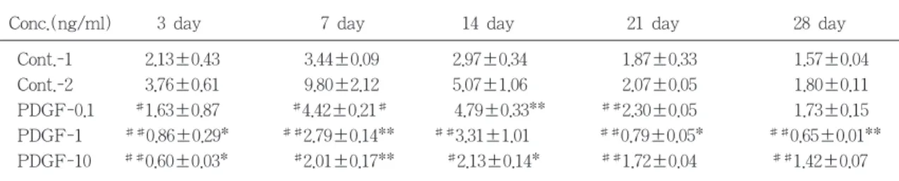
Conc.(ng/ml) 3 day 7 day 14 day 21 day 28 day
Cont.-1 2.13±0.43 3.44±0.09 2.97±0.34 1.87±0.33 1.57±0.04
Cont.-2 3.76±0.61 9.80±2.12 5.07±1.06 2.07±0.05 1.80±0.11
PDGF-0.1 #1.63±0.87 #4.42±0.21# 4.79±0.33** ##2.30±0.05 1.73±0.15
PDGF-1 ##0.86±0.29* ##2.79±0.14** ##3.31±1.01 ##0.79±0.05* ##0.65±0.01**
PDGF-10 ##0.60±0.03* #2.01±0.17** #2.13±0.14* ##1.72±0.04 ##1.42±0.07
MC3T3-E1 cells were seeded at 1×10⁴ cells/ml in alpha-minimum essential medium containing 10% fetal bovine serum, 10 mM β-glycerophosphate and 50㎍/ml of ascorbic acid. Before 48 hours of indicated time, medium were changed with serum free medium containing 10 mM β-glycerophosphate and 50㎍/ml of ascorbic acid.. After 24 hours, indicated amounts of platelet derived growth factor were added. Alkaline phosphatase activity were measured as described in materials and methods. Each value represents the mean and S.D. of three determinants.
* : significantly different from control-1 value in Dose resoponse effect (P < 0.05)
** : significantly different from control-1 value in Dose response effect (P < 0.01)
# : significantly different from control-2 value in Dose resoponse effect (P < 0.05)
## : significantly different from control-2 value in Dose response effect (P < 0.01)
표 1 Time and Dose-response effect of Platelet-Derived growth factor-BB on Alkaline Phosphatase activity by MC3T3-E1 cells cultured for 3, 7, 14, 21, 28 days
성은 농도의존적으로 감소하였는데 대조군-1 의 2.13±0.43과 비교시 PDGF 0.1 ng/ml 적용 군은 1.63±0.87로써 ALP활성의 미약한 감소 를 보였으나 통계학적 유의성은 없었고, 1ng/ml 적용군의 0.86±0.29와 10ng/ml 적용 군의 0.60±0.03에서는 대조군-1에 비해 통계 적으로 유의성있는 감소를 보였다(P<0.05).
대조군-2와 비교시 0.1ng/ml의 1.63±0.87, 1ng/ml의 0.86±0.29, 10 ng/ml 적용군의 0.60
±0.03 모두에서 통계적으로 대조군-2의 3.76
±0.61에 비해 유의성있는 감소를 보였다 (P<0.05, P<0.01).
7일군에서 대조군-1의 3.44±0.09에 비교시 PDGF 0.1ng/ml 적용군에서 4.42±0.21로 ALP 활성도가 증가했다가, 1ng/ml적용군에 서는 2.79±0.14, 10ng/ml 적용군에서 2.01±
0.17로 나타나 농도의존적으로 감소하였으며 모든 군에서 대조군-1에 비해 통계학적 유의 성을 보였고 대조군-2의 9.80±2.12와 비교시
PDGF 0.1, 1, 10 ng/ml 적용군 모두에서 대조 군-2에 비해 통계학적으로 유의성있는 농도 의존적인 감소를 보였다(P<0.05, P<0.01).
14일군에서 대조군-1의 2.97±0.34에 비해 PDGF 0.1ng/ml적용군에서 4.79±0.33로 ALP 활성도가 증가했다가 1ng/ml 적용군에서는 3.31±1.01로 감소했으나 여전히 대조군-1에 비해서는 높은 수치를 보였으며 10ng/ml 적 용군에서는 2.13±0.14로써 대조군-1보다 더 낮은 수치로 감소했으며 1ng/ml 적용군을 제 외한 군에서 통계학적 유의성을 보였다.
(P<0.05, P<0.01) 대조군-2의 5.07±1.06과 비 교시 PDGF의 농도증가에 대해 ALP 활성도 는 농도의존적으로 감소하였는데 PDGF 10ng/ml 적용군에서만 통계학적으로 유의성 있는 감소를 보였다(P<0.05).
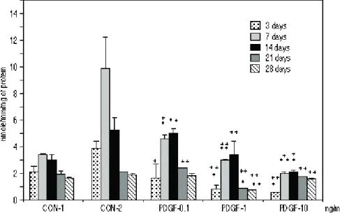
21일군에서 대조군-1의 1.87±0.33에 비해 PDGF 0.1 ng/ml 적용군에서 2.30±0.05로써 ALP 활성도가 증가했다가 1 ng/ml 적용군에 그림 1 Time-response effect of 0.1, 1, 10 ng/ml of Platelet-Derived Growth factor on alkaline phosphatase activity by
MC3T3-E1 cells cultured for 3, 7, 14, 21, 28 days.
* : significantly different from the value of Day 3 group (P < 0.05)
** : significantly different from the value of Day 3 group (P < 0.01)
3 7 14 21 28 days
nmole/min/mg of Protein
12
10
8
6
4
2
0
CON1 CON2 PDGF0.1 PDGF1 PDGF10
서 0.79±0.05로 감소하고, 10 ng/ml 적용군에 서 1.72±0.04로 다시 증가하였으나 대조군에 비해서는 더 낮은 활성도를 보였고 1 ng/ml 적용군에서만 통계학적 유의성이 있는 감소 를 보였다(P<0.05). 대조군-2의 2.07±0.05와 비교시 PDGF 0.1 ng/ml 적용군에서 ALP 활 성도가 증가했다가 1 ng/ml 적용군에서 감소 하였으며, 10 ng/ml 적용군에서 다시 증가하 지만 대조군에 비해서는 더 낮은 활성도를 보였고 모든 군에서 통계학적 유의성이 있었 다(P<0.01).
28일군에서는 대조군-1의 1.57±0.04에 비해 PDGF 0.1 ng/ml 적용군에서 1.73±0.15으로 ALP 활성도가 증가하다가 1 ng/ml 적용군에 서 0.65±0.01로 감소하여, 10 ng/ml 적용군에 서 1.42±0.07으로 다시 증가하였으나 대조군 에 비해서는 여전히 낮은 활성도를 보였고 1 ng/ml 적용군에서만 통계학적으로 유의성있
는 감소를 보였다. 대조군-2의 1.80±0.11과 비 교시 PDGF 0.1 ng/ml 적용군과 1 ng/ml 적 용군에서 적용농도에 대해 ALP 활성도가 농 도의존적으로 감소하다가 10 ng/ml 적용군에 서 약간 증가하지만 대조군-2에 비해서는 여 전히 낮은 활성도를 보였으며 0.1 ng/ml 적용 군을 제외한 모든 군에서 통계학적 유의성을 보였다(P<0.01)(표 1, 그림 2 참조).
III. 총괄 및 고찰
치주조직재생에 관여하는 치주조직은 치은, 치주 인대, 치근 백악질과 치조골로 되어 있 으며1), 이 중 골조직의 재형성은 골흡수와 골 형성의 복합된 과정으로 일어나고, 이러한 활 동은 골기질과 연관되어 골흡수동안 분비되 는 polypeptide계 성장인자의 영향을 받게 된
다34, 35). 여러가지 호르몬과 PDGF와 EGF같
그림 2 Dose-response effect of 0.1, 1, 10 ng/ml of Platelet-Derived Growth factor on alkaline phosphatase activity by MC3T3-E1 cells cultured for 3, 7, 14, 21, 28 days.
* : significantly different from control-1 value in Dose resoponse effect (P < 0.05)
** : significantly different from control-1 value in Dose response effect (P < 0.01) + : significantly different from control-2 value in Dose resoponse effect (P < 0.05) ++ : significantly different from control-2 value in Dose response effect (P < 0.01)
은 전신적, 국소적 성장인자는 in vivo와 in vitro에서의 연구에서 골성장에 영향을 끼치는 것으로 알려져 있다36, 37). 이러한 성장인자 가 운데 PDGF는 골세포 배양에서 세포의 복제 와 분화기능을 조절하는 것으로 알려져 있다
38, 39).
PDGF의 골세포에 대한 영향으로 Canalis 등35)은 PDGF-AA 또는 -BB 0.3-3.3nM로 골 세포를 처치한 경우 PDGF-A mRNA가 그 양과 비례해서 증가하는 양상을 보였다고 보 고하였고, Hanks 등40)은 배양한 쥐의 두개골 세포에 PDGF를 적용한 실험에서 새로운 단 백질의 형성이 자극되는 것으로 보아 상해부 위에서 혈관밖으로 나온 혈액내에 존재하는 PDGF가 정상적인 골막하에서 골의 축적을 자극할 수 있다고 보고하였으며, Centrella 등
15)은 PDGF-BB를 태생쥐의 parietal bone에서 채취한 골아세포에 적용시 핵산, 교원성 단백 질 및 비교원성 단백질의 합성을 증가시킴을 보고하였다.
Pfeilschifter 등41)은 태생쥐의 두개골을 PDGF로 처치한 경우 골아유사세포의 증식을 증가시킴으로써 간접적으로 교원성 단백질의 합성을 증가시키는 효과를 가져오지만, 교원 성 단백질의 분해에 대해서는 직접적인 자극 효과를 가진다고 보고하였다. Canalis 등38)의 연구에서 PDGF는 골기질의 형성을 증가시키 지 않는 것으로 보고되었고, Centrella 등15)의 연구에서는 태생기 쥐의 골에서 채취한 골아 세포에 3nM PDGF-BB, -AB 적용시 알칼린 인산효소활성도가 대조군에 비해 현저히 감 소하였으나, PDGF-AA에 의한 감소는 통계 상 유의성이 없는 것으로 나타났다.
이처럼 PDGF의 골조직에 대한 영향은 여 러 연구에서 다소 상반된 견해를 보이고 있 어, 본 연구에서는 PDGF-BB의 적용이 골세 포의 분화에 미치는 영향을 알아보고자 다양 한 적용농도와 적용시간에 따른 MC3T3-E1 세포의 ALP활성도와 bone nodule의 형성을
관찰해 보고자 하였다.
본 연구에서 MC3T3-E1 세포에 대한 PDGF-BB의 영향을 알아보기 위해 농도별, 시간경과에 따른 골결절 형성을 측정해 본 결과 MC3T3-E1 세포는 대조군과 실험군에 서 공히 21일경에 골결절의 형성을 보였으며, 28일경에는 대조군보다 실험군에서 현저히 많은 bone nodule의 형성을 보였다. 이러한 실험결과는 PDGF를 첨가하지 않은 신생쥐의 두개골에서 추출한 MC3T3-E1세포에서 21일 경에 bone nodule의 형성이 관찰되었다는 Hiroko Sudo 등의 보고와42) 일치하였고, 동일 한 세포의 배양시 16일경부터 bone nodule이 관찰되었으며 그 후 30일경까지 시간의 경과 에 따라 nodule의 수와 크기가 차츰 증가했다 는 최 등43)의 보고와는 약간 다른 양상을 보 이고 있다.
ALP는 칼슘과 인 대사에서 관여하는 효소 로서 정확한 기능은 알려져있지 않지만 Robinson 등44)은 유기인산 기질에서 무기인산 을 분리해 낼 수 있는 효소라고 보고하였으 며, Siffert31)는 골형성 이전의 세포대사와 칼 슘과 인 이온이 결정화되기 이전의 골기질 형성에 주로 관여한다고 보고하였고, Stein 등
45)은 골세포의 표지인자로서 ALP활성도를 측정해 보아야 하며 높은 ALP활성도는 골세 포 분화의 표지인자라고 보고하였다.
본 실험에서 시간 경과에 따른 ALP활성도 를 측정하였던 바 대조군에서는 공히 7일째 까지 ALP활성도가 증가하다가 그 이후 감소 하는 양상을 보였으며, 실험군에서는 0.1, 1, 10ng/ml 투여군 모두에서 14일째까지 증가하 다가 그 후 낮은 활성도를 보였다. 이는 오 등46)의 연구에서 7일째보다 14일째에 높은 ALP활성도를 나타낸 것과 서 등47)과 Nojima 등48) 의 연구에서 치주인대세포의 ALP활성 도가 시간이 경과함에 따라 증가했다는 결과 와 일치한다고 볼 수 있으며, MC3T3-E1 세 포의 ALP활성도는 처음 며칠동안 증가하여
10일경에 최고치에 도달하였으며, 그 후에는 차츰 감소하였으나 처음 4일째보다는 높은 수치를 유지하였다는 최 등42)의 연구와도 일 치한다. 이로 보아 ALP활성도는 osteocalcin의 발현과 bone nodule 형성전에 최고치를 나타 냄을 알 수 있다. 본 실험에서 실험군이 대조 군보다 ALP활성도의 최고치가 늦게 나타난 결과는 PDGF-BB의 영향에 의해 골세포 분 화 초기에 그 활성의 발현이 억제된 결과로 사료되는데, PDGF-BB가 단순히 ALP활성만 을 지연시킨 결과인지 다른 성장인자나 다른 골기질 단백질에 영향을 미친 결과인지에 대 해서는 본 실험에서는 알 수 없었다.
대조군-2와 비교시 PDGF-BB의 농도에 따 른 영향은 농도가 증가함에 따라 ALP활성도 가 감 소 하 는 결 과 를 보 이 며 , 이 것 은 Centrella36) 등이 태생기 쥐의 골에서 채취한 골아세포에 3nM의 PDGF-BB, -AB를 적용한 경우 알칼린인산효소활성도가 대조군에 비해 현저히 감소하였다는 보고와 일치하며, 그 원 인으로는 Canalis 등49)이 PDGF-BB를 배양한 쥐의 골세포에 투여한 경우 Insulin-like growth factor의 합성을 억제시킨 결과와 유 사하게 PDGF가 골아세포에 작용하는 다른 다양한 성장인자의 효과를 조절하기 때문인 것으로 사료된다. 본 실험에서 PDGF-BB를 투여한 군에서 ALP활성도는 14일째에 최고 치를 나타내고 그 이후 차츰 감소하는 양상 을 보였으며, bone nodule의 형성은 21일째부 터 나타나며 그 이후 증가하는 양상을 보이 는 것으로 보아 ALP활성은 bone nodule 형 성의 초기에만 관여하는 것으로 사료된다.
이러한 PDGF-BB의 영향은 다른 골기질 단백질이나 성장인자에 직접적으로 영향을 끼치거나 PDGF-BB에 영향을 받은 ALP가 다른 성장인자나 골기질 단백질에 영향을 끼 친 결과로 사료되며, 본 실험에서 PDGF-BB 의 골세포에 대한 효과를 토대로, 다른 성장 인자와 병용시 골세포의 분화와 증식에 대한
효과와 다양한 골기질 단백질 발현에 대한 연구가 더 필요하리라고 사료된다.
IV. 결론
치주조직의 재생을 위해서는 골조직의 재생 이 필수적이라 할 수 있으며, 본 연구에서는 골세포에 대한 성장인자의 영향을 관찰하였 다. 중배엽세포를 조절하는 성장인자 중의 하 나인 혈소판유래성장인자(Platelet-Derived Growth Factor, 이하 PDGF-AA, BB로 표기) 는 폴리펩타이드계 성장인자로써 골조직을 포함한 다양한 세포들에 대해 증식, 이주 및 기질합성에 촉진효과가 있다고 밝혀지고 있 어, 본 연구에서는 배양된 MC3T3-E1 cell에 혈소판유래성장인자를 농도별로 주입해서 골 결절 형성능과 골형성세포로의 분화에 대한 표식인자로 알칼린인산효소활성도를 알아봄 으로써 혈소판유래성장인자가 골아유사세포 의 분화에 미치는 영향을 규명하고자 본 실 험을 시행하였다.
PDGF를 주입시키지 않은 군을 대조군으로 하고, PDGF를 각각 0.1, 1, 10ng/ml로 주입시 킨 군을 실험군으로하여 3, 7, 14, 21, 28일째 에 골결절 형성과 알칼린인산효소활성도를 측정하여 다음과 같은 결과를 얻었다.
1. 골결절 형성은 대조군과 모든 실험군에서 ALP활성도가 최고치 이후 21일째에 관찰 되었고, 28일째는 모든 실험군에서 대조군 보다 많은 골결절 형성이 관찰되었다.
2. 대조군-1과 비교시 PDGF 0.1ng/ml군에 서는 ALP활성도가 증가양상을 보였으 며, 1, 10ng/ml군에서는 감소되는 양상을 보였고(p < 0.05, p < 0.01), 대조군-2와 비교시 PDGF 0.1ng/ml, 21일째를 제외한 모든 실험군에서 ALP 활성도가 감소되 는 양상을 나타내었다(p < 0.05, p <
0.01).
3. 시간 경과에 따른 효과에서 모든 실험군 에서 14일째까지 현저한 ALP 활성도 증 가를 보였으며 이후 감소하는 양상을 보 였고(p < 0.05, p < 0.01), 농도변화에 따 른 효과에서 PDGF의 농도가 증가할수록 14일까지 ALP 활성도가 감소하다가 21 일이후 1ng/ml에서 가장 낮게 나타났으 며, 대조군은 7일째, 실험군은 0.1ng/ml군 의 14일째 ALP 활성도가 가장 높게 나 타났다.
V. 참고문헌
1. 서조영 외 17인 : 치주과학 지영문화사, pp6-59, 1992.
2. Maghji, S.: Bone remodeling, Br. Dent.
J.,172 : 235-242, 1992
3 Terranova, V. P. and Wikesjo, U. M. E.
: Extracellular matrices and polypeptide growth factors as mediators of functions of cell of the periodontium, J.
Periodontol., 58 : 371-380, 1987.
4. Graves, D. T. and Cochran, D. L. : Mesenchymal cell growth factors, Crit.
Rev. Oral Biol. Med., 1 : 17-36, 1990.
5. Ross, R., Raines, E. W. and Bowen- Popo, F. : The biology of platelet- derived growth factor, Cell, 46 : 155- 169, 1986.
6. Stiles, C. D., Capone, G. T., Scher, C.
D., Antoniades, H. N., Van Wyk, J. J.,
& Pledger, W. J. : The molecular biology of platelet-derived growth factor, Cell, 33 : 653-659, 1983.
7. Heldin, C. H., Backstrom, G. and Ostman, A. : Binding of different dimeric forms of PDGF to human fibroblast evidence for two separate receptor type, EMBO J., 7 : 1387-1393,
1988.
8. Hammacher, A., Hellman, U. and Johnsson, A. : A major part of PDGF purified form human platelet is a heterodimer of one A and one B chain, J. Biol. Chem., 263 : 16493-16498, 1988.
9. Hawiger, J : Platelet secretory pathway: An overview. Method.
Enzyme., 169:191-195, 1989
10. Rapploee, D. A., Mark, D. and Banda, M. J. : Wound macrophages express TGF-alpha and other growth factors in vivo: Analysis of mRNA phenotype, Science, 241: 707-712, 1988
11. Antoniades, H. N., Galanopoulas, T. and Neville-Golden, T. : Injury induces in vivo expression of platelet-derived growth factor(PDGF) and PDGF receptor mRNAs in skin epithelial cells and PDGF mRNA in connective tissue fibroblast, Proc. Natl. Acad. Sci, USA., 88: 565-569, 1991
12. Sitarus, N. M., Sariban, E. and pantagis, P.: Human iliac artery endothelial cells express both genes encoding the chains of platelet-derived growth factor(PDGF) and synthesize PDGF-like mitogen, J.
Cell. Physiol., 132: 376-380, 1987
13. Hauschka, P.C., Mavrakos, A. E., lafrati, M. D., Doleman, S. E. and klagsbrun, M. : Growth factors in bone matrix, J.
Biol. Chem., 261: 12665-12674, 1986 14. Betsholtz, C., Johnson, A., Heldin, C. H.,
Westenmark, B., Lind, P., Urdea, M.
S.,Eddy, R., Shows, T. B., Philpott, K., Mellor, A. L., Knott. T. J. & Scott,J. : cDNA sequence and chromosomal localization of human platelet-derived growth factor a-chain and its expression
in tumor cell lines, Nature, 320 : 695- 699, 1986.
15. Centrella, M., McCarthy, T. L., Kusmik, W. F. & Canalis, E. : Relative binding and biochemical effects of heterodimeric and homodimeric isoforms of platelet- derived growth factor in osteoblast- enriched cultures from fetal rat bone, J.
Cell. Physiol., 147 : 420-426, 1991.
16. Kohler, N. & Lipton, A : Platelets as a source of fibroblast growth-promoting activity, Exp. Cell Res., 87 : 297, 1974 17. Ross, R., Glomset, J. A., Kariya, B. &
Harker, L. : Platelet dependent serum factor that stimulates the proliferation of arterial smooth muscle cells in vitro, Proc. Natl. Acad. Sci. USA, 71 : 1207, 1974
18. Antoniades, H. N., Pantazis, P. &
Owen, A. E. : Platelet-derived growth factor and malignant transformation, Biochem. Pharm., 33 : 2823, 1984 19. Lynch, S. E., Nixon, J. C., Colvin, R. B.
& Antoniades, H. N. : Role of platelet- derived growth factor in wound healing
; Synergistic effects with other growth factors, Proc. Natl. Acad. Sci. USA, 84 : 7696-7700, 1987.
20. N. Matsuda, W-L. Lin, N. M. Kumar, M. I. Cho, & R. J. Genco. : Mitogenic, chemotactic and synthetic responses of rat periodontal ligament fibroblastic cells to polypeptide growth factors in vitro, J.
Periodontol., 63: 515-525, 1992
21. Lynch SE, Ruiz de Castilla G, Williams RC : Injury induces in vivo expression of PDGF and PDGF receptor mRNAs in skin epithelial cells and PDGF mRNA in connective tissue fibroblasts,
Proc. Natl. Acad. Sci., USA, 88: 565- 569, 1991
22. Moon-Il Cho, Wen-Lang Lin, & Robert J. Genco : PDGF-modulated guided tissue regenerative therapy, J.
Periodontol., 66: 522-530, 1995
23. Centrella, M., McCarthy, TL., and Canalis, E. : Platelet-derived growth factor enhances deoxyribonucleic acid and collagen synthesis in osteoblast- enriched cultures from fetal rat parietal bone, Endocrinology, 125 : 13-19, 1989.
24. Canalis, E., McCarthy, T., Centrella, M.
: Effects of platelet-derived growth factor on bone formation in vitro, J. Cell.
Physio., 140 : 530-537, 1989
25. Cochran, D. L., Rouse, C. A., Lynch, S.
E. & Graves, D. T. : Effects of platelet-derived growth factor isoforms on calcium release from neonatal mouse calvariae, Bone, 14 : 53-58, 1993.
26. Hock. J. M. and Canalis, E. : Platelet- derived growth factor enhances bone cell replication, but not differentiated function of osteoblasts, Endocrinology, 134 : 1423-1428, 1994.
27. Centrella, M., McCarthy, T. L., Kusmik, W. F., & Canalis, E. : Relative binding and biochemical effects of heterodimeric and homodimeric isoforms of PDGF in osteoblast-enriched cultures from fetal rat bone, J. of Cell. Physio., 147: 420- 426, 1991
28. Lynch et al. : The effect of systemically administered PDGF-BB on the rodent skeleton, J. Bone Mineral.
Res., 11: 238-247, 1996
29. Andrew, J. G., Hoyland, J. A., Freemont, A. J. & Marsh, D. R. :
PDGF expression in normally healing human fractures, Bone, 16: 455-460, 1995
30. Walther M. M., Kragel, P. J., Trahan, E., Venzon, D., Blair, H. C., Schlesinger, P. H., Jamai-Dow, C., Ewing, M. W., Myers, C. E., & Linehan, W. M. : Sumarin inhibits bone resorption and reduces osteoblast number in a neonatal mouse calvarial bone resorption assay, Endocrinology, 131 : 2263-2270, 1992.
31. Siffert, R. S. : The role of ALPase in osteogenesis, J. Exp. Med., 93: 415-425, 1951
32. Bessay O.A., Lowry O.H., Brock M.J. : A Method for the rapid determination of alkaline phosphatase with fibecubic millimeters of serum, J. Biol. Chem, 164:
321-329, 1946
33. Lowry O.B., Rosenbrough M.J., Farr A.L., Rebar R.W. : Protein measurement with folin phenol reagent, J. Bone Joint Chem, 193: 255-260, 1951 34. Pierce G. F., Mustoe, T. A., Altrock, B.
W., Deuel, T. F., & Thomason, A. : Role of platelet-derived growth factor in wound healing, J. Cell. Biochem, 45 : 319-326, 1991.
35. Canalis, E., McCarthy, T., Centrella, M.
: Growth factors and the regulation of bone remodeling, J. Clin. Invest., 81 : 277, 1988.
36. Hauschka, P.C., Mavrakos, A. E., Iafrati, M. D., Doleman, S. E., & Klagsbrun, M. : Growth factors in bone matrix ; isolation of multiple types by affinity chromatography on heparin-sepharose, J.
Biol. Chem., 261 : 12665, 1986.
37. Raisz, L.G. : Studies on bone formation
and resorption in vitro, Horm. Res., 20 : 22, 1984.
38. Canalis, E., Lian, J. B. : Effect of growth factors on bone cell replication and differentiation, Clin. Orthop. Rel.
Res., 193 : 246, 1985.
39. Centrella, M., MaCarthy, TL., Canalis, E. : Mitogenesis in fetal rat bone cells simultaneously exposed to transforming growth factor β and other growth regulators, FASEB J., 1 : 312, 1987.
40. Hanks, CT., Kim, J-S., Edwards, CA. : Growth control of cultured rat calvarium cells by platelet-derived growth factor, J.
Oral. Pathol., 15 : 476-483, 1986.
41. Pfeilschifter, J., Oechsner, M., Naumann, A., Gronwald, R. G. K., Minne, H. W.,
& Ziegler. R. : Stimulation of bone matrix apposition in vitro by local growth factors ; a comparison between insulin-like growth factor I, platelet- derived growth factor, and transforming growth factor β, Endocrinology, 127 : 69-75, 1990.
42. Hiroko Sudo, Hiro-Ari Kodama, Yuji Amagai, Shigehisa yamamoto, & Shiro Kasai : In vitro differentiation and calcification in a new clonal osteogenic cell line derived fromnewborn mouse calvaria, J. of Cell Biology, 96 : 191- 198,1983
43. Je-Yong Choi, Byung-Heon Lee, Keun- Bae Song, Rang-Woon Park, In-San Kim, Kun-Young Sohn, Joon-Seung Jo,
& Hyun-Mo Ryoo : Expression patterns of bone-related proteins during osteoblastic differentiation in MC3T3-E1 cells, J. Cell. Biochem., 61 : 609-618, 1996.
44. Robinson, R. : XXXIII. The possible significance of hexosephos-phoric esters in ossification, Biochem. J., 111 : 286- 293, 1923
45. Stein, G. S., Lian, J. B. and Owen, T.
A. : Relationship of cell growth to the regulation of tissue-specific gene expression during osteoblast differenti- tion, FASER. J., 4 : 82-94, 1990.
46. 오상덕, 이재목, 서조영 : Platelet- derived growth factor-AA, BB가 치주 인 대세포의 세포활성에 미치는 영향에 대한 연구, 대한치주과학회지, 24 : 303- 320, 1994
47. 서조영, 최재용, 유현모, 박준봉, 조준승
: 치주인대세포와 치은섬유아세포의 성 상에 관한 비교, 대한구강생물학회지, 15 :14-27, 1991
48. Nojima, N., Kobayachi, M., Shionomo, H., Takanashi, N., Sudo, T. and H- asegawa, K. : Fibroblastic cells derived from boving periodontal ligaments have the phenotypes of osteoblast, J.
Periodont. Res., 25 : 179-185, 1990.
49. Canalis, E., Pash, J., Gabbitas, B., Rydziel, S., & Varghese, S. : Growth factors regulate the synthesis of insulin- like growth factor-I in bone cell cultures, Endocrinology, 133 : 33-38, 1993.
사진부도설명
그림 A & B 3 days control & experimental group. Photomicrograph shows no bone nodules.( × 40)
그림 C & D 7 days control & experimental group. Photomicrograph shows no evidence of bone nodules.(× 40)
그림 E & F 14 days control & experimental group. Photomicrograph shows no bone nodules.( × 40)
그림 G & H 21 days control & experimental group. Photomicrograph shows bone nodules both control & experimental group.(× 40)
그림 I & J 28 days control & Experimental group. Photomicrograph shows much more bone nodules in experimental group than control group.(× 40)
# c; control group e; experimental group
사진부도( I )
사진부도( II )
-Abstract-
The Effects of PDGF-BB on the ALP Activity of MC3T3-E1 Cells
Kyung-Hee Lee*, Jae-Mok Lee*, Byung-Ju Choi**, Hyun-Mo Yu***, Jo-Young 3Suh*
Department of Periodontology, School of Dentistry, Kyungpook National University Taegu, Korea*
Department of Dental Pharmacology, School of Dentistry, Kyungpook National University, Taegu, Korea**
Department of Oral Biochemistry, School of Dentistry, Kyungpook National University, Taegu, Korea***
The ultimate aim of periodontal treatment is periodontal regeneration, which necessiates the regeneration of bone tissues. This paper investigated the effect of growth factor on bone cells.
Platelet-derived growth factor(PDGF) is the one of the polypeptide growth factor that has been reported as a biological mediator which regulates activities of the cell proliferation, migration and metabolism of undifferentiated mesenchymal cells. The purpose of this study is to evaluate the effects of PDGF on bone nodule formation and ALP activity of MC3T3-E1 cells.
Cells were seeded at 1×10⁵cells/well in alpha-modified eagle medium containing 10% fetal bovine serum, 10ml beta-glycerophosphate and 50μg/ml of ascorbic acid. PDGF 0, 0.1, 1, 10 ng/ml were added to the cells at a confluent state and cultured for 3, 7, 14, 21, 28 days. We examined bone nodule formation and alkaline phosphatase activity.
The results were as follows :
There were bone nodule formation at day 21 both in control and all the experimental groups, and at day 28, all the experimental groups showed much more bone nodules than control groups.
Compared to control-1 group, ALP activity was increased in PDGF 0.1ng/ml group and was decreased in 1,10ng/ml PDGF treated groups.(P< 0.05, P< 0.01)
Compared to control-2, ALP activity was decreased in all the experimental groups except PDGF 0.1ng/ml in 21 day group.
In the time-response effect, ALP activity was increased by the day 14 in all the experimental groups and thereafter ALP activity was decreased.(P<0.05, P< 0.01) In the dose-response effect, ALP activity was decreased as the dose of PDGF was increased, and after 21 day ALP activity was lowest in 1 ng/ml group, ALP activity was highest in the day 7 in control group and 0.1 ng/ml, 14 day experimental group.
In conclusion, PDGF is considered more effective in the proliferation than differentiation of osteoblast-like cells, and it may be useful to study the combined effect of PDGF and other growth factors on osteoblast-like cells.