I. 서론
염증성 질환인 치주염은 치태 세균에 의한 직 접적인 작용 또는 세균에 대한 염증 및 면역반 응에 의하여 치주조직이 파괴되는 질환으로서 치아를 지지하고 있는 치조골의 파괴가 주된 증상이다1 ). 따라서 골조직의 재생은 치주치료 의 이상적인 목표로서 골조직을 재생시키기 위 한 많은 연구가 진행되어 왔다2 ). 특히 파괴된 골의 재생을 목적으로 조골세포분화를 유도하 는 성장인자에 대한 관심이 높아져 bone mor-phogenetic protein(BMP)에 대한 연구가 활 발히 진행되고 있다3 ). BMP는 골 및 상아질에 존재하는 강력한 골흡수 유도인자로 인정을 받 았으며4 , 5 ) 동물실험에서도 치주염시 야기되는 치조골재생에 효과가 있는 것으로 보고되었으 나6 , 7 )아직 임상에 적용되지는 않고 있다. 한방에서 오래전 부터 사용되어 온 녹용은 숫 컷 사슴의 갓자란 뿔을 가공하여 말린 것으로 의방류취, 명의별록, 약성론 및 본초경소론에서 는 노화방지, 다뇨증, 피부 소양감, 허리와 등의 통증에 효능이 있는 것으로 기록되어 있다8 ). 사 슴의 뿔은 몇 해에 한번 씩 새로 나며 이때 섬유 성조직과 연골조직 그리고 맥관들로 이루어져 있는 유년조직이 자라나오게 되며 밑부분이 차 츰 연골로 변하고 나중에는 석회화 과정을 거 쳐 단단한 뼈조직으로 성장하게 된다. 이와 같 이 사슴뿔은 절단 후 다시 성장하므로 포유동 물의 골격의 성장 및 분화기전을 연구하는데 좋은 모델로 제시되었다9 , 1 0 ). 사슴의 뿔은 계속 성장하며 일부약전에서 녹용이 이와 뼈를 튼튼 하게 하는 것으로 기록되어 있는 것으로 보아8 ) 녹용에는 골의 성장 및 석회화를 조절하는 성 분이 함유되어 있을 것으로 추정된다. 현재까지 사슴뿔의 성장 및 석회화시 골단백기질의 발현 및 호르몬변화에 대한 연구가 진행되어 있을 뿐1 1 - 1 6 ) 실제로 녹용에 골형성에 관여하는 조 골세포의 분화를 조절할 수 있는 성분이 함유 되어 있는지, 있다면 어떤 성분에 의한 것인지 밝혀져 있지 않다. 따라서 치주치료에 활용이 될 수 있는 새로운 골형성 유도물질의 개발을 목적으로 본 연구에서는 일차적으로 유기용매 로 녹용 추출물을 분리한 후 이들 추출물의 조 골세포분화능을 연구하여 녹용의 치주 치료제 로서의 개발 가능성을 평가하였다.녹용 추출물에 의한 M C 3 T 3세포의 분화 촉진
유윤정
1・이현정
1・임소형
2・강정화
1・이은희
2・옥승호
1・최봉규
1・전길자
3 1연세대학교 치과대학 구강생물학교실 2이화여자대학교 분자생명과학부-화학생물 3이화여자대학교 화학과 대한치주과학회지 : Vol. 30, No. 4, 2000 * 본연구는 2 0 0 0년연세학술연구비지원으로실행하였습니다. * 본연구의일부는학술진흥재단연구비( K R F - 9 9 - 0 1 5 - D I 0 0 7 2 )에 의해수행되었습니다.II. 실험재료 및 방법
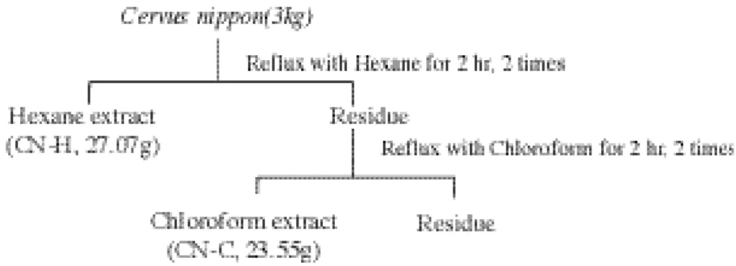
1. 녹용에서 조골세포분화 유도물질의 분리 녹용은 시중에서 판매되는 대만산 꽃사슴 (Cervus nippon)을 사용하였다. 녹용(3 ㎏)을 잘게 부순 뒤 녹용이 잠길 정도의 n- h e x a n e을 넣고 4 0℃에서 2시간 동안 증류장치로 가열하 여 추출하고 이를 여과하였다. 이 여과액을 감 압하여 용매를 제거하고 hexane 추출물( C N -H) 27.07g을 얻었다. 동일한 방법으로 잔사를 c h l o r o f o r m으로 추출하여 chloroform 추출물 (CN-C) 23.55g을 얻었다. 이렇게 얻은 추출 물들은 건조하여 보관하였으며 d i m e t h y l s u l-foxide 및 ethyl alcohol 혼합용액으로 1 ㎎/㎖ 의 농도로 용해하여 실험에 사용하였다( F i g u r e 1) 2. 조골세포의 분리 및 배양 녹용 추출물에 의한 조골세포분화능은 전구 조골세포주인 MC3T3 세포에서 측정하였다. MC3T3 세포는 10% 우태아혈청이 함유된α -MEM 배지에서 배양하여 사용하였다. 세포를 세포배양배지 5 ml당 8 x 10⁵ 되게 6 0 - m m d i s h에 분주하여 5 % CO2가 유지되는 3 7℃ 세 포배양기에서 배양하였다. 세포가 단층을 형성 한 후 대조군에서는 50 ㎍/㎖ a s c o r b i c acid(AA), 10 mM β- g l y c e r o p h o s p h a t e ( G P ) 가 함유된 α-MEM 배지(이하 조골세포 배양 배지라 함)에서 배양하였고, 실험군에서는 위 의 성분과 CN-C, CN-H가 각각 일정한 농도 (10 ㎍/㎖)로 함유된 배지에서 1 6일 간 배양한 후 아래와 같이 Von Kossa 염색을 실시하여 이들 녹용 추출물에 의하여 형성된 석회화 결 절을 관찰하였으며 R N A를 분리하여 N o r t h e r n b l o t으로 조골세포의 분화 표식인자인 a l k a l i n e phosphatase(ALP), bone sialoprotein(BSP) 및 o s t e o c a l c i n ( O C )의 발현 정도를 확인하였다.
3. mRNA 분리 및 Northern blot
녹용 추출물로 처리한 M C 3 T 3세포로부터 Trizol 용액(GIBCO BRL, USA)을 이용하여 R N A를 분리하였다. 분리한 RNA 20 ㎍를 f o r m a l d e h y d e를 함유한 1% agarose gel상에 서 전기영동한 후 Nylon plus membrane에 전 이시킨 후 ultraviolet radiation으로 R N A를 고 정시켰다. RNA가 부착된 막을 t u b e에 넣고 hybridization buffer(0.1㎎/㎖의 s a l m o n sperm DNA가 함유된 50% formamide/5x Denhardt's 용액/5xSSC/0.5% SDS 용액)를 가하고 4 2℃가 유지되는 hybrid minihy-bridization oven에서 3 0분간 p r e h y b r i d를 실 시하였다. 그 후 동위원소로 표지화된 A L P , BSP 및 OC 유전자 표식자를 첨가하여 4 2℃에 서 1 5시간 h y b r i d i z a t i o n을 실시하였다. RNA 표식자는 아래와 같은 c D N A를 이용하여 [3 2P]dCTP(3,000 Ci/mmol, Dupont NEN Research Products, Boston, MA, USA)로 표 지화 하였으며 random primed DNA labeling kit(GIBCO BRL, USA)를 이용하여 합성하였 다. 2.4kb의 rat ALP cDNA insert; 1.165 kbp 의 rat BSP cDNA insert; 520bp의 OC cDNA insert. Nylon membrane을 세척액으로 세척 하고 이를 - 7 0℃에서 Kodak X-OMAT film 에 감광시킨 후 현상하였다. 전기영동한 R N A 의 농도는 G A P D H를 기준으로 분석하였다.
4. Von Kossa staining
녹용 추출물로 처리한 M C 3 T 3세포는 10 % neutral formaldehyde용액으로 고정한 후 2 . 5 % silver nitrate용액에서 3 0분간 처리하여 세 척하였다. 세포를 다시 sodium carbonate f o r m a l d e h y d e용액에서 2 - 3분간 처리하고 세 척하여 육안으로 검은색의 석회화 결절의 형성
여부를 대조군과 비교하여 관찰하였다.
III. 결과
1. 녹용추출물이 석회화결절형성에 미치는 영향
MC3T3 세포를 녹용에서 추출한 CN-H 또
Figure 1. Schematic diagram of the extraction procedure from deer antler
Figure 2. Phase contrast micrographs of MC3T3 cells cultured with extracts of deer antler. MC3T3 cells were inoculated into 60-mm dishes and cultured in the absence(A) or presence of extracts of deer antler[CN-H(B), CN-C(C)] with ascorbic acid(50㎍/㎖) and β- g l y
c-Figure 3. Effects of deer antler extracts on bone nodule formation in MC3T3 cell. MC3T3 cells were inoculated into 60-mm dishes and cultured in the absence(A) or presence of extracts of deer antler[CN-H(B), CN-C(C)] with ascorbic acid(50㎍/㎖) and β- g l y c e r o p h o s p h a t e ( 1 0 mM) for 16 days. Mineralization nodule were stained by the Von Kossa technique and
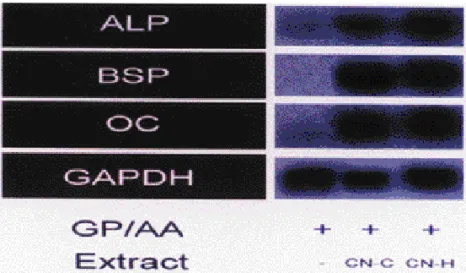
는 C N - C가 함유된 배지에서 배양하면서 위상 차 현미경상에서 석회화결절의 형성을 관찰하 였다. 배양 1 6일 후 CN-H 또는 C N - C가 함유 된 경우에서 모두 석회화결절의 형성을 관찰할 수 있었으나 시료가 첨가되지 않은 대조군에서 는 석회화결절이 나타나지 않았다(Figure 2). 또한 이를 Von Kossa 염색을 통하여 확인한 결과 위상차 현미경상에서 관찰한 결과와 동일 하게 녹용추출물로 처리하지 않은 대조군에서 는 석회화결절이 나타나지 않았으며, 녹용추출 물로 처리한 세포에서 검은색의 석회화 결절이 생성되는 것을 확인할 수 있었다(Figure 3). 2. 녹용추출물이 골기질 단백질 mRNA 발 현에 미치는 영향 각 녹용 추출물에 의한 골형성능을 재확인하 기 위하여 M C 3 T 3세포를 각 녹용추출물이 함 유된 배지에서 배양한 후 m R N A를 분리하여 골기질 단백질, 즉 ALP, BSP 및 OC mRNA 발 현정도를 대조군과 비교하여 분석하였다. 대조 군에서는 ALP 및 O C가 약하게 발현이 되었으 며 B S P는 발현되지 않았다. 각각의 녹용추출물 로 처리한 경우 ALP, BSP 및 OC mRNA 발현 이 대조군에 비하여 증가하였다(Figure 4).
IV. 고찰
인간에서의 골형성은 조골세포에 의하여 유 도되며, 조골세포의 분화는 B M P4 , 5 ) ,i n s u l i n e -like growth factor(IGF)1 7 ), melatonin1 8 ) 및 e s t r a d i o l1 9 )과 같은 다양한 성장인자들에 의하 여 조절된다. 사슴뿔의 성장시 조골세포의 분화 표식인자인 ALP 및 O C의 농도가 증가하며 9 , 1 2 , 1 3 ), 골형성유도 인자인 B M P가 발현되며2 0 ) m e l a t o n i n의 생성도 증가하는1 1 ) 것으로 보고 되었다. 또한 난소가 제거된 사슴에 e s t r a d i o l 투여시 뿔의 성장이 촉진되며1 4 )뿔에서 분리된 세포를 I G F로 처리한 경우 A L P의 발현 및 증 식능이 증가하는 것으로 보고되었다1 5 , 1 6 ). 이와 같이 사슴뿔의 성장시 골형성 조절인자의 생성 또는 조골세포분화 표식인자의 발현의 증가는 사슴뿌리의 성장 및 석회화가 사람뼈의 형성과 유사한 기전에 의하여 조절될 수 있음을 시사 하며 아울러 녹용에 골형성에 관여하는 성분이Figure 4. Effects of deer antler extracts on mRNA expression of ALP, BSP and OC in MC3T3 cells. MC3T3 cells were inoculated into 60-mm dishes and cultured in the absence or presence of extracts of deer antler(CN-H, CN-C) with ascorbic acid(50㎍/㎖) and β- g l y c e r o p h o s-phate(10mM) for 16 days. After RNA isolation, ALP, BSP and OC were quantified relative to that of the GAPDH gene by Northern hybridization.
들어 있을 가능성을 제시한다. 녹용추출물에 의한 조골세포 분화능을 관찰 하기 위하여 본 연구에서는 마우스 두개골세포 에서 분리한 MC3T3 세포를 사용하였다. M C 3 T 3는 ALP, BSP 및 O C를 발현하지 않는 전구조골세포주로서 ascorbic aicd(AA) 및 β-glycerophosphate(BP) 함유 배지에서 배 양할 경우 2 내지 3 주후 조골세포로 분화하여 시험관내에서 석회화결절을 형성한다1 8 , 2 1 ). 이 렇게 M C 3 T 3세포가 분화하여 형성한 석회화 결절은 골조직과 유사한 구조를 지니고 있음이 입증이 되어 시험관내에서 물질의 골형성능을 평가하기 위하여 M C 3 T 3가 많이 사용되어 왔 다2 2 ). 본 연구에서도 AA 및 G P가 함유된 기본 배지에서 MC3T3 세포를 배양하여 석회화 결 절의 형성능을 평가하여 녹용추출물에 의한 조 골세포분화능을 평가하였다. 골조직 성분 중 citric aicd의 대부분은 c a l c i u m과 결합된 형태 즉 calcium citrophosphate 상태로 존재하므로 calcium citrophosphate를 특이적으로 염색하 는 Von Kossa 염색법으로 석회화결절형성능 을 확인하였다2 3 ). MC3T3 세포는 GP 및 A A만 함유된 배지에서 1 6일간 배양한 경우에는 석회 화결절을 형성하지 않았으나 동일한 세포를 CN-C 또는 C N - H로 처리한 경우에는 많은 수의 석회화결절을 형성하였다. 조골세포의 분화는 골기질단백질의 발현과 밀접한 관련이 있다2 4 , 2 5 ). 골기질단백질 중 collagen, ALP 및 o s t e o p o n t i n은 분화초기에 발현되며 BSP 및 O C는 성숙된 조골세포에서 만 발현이 된다2 5 ). 이 중 A L P는 유기인산 에스 테르를 가수분해하여 골형성이 일어나는 부위 의 인산이온의 농도를 증가시키며2 6 ) B S P는 RGD sequence 및 glutamate rich sequence 를 함유하고 있어 골조직세포의 석회화기질 부 착에 관여할 뿐만 아니라 h y d r o x y a p a t i t e crystal 형성에도 중요한 역할을 하는 것으로 생각되고 있다2 7 - 3 3 ). 또한 O C는 아직 기능은 확실히 밝혀져 있지 않으나 골이나 상아질을 형성하는 세포에서만 발현되어 골형성에 있어 서의 역할의 중요성이 강조되어 왔다. 본 연구 에서 CN-C 및 C N - H는 석화화결절의 형성을 촉진할 뿐 만 아니라 실제로 석회화 과정에 중 요한 역할을 하는 이들 골기질단백질 즉 A L P , BSP 및 O C의 발현을 증가시키는 것으로 나타 났으며 이는 녹용의 CN-C 및 C N - H에 조골 세포의 분화를 유도하는 물질이 함유되어 있음 을 시사한다. 녹용에는 다당체, 단백질, 지질 및 무기물 등 다양한 성분이 함유되어 있으며 지질성분으로 는 prostagalndin 유사물질, phospholipid, glycolipid 및 g a n g l i o s i d e가 함유되어 있다 3 4 , 3 5 ). 이중 다당체는 궤양 예방효과 있으며3 6 ), 6 8개의 아미노산으로 이루어진 단백질은 항염 작용이 있는 것으로3 7 ), 또한 정제하지 않은 녹 용추출물은 노화방지효과가 있으며3 8 ) 녹용의 ethanol 추출물은 식균세포의 식균작용을 촉진 시키며3 9 ), 당뇨병시 증가되는 혈당을 감소시키 며4 0 ), 소수성물질은 Candida albicans의 균사 형성을 선택적으로 억제하는 것으로 보고되었 다4 1 ). 이와 같이 아직 그 구조가 확실히 밝혀지 지 않았으나 녹용은 다양한 효능을 나타내는 성분을 함유하고 있다. 본 연구에서는 처음으로 녹용의 조골세포의 분화능을 입증하였으며 chloroform 및 h e x a n e에 용해되는 성분이 조 골세포분화능을 나타내었다. chloroform 및 h e x a n e에는 단백성분 및 극성 다당체 보다는 비극성 성분이 주로 용해되므로 녹용추출물에 의한 조골세포분화능은 비극성 성분에 의하여 유도되었을 것으로 추정이 되나 성분에 대해서 는 정확한 분석이 필요할 것으로 사료된다. 치 주치료의 목표는 원인이 되는 치태의 기계적 제거와 더불어, 치주인대세포와 골조직세포의 증식 및 분화를 도모하여, 손상된 조직의 회복 을 이루는 것이다4 2 ). 특히 치조골 흡수에 의한 치아의 상실은 치주염의 주요한 증상이므로 치 주 치료시 치조골의 회복은 건강한 치아를 유 지하는데 중요한 요인이다. 따라서 앞으로 조골
세포분화촉진능이 있는 녹용성분이 밝혀지며 또한 밝혀진 물질의 생체내 골형성 촉진능이 입증이 된다면 이는 치주치료에 활용될 가능성 이 높을 것으로 사료된다.
V. 결론
사슴의 뿔은 계속 성장하며 일부약전에서 녹 용이 이와 뼈를 튼튼하게 하는 것으로 기록되 어 있는 것으로 보아 녹용에는 골의 성장 및 석 회화를 조절하는 성분이 함유되어 있을 것으로 추정된다. 따라서 치조골 파괴가 야기되는 치주 치료에 활용할 수 있는 새로운 골형성 유도물 질의 개발을 최종 목적으로 본 연구에서는 일 차적으로 녹용의 조골세포분화 유도능을 평가 하였다. 녹용으로부터 chloroform 추출물 (CN-C) 및 hexane 추출물( C N - H )을 분리한 후 이들 추출물이 전구조골세포주인 M C 3 T 3 세포의 석회화결절형성 및 골기질단백물질 (ALP, BSP 및 O C )의 발현에 미치는 영향을 평가하였으며, MC3T3세포의 석회화결절 형성 능은 칼슘( C a2 +)이 침착된 부위만 특이적으로 염색하는 Von Kossa염색법으로, 골기질 단백 물질의 발현은 Northern blot로 확인하였다. 녹 용추출물로 처리하지 않은 M C 3 T 3세포와 비 교한 경우 CN-C 및 C N - H는 각각 M C 3 T 3 세포의 석회화결절 형성능을 증가시켰으며 또 한 ALP, BSP 및 OC mRNA의 발현을 증가시 켰다. 이와 같은 결과는 녹용의 CNH 및 C N -C가 조골세포에 의한 골형성능을 증가시킬 수 있음을 시사한다.VII. 참고문헌
1 . Page RC, Offenbacher S, Schroeder HE, Seymour GJ, Kornman KS: Advances in the pathogenesis of periodontitis: Summary of developments, clinical implications and future directions.Periodontol 2000 14: 216-248, 1997.
2 . Cochran DL, Wozney JM: Biological mediators for periodontal regeneration. Periodontol 2000 19: 40-58, 1999.
3 . Ripamonti U, Reddi AH: Tissue engineering, morphogenesis, and regen-eration of the periodontal tissues by bone morphogenetic proteins. Crit. Rev.
Oral Biol. Med. 8: 154-163. 1997.
4 . Barboza E, Caula A, Machado F: Potential of recombinant human bone morphogenetic protein-2 in bone regeneration. Implant Dent. 8: 360-367, 1 9 9 9 .
5 . Asahina I, Sampath TK, Nishimura I, Hauschka PV: Human osteogenic pro-tein-1 induces both chondroblastic and osteoblastic differentiation of osteo-progenitor cells derived from newborn rat calvaria. J. Cell. Biol. 123: 921-933, 1993.
6 . Giannobile WV, Ryan S, Shih MS, Su DL, Kaplan PL, Chan TC: Recombinant human osteogenic protein-1(OP-1) stimulates periodontal wound healing in class III furcation defects. J .
P e r i o d o n t o l . 69: 129-137, 1998.
7 . Kinoshita A, Oda S, Takahashi K, Yokota S, Ishikawa I: Periodontal regeneration by application of recombi-nant human bone morphogenetic pro-tein-2 to horizontal circumferential defects created by experimental peri-odontitis in beagle dogs. J. Periodontol. 68: 103-109, 1997.
8 . 권혁세: 만병을 다스리는 민간약술
88, 제1판, 하나로, 서울, pp.172-187, 1 9 9 7
CM: Circulating levels of 1,25 dihydrox-yvitamin D, alkaline phosphatase, hydroxyproline, and osteocalcin associ-ated with antler growth in white-tailed deer. Acta. Endocrinol. 118: 407-414, 1988.
1 0 . Bubenik GA, Sempere AJ, Hamr J: Developing antlers, a model for endocrine regulation of bone growth. Concentration gradient of T3, T4, and alkaline phosphatase in the antlers, jugular, and the sapherous veins. C a l c i f .
Tissue Int. 41: 38-43, 1987.
1 1 . Bubenik GA, Smith PS: Circardian and circannual rhythms of melatonin in plasma of male white-tailed deer and the effect of oral administration of mela-tonin. J. Exp. Zool. 241: 81-89, 1987. 1 2 . Eiben B, Scharla S, Fischer K,
Schmidt-Gayk H: Seasonal variations of serum 1,25-dihydroxyvitamin D3 a n d alkaline phosphatase in relation to the antler formation in the fallow deer(D a m a
d a m a I.) Acta. Endocrinol. 107:
141-144, 1984.
1 3 . Barling PM, Gupta DK, Lim CE: Involvement of phosphodiesterase I in mineralization: histochemical studies using antler from red deer(Cervus ela
-p h u s) as a model. Calcif. tissue Int. 6 5 :
384-389, 1999.
1 4 . Lincoln GA, Tyler NJ: Role of oestradiol in the regulation of the sea-sonal antler cycle in female reindeer,
Rangifer tarandus. J. Reprod. Fertil. 1 1 5 :
167-174, 1999.
1 5 . Price JS, Oyajobi BO, Oreffo RO, Russell RG: Cells cultured from the growing tip of red deer antler express
alkaline phosphatase and proliferate in response to insulin-like growth factor-I. J. Endocrinol. 143: R9-16, 1994. 1 6 . Li C, Littlejohn RP, Suttie JM:
Effects of insulin-like growth factor 1 and testosterone on the proliferation of antlergenic cells in vitro. J. Exp. Zool. 284: 82-90, 1999.
1 7 . Ohlsson C, Vidal O: Effects of growth hormone and insulin-like growth factors on human osteoblasts. Eur. J.
Clin. Invest. 28: 184-186, 1998.
1 8 . Roth JA, Kim BG, Lin WL, Cho MI: Melatonin promotes osteoblast differen-tiation and bone formation. J. Biol. Chem. 274: 22041-22047, 1999.
1 9 . Ernst M, Heath JK, Schmid C, Froesch RE, Rodan GA: Evidence for a direct effect of estrogen on bone cells in vitro. J. Steroid. Biochem. 34: 279-284, 1989.
2 0 . Feng JQ, Chen D, Esparza J, Harris MA, Mundy GR Harris SE: Deer antler tissue contains two types of bone mor-phogenetic protein 4 mRNA trranscripts.
Biochim. Biophys. Acta 1263: 163-168,
1 9 9 5 .
2 1 . Quarles LD, Yohay DA, Lever LW, Caton R, Wenstrup RJ: Distinct prolifer-ative and differentiated stages of murine MC3T3-E1 cells in culture: an in vitro model of osteoblast development. J .
Bone Min. Res. 7: 683-692, 1992.
2 2 . Sudo HA, Kodama HA, Amagai Y, Yamamoto S, Kasai S: In vitro d i f f e r e n-tiation and calcification in a new clonal osteogenic cell line derived from new-born mouse calvaria. J. Cell. Biol. 96: 191-198, 1983.
2 3 . Bills CE, Eisenberg H, Pallante SL: Complexes of organic acids with calcium phosphate: The von kossa stain as a clue to the composition of bone mineral.
Hopkins Med. J. 128: 194-207, 1971
2 4 . Lian JB, Stein GS: Concepts of osteoblast growth and differentiation: Basis for modulation of bone cell devel-opment and tissue formation. Crit. Rev.
Oral Biol. Med. 3 : 269-305, 1992.
2 5 . Yao KL, Todescan R, Sodek J: Temporal changes in matrix protein synthesis and mRNA expression during mineralized tissue formation by adult rat
bone marrow cells in culture. J. Bone
Min. Res. 9: 231-240, 1994.
2 6 . Beertsen W, Theo VDB: Calcification of dental collagen by cul-tured rabbit periosteum: The role of alkalaine phosphatase. M a t r i x 9: 159-171, 1989.
2 7 . Fisher LW, McBride OW, Termine JD, Young MF: Human bone sialoprotein. Deduced protein sequence and chromo-somal localization. J. Biol. Chem. 2 6 5 : 2347-2351, 1990.
2 8 . Oldberg A, Franzen A, Heinegard D: The primary structure of a
cell-binding bone sialoprotein. J. Biol. Chem. 263: 19430-19432, 1988.
2 9 . Shapiro HS, Chen J, Wrana JL, Zhang Q, Blum M, Sodek J: Characterization of porcine bone sialoprotein: primary structure and cellular expression. M a t r i x 13: 431-440, 1 9 9 3 .
3 0 . Hunter GK, Goldberg HA: Nucleation of hydroxyapatite by bone sialoprotein. P r o c .
Natl. Acad. Sci. USA 90: 8562-8565, 1993.
3 1 . Hunter GK, Goldberg HA: Modulation of crystal formation by bone phosphoprotein : role of glutamic acid-rich sequences in the nucleation of hydroxyapatitie by bone sialo-protein. Biochem. J. 302: 175-179, 1994.
3 2 . Sodek J, Chen J, Kasugai S, Nagata T, Zhang Q, Mckee MD, Nanci A: Elucidation the functions of bone sialoprotein and osteopontin in bone formation, pp.297-306, In: H. Slavkin and P. Price(ed), Chemistry and biology of mineralized tissues. B.V Elsevier Science Publishers, 1992.
3 3 . Butler WT: Sialoproteins of bone and dentin. J. Biol. Buccale. 19: 83-89, 1991. 3 4 . Ivankina NF, Isay SV, Busarova NG, Mischenko TY: Prostaglandin-like activity,
fatty acid and Phospholipid composition of Sika deer(Cervus nippon) antlers at different growth stages. Comp. Biochem. Physiol. 106: 159-162, 1993.
3 5 . Jhon GJ, Park SY, Han SY, Lee S, Kim Y, Chang YS: Studies of the chemical struc-ture of gangliosides in Deer Antler, Cervus nippon. C h e m . Pharm. Bull. 47: 123-127, 1 9 9 9 .
3 6 . Wang BX, Liu AJ, Cheng XJ, Wang QG, Wei GR, Cui JC : Anti-ulcer action of the polysaccharides isolated from pilose antler. Yao Hsueh Hsueh Pao 20: 321-325, 1985. 3 7 . Zhang ZQ, Wang Y, Zhang H, Zhang W, Zhang Y, Wang BX: Anti-inflammatory
effects of pilose antler peptide. C h u n g Kuo Yao Li Hsueh Pao 15: 282-284, 1994.
3 8 . Son NW, Shin MK, Lee HI: The effects of deer antler on the neuro response of starved mice. Korean J. Oriental Medicine 7: 174-183, 1986.
3 9 . Suh JS, Eun JS, So JN, Seo JT, Jhon GJ: Phagocytic activity of ethyl alcohol fraction of deer antler in murine periodontal macrophage. Biol. Pharm. Bull. 22: 932-935, 1999. 4 0 . Cho HJ, Jhon GJ: Changes of gangliosides metabolism in streptozotocin-induced
diabetic rats and effect of deer antler J.Appl.Pharm. 2: 223-228, 1994.
4 1 . Park HS, Jhon GJ, Choi W: Deer antler extract selectively suppresses hyphal growth in dimorphic fungus, Candida albicans. J. Microbiol. Biothechnol. 8: 291-294, 1998.
4 2 . Takata T: Oral wound healing concepts in periodontology. Curr. Opin. Periodontol. 119-127, 1994.
A b s t r a c t
-The Effects of Deer( C e r v u s
n i p p o n ) Antler Extracts on
Differentiation of MC3T3
Cells
Yun-Jung Yoo1, Hyun Jung Lee1, So hyung L i m2, Jung-Hwa Kang1,
Yin Ji Li2, Seung-Ho Ohk1, Bong-Kyu C h o i1, and Gil Ja Jhon3
1Dept. of Oral Biology, College of Dentistry, Yonsei University
2Dept. of Molecular Life Science-Chemical B i o l o g y
3Dept. of Chemistry, Ewha Womans U n i v e r s i t y
Deer antler has been widely prescribed in Chinese and Korean pharmacology. Although there have been several reports concerning the effects of deer antler, such as anti-aging action, anti-inflammatory activity, antifungal action and regulatory activity of the level of glucose, the effect on bone has not determined yet. The purpose of this study was to examine the effect of deer antler on osteoblast differentiation. Hexane extract(CN-H) and chloroform extract(CN-C) were acquired from deer a n t l e r (Cervus nippon) and MC3T3-E1 pre-osteoblasts were cultured in the pres-ence or abspres-ence of each extract. Osteoblast differentiation was estimated with the for-mation of mineralized nodules and the
mRNA expression of alkaline phos-phatase(ALP), osteocalcin(OC) and bone sialoprotein(BSP) which are markers of osteoblast differentiation. Non-treated group did not show mineralized nodule. CN-C or CN-H-treated group showed minerlaized nodules in 16 days. In northern blot analysis, CN-C or CN-H-treated group showed the elevated expression of ALP, BSP and OC in 16 days. These results suggest the possibility to develop deer antler as a bone regenerative agent in peri-odontal therapy by showing the stimulating activity of deer antler on differentiation of o s t e o b l a s t .
Key words: Deer antler extract, MC3T3, Osteoblast differentiation, bone regenera-tive agent